

Boron doped diamond thin films for the electrochemical detection of SARS-CoV-2 S1 protein
- PMID: 34334952
- PMCID: PMC8316675
- DOI: 10.1016/j.diamond.2021.108542
Boron doped diamond thin films for the electrochemical detection of SARS-CoV-2 S1 protein
Abstract
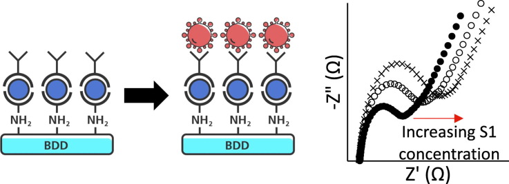
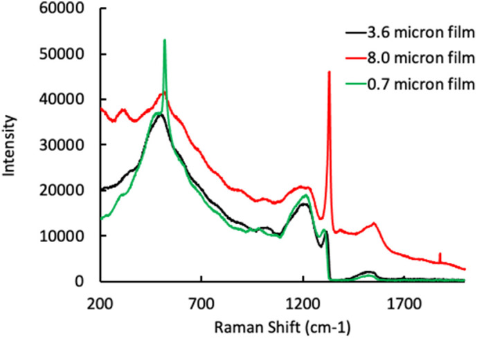
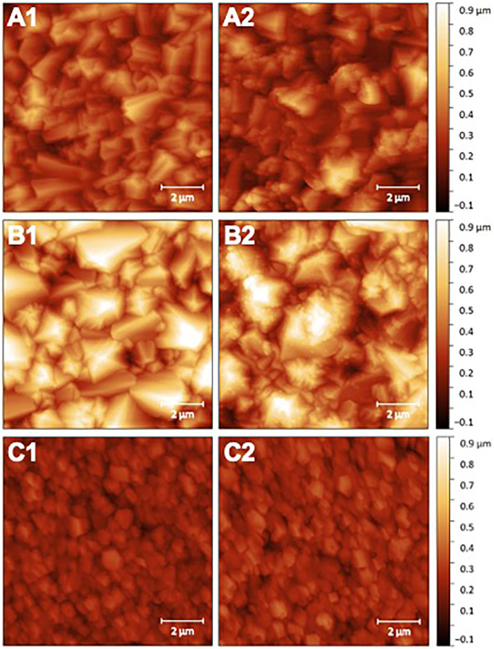
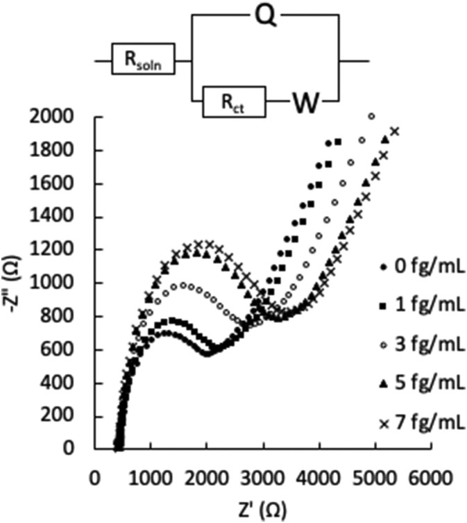
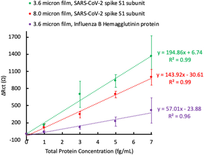
Amidst a global pandemic, a precise and widely accessible rapid detection method is needed for accurate diagnosis and contact tracing. The lack of this technology was exposed through the outbreak of SARS-CoV-2 beginning in 2019. This study sets the foundation for the development of a boron doped diamond (BDD)-based impedimetric sensor. While specifically developed for use in the detection of SARS-CoV-2, this technology uses principles that could be adapted to detect other viruses in the future. Boron doped polycrystalline diamond electrodes were functionalized with a biotin-streptavidin linker complex and biotinylated anti-SARS-CoV-2 S1 antibodies. Electrodes were then incubated with the S1 subunit of the SARS-CoV-2 spike surface protein, and an electrical response was recorded using the changes to the electrode's charge transfer resistance (Rct), measured through electrochemical impedance spectroscopy (EIS). Detectable changes in the Rct were observed after 5-min incubation periods with S1 subunit concentrations as low as 1 fg/mL. Incubation with Influenza-B Hemagglutinin protein resulted in minimal change to the Rct, indicating specificity of the BDD electrode for the S1 subunit of SARS-CoV-2. Detection of the S1 subunit in a complex (cell culture) medium was also demonstrated by modifying the EIS protocol to minimize the effects of sample matrix binding. BDD films of varying surface morphologies were investigated, and material characterization was used to give insight into the microstructure-performance relationship of the BDD sensing surface.
Keywords: Biosensor; Boron doped diamond; Impedimetric sensor; SARS-CoV-2.
© 2021 Elsevier B.V. All rights reserved.
Conflict of interest statement
The authors declare that they have no known competing financial interests or personal relationships that could have appeared to influence the work reported in this paper.
Figures
Similar articles
-
Impedimetric Detection Based on Label-Free Immunoassay Developed for Targeting Spike S1 Protein of SARS-CoV-2.Diagnostics (Basel). 2022 Aug 17;12(8):1992. doi: 10.3390/diagnostics12081992. Diagnostics (Basel). 2022. PMID: 36010342 Free PMC article.
-
Polycrystalline boron-doped diamond-based electrochemical biosensor for simultaneous detection of dopamine and melatonin.Anal Chim Acta. 2020 Oct 23;1135:73-82. doi: 10.1016/j.aca.2020.08.042. Epub 2020 Aug 27. Anal Chim Acta. 2020. PMID: 33070861
-
Thickness Effects on Boron Doping and Electrochemical Properties of Boron-Doped Diamond Film.Molecules. 2023 Mar 21;28(6):2829. doi: 10.3390/molecules28062829. Molecules. 2023. PMID: 36985800 Free PMC article.
-
Boron-doped diamond electrode: synthesis, characterization, functionalization and analytical applications.Analyst. 2009 Oct;134(10):1965-79. doi: 10.1039/b910206j. Epub 2009 Aug 7. Analyst. 2009. PMID: 19768202 Review.
-
Boron-Doped Diamond Electrodes for Toxins Sensing in Environmental Samples-A Review.Sensors (Basel). 2025 Apr 7;25(7):2339. doi: 10.3390/s25072339. Sensors (Basel). 2025. PMID: 40218850 Free PMC article. Review.
Cited by
-
Hybrid Impedimetric Biosensors for Express Protein Markers Detection.Micromachines (Basel). 2024 Jan 25;15(2):181. doi: 10.3390/mi15020181. Micromachines (Basel). 2024. PMID: 38398911 Free PMC article. Review.
-
Peptide-based simple detection of SARS-CoV-2 with electrochemical readout.Anal Chim Acta. 2022 May 1;1205:339739. doi: 10.1016/j.aca.2022.339739. Epub 2022 Mar 21. Anal Chim Acta. 2022. PMID: 35414399 Free PMC article.
-
Electrochemical and Bioelectrochemical Sensing Platforms for Diagnostics of COVID-19.Biosensors (Basel). 2023 Mar 3;13(3):336. doi: 10.3390/bios13030336. Biosensors (Basel). 2023. PMID: 36979548 Free PMC article. Review.
-
Electrochemical Biosensor for the Determination of Specific Antibodies against SARS-CoV-2 Spike Protein.Int J Mol Sci. 2022 Dec 31;24(1):718. doi: 10.3390/ijms24010718. Int J Mol Sci. 2022. PMID: 36614164 Free PMC article.
-
Impedimetric Detection Based on Label-Free Immunoassay Developed for Targeting Spike S1 Protein of SARS-CoV-2.Diagnostics (Basel). 2022 Aug 17;12(8):1992. doi: 10.3390/diagnostics12081992. Diagnostics (Basel). 2022. PMID: 36010342 Free PMC article.
References
-
- WHO . “Novel Coronavirus (2019-nCoV) Situation Report - 1”, WHO Bull., no. JANUARY. 2020. pp. 1–7.https://www.who.int/emergencies/diseases/novel-coronavirus-2019/situatio...
-
- Bhat T.A., Kalathil S.G., Bogner P.N., Blount B.C., Goniewicz M.L., Thanavala Y.M., Zou L., Ruan F., Huang M., Liang L., Huang H., Hong Z., Yu J., Kang M., Song Y., Xia J., Guo Q., Song T., He J., Yen H.-L., Peiris M., Wu J. SARS-CoV-2 viral load in upper respiratory specimens of infected patients. N. Engl. J. Med. 2020;382(12):1177–1179. doi: 10.1056/NEJMc2001737. - DOI - PMC - PubMed
-
- Wang D., Hu B., Hu C., Zhu F., Liu X., Zhang J., Wang B., Xiang H., Cheng Z., Xiong Y., Zhao Y., Li Y., Wang X., Peng Z. Clinical characteristics of 138 hospitalized patients with 2019 novel coronavirus-infected pneumonia in Wuhan, China. JAMA - J. Am. Med. Assoc. 2020;323(11):1061–1069. doi: 10.1001/jama.2020.1585. - DOI - PMC - PubMed
LinkOut - more resources
Full Text Sources
Miscellaneous