. 2021 Jul 24:16:5039-5052.
doi: 10.2147/IJN.S316388. eCollection 2021.

Therapeutic Effect of IL-4 Receptor-Targeting Pro-Apoptotic Peptide (AP1-ELP-KLAK) in Glioblastoma Tumor Model

Affiliations

Therapeutic Effect of IL-4 Receptor-Targeting Pro-Apoptotic Peptide (AP1-ELP-KLAK) in Glioblastoma Tumor Model

Vijaya Sarangthem et al. Int J Nanomedicine. .

Abstract

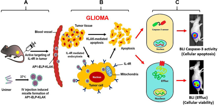
Background: Thermal-responsive self-assembled elastin-like polypeptide (ELP)-based nanoparticles are an emerging platform for controlled delivery of therapeutic peptides, proteins and small molecular drugs. The antitumor effect of bioengineered chimeric polypeptide AP1-ELP-KLAK containing an interleukin-4 receptor (IL-4R) targeting peptide and pro-apoptotic peptide (KLAKLAK) was evaluated in glioblastoma (GBM) in vitro and in vivo.

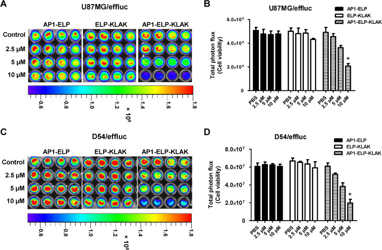
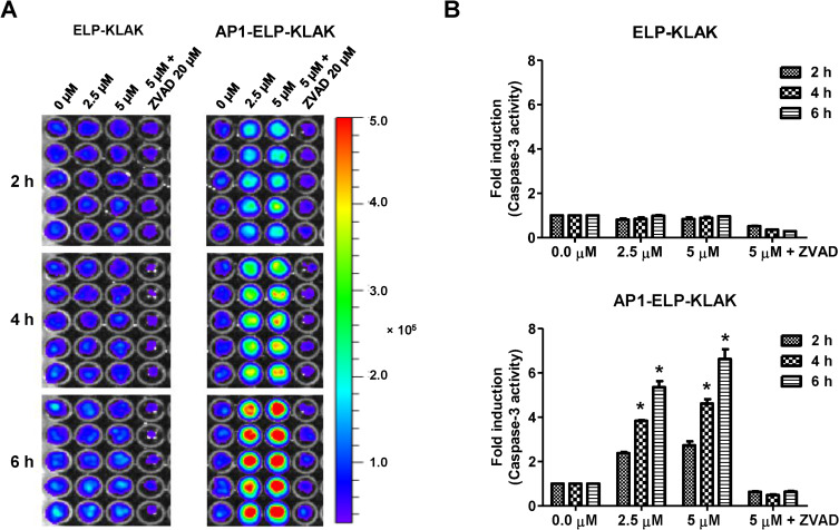
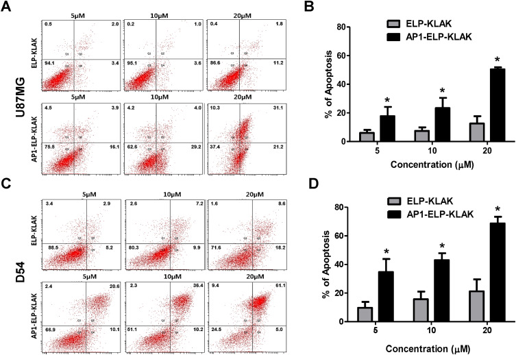
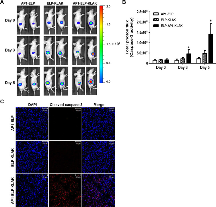
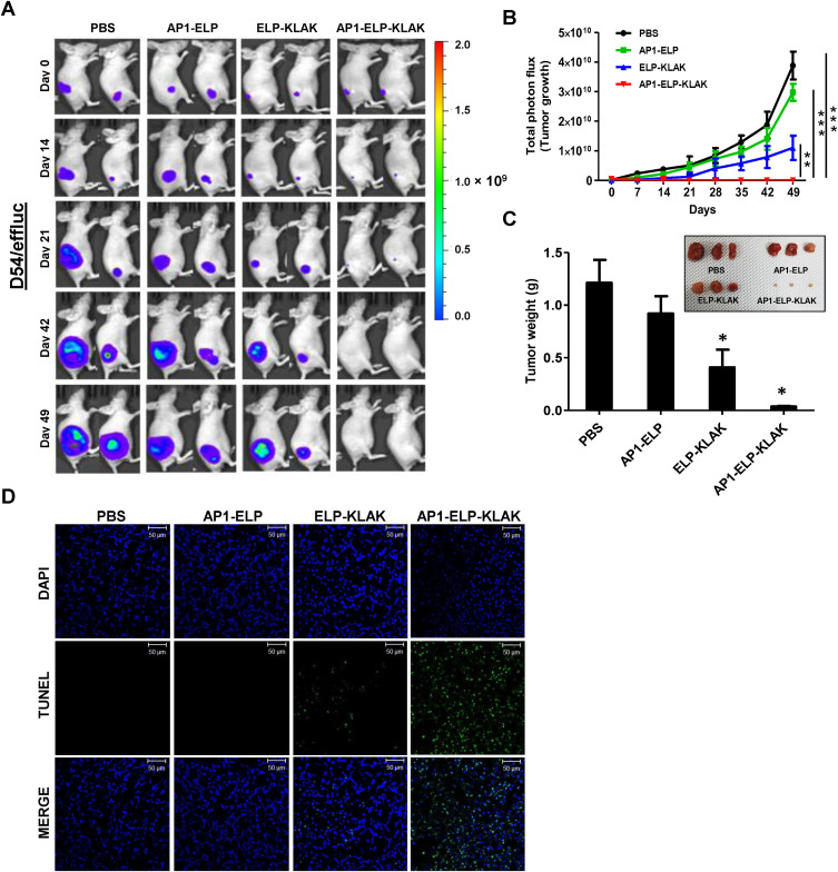
Methods and results: Herein, the therapeutic effect of AP1-ELP-KLAK was tested in advanced, and less curable glioblastoma cells with higher expression of IL-4R. Glioblastoma cell lines stably expressing different reporter systems i.e., caspase-3 sensor (surrogate marker for cellular apoptosis) or effluc/enhanced firefly luciferase (cellular viability) were established to measure cell death non-invasively. Bioluminescence imaging (BLI) of D54/effluc and U97MG/effluc treated with AP1-ELP-KLAK exhibited higher cell death up to 2~3-fold than the control. Treatment with AP1-ELP-KLAK resulted in time-dependent increase of caspase-3 sensor BLI activity in D54/C cells and D54/C tumor-bearing mice. Intravenous injection of AP1-ELP-KLAK dramatically reduced tumor growth by inducing cellular apoptosis in D54/effluc tumor-bearing mice. Further, the immuno-histological examination of the excised tumor tissue confirmed the presence of apoptotic cells as well as caspase-3 activation.

Conclusion: Collectively, AP1-ELP-KLAK effectively induced cellular apoptosis of glioblastoma cells and non-invasive imaging provides a window for real-time monitoring of anti-tumor effect with the provision of improving therapeutic efficacy in a glioblastoma mice model.

Keywords: ELP; IL-4 receptor; apoptosis; caspase-sensor; glioblastoma; tumor targeting.


Conflict of interest statement

All authors declare no conflicts of interest in this work.

Figures

Figure 1
Figure 1
Schematic diagram for non-invasive imaging of AP1-ELP-KLAK induced apoptosis in glioblastoma mouse model. (A) Systemic administration of AP1-ELP-KLAK in glioblastoma bearing mice. (B) Uptake of AP1-ELP-KLAK by glioma via IL-4R receptor mediated endocytosis and EPR effect, causing massive apoptosis of tumor tissue. (C) Real-time monitoring of apoptosis by caspase-3 sensor and cellular viability by effluc reporter system stably expressed in glioblastoma cells.
Figure 2
Figure 2
Characterization of ELP-KLAK and AP1-ELP-KLAK. (A) The structure of ELP-KLAK and AP1-ELP-KLAK along with coding sequence of the polypeptides. Both the polypeptides underwent phase transition and retained as nanoparticle-like structures at 37°C. Cryo-TEM of self-assembled ELP-KLAK and AP1-ELP-KLAK. Scale bar, 100 nm. (B) ELP-KLAK and (C) AP1-ELP-KLAK, SDS-PAGE (10%) of polypeptides purified using ITC. After 3rd ITC single band for each polypeptide were observed indicating high level of purity. Molecular weight (in KDa) of the polypeptides were indicated on the right.
Figure 3
Figure 3
Assessment of IL-4R expression and cellular uptake assay. (A) Representative histogram of IL-4R expression analyzed through flow cytometry after staining with anti-IL-4Rα antibody. (B) Percentage of IL-4R expression in different cancer cells was analyzed by comparison with respective IgG isotype control (n = 3). *P<0.05, (Student’s t-test). Cellular uptake and internalization of fluorescently labeled polypeptides by (C) U87MG and (D) D54 at 37°C. Cell membrane stained with WGA Alexa 594 and nuclei stained with Hoechst. Scale bar, 20 μm.
Figure 4
Figure 4
AP1-ELP-KLAK induced cytotoxicity in glioblastoma cells. (A and C) Luciferase activity of U87MG/effluc cells and (B and D) D54/effluc cells were determined after adding 3 µL D-luciferin (30 mg mL−1) in each well, and then the bioluminescence signals were measured using an IVIS Lumina III in vivo imaging system. Data are represented as mean ± s.d. (n = 5). *P<0.05 (one-way ANOVA).
Figure 5
Figure 5
Analysis of caspase activation mediated apoptosis by AP1-ELP-KLAK in D54/C cells. (A) Bioluminescence imaging (BLI) activity of the caspase-3 sensor was measured following the treatment of different concentration apoptotic peptides with or without Z-VAD (caspase inhibitor) to D54/C cells. BLIs were also obtained using an IVIS imaging system post treatment. (B) BLI activity was plotted as the fold induction caspase activation over values obtained from cells treated with PBS only. Experiments were performed at least in triplicate, and mean values ± s.d. were plotted. The intergroup variation was measured by one-way ANOVA, followed by Bonferroni post hoc test (*P<0.05).
Figure 6
Figure 6
AP1-ELP-KLAK induced apoptosis in glioblastoma cells. (A and C) U87MG/effluc cells, (B and D) D54/effluc cells. The percentage of apoptotic cells stained with annexin V conjugated to green-fluorescent FITC dye and dead cells with propidium iodide (PI) were measured by flow cytometry. Representative data of three independent experiments, mean ± s.d. (n = 5). Percentage of apoptosis significantly increased in AP1-ELP-KLAK treated cells at all given concentrations. *P<0.05 (one-way ANOVA).
Figure 7
Figure 7
Non-invasive imaging of caspase activation in vivo. (A) BLI of caspase-3 activity after the IV injection of either ELP-KLAK and AP1-ELP-KLAK in D54/C tumor–bearing mice. (B) Quantification of caspase-3 sensor BLI activity. Experiments were conducted in triplicate, and the mean values ± s.d. were plotted. Significant increase in caspase-3 activity was observed in AP1-ELP-KLAK treated mice at all time points, *P<0.01 (two-way ANOVA). (C) At day 6, mice were sacrificed and ex vivo staining of excised tumor tissues were done with anti-cleaved-Caspase-3 (red), followed by counter staining with DAPI for nuclei (blue). Scale bar, 50 μm.
Figure 8
Figure 8
Anti-tumor effect of AP1-ELP-KLAK in glioma bearing mice. (A and B) D54/effluc bearing mice were intravenously (IV) injected with 150 mg kg−1 of respective polypeptides and control daily for 8 days. BLI (surrogate marker of tumor growth) were obtained after injection of d-luciferin at indicated time (n = 10). A significant reduction of BLI of effluc was obtained in AP1-ELP-KLAK treated mice, revealing a dramatic inhibition of glioma tumor growth, while no such BLI reduction was seen in ELP-KLAK treated mice, ***P<0.001, significant difference for AP1-ELP-KLAK compared with PBS, AP1-ELP; **P<0.01, significant difference for AP1-ELP-KLAK compared with ELP-KLAK (two-way ANOVA). (C) The excised tumor weights obtained at the end of therapy were significantly reduced in AP1-ELP-KLAK treated mice but not in control and ELP-KLAK groups. Data are represented as mean ± s.d. (n = 10). *P<0.01 (Student’s t-test). (D) Excised tumor tissues showing TUNEL positive apoptotic cells (green) and (blue), as nuclei; Scale bar, 50 μm.

Similar articles

Cited by

References

    1. Jain RK. Delivery of molecular and cellular medicine to solid tumors. Adv Drug Deliv Rev. 2001;46:149–168. doi:10.1016/s0169-409x(00)00131-9 - DOI - PubMed
    1. Moses MA, Brem H, Langer R. Advancing the field of drug delivery: taking aim at cancer. Cancer Cell. 2003;4(5):337–341. doi:10.1016/s1535-6108(03)00276-9 - DOI - PubMed
    1. Allen TM, Cullis PR. Drug delivery systems: entering the mainstream. Science. 2004;303(5665):1818–1822. doi:10.1126/science.1095833 - DOI - PubMed
    1. Frandsen JL, Ghandehari H. Recombinant protein-based polymers for advanced drug delivery. Chem Soc Rev. 2012;41(7):2696–2706. doi:10.1039/c2cs15303c - DOI - PubMed
    1. Meyer DE, Chilkoti A. Genetically encoded synthesis of protein-based polymers with precisely specified molecular weight and sequence by recursive directional ligation: examples from the elastin-like polypeptide system. Biomacromolecules. 2002;3(2):357–367. doi:10.1021/bm015630n - DOI - PubMed