. 2021 Mar 23:14:17562864211004318.
doi: 10.1177/17562864211004318. eCollection 2021.

Diagnosing small fiber neuropathy in clinical practice: a deep phenotyping study

Affiliations

Diagnosing small fiber neuropathy in clinical practice: a deep phenotyping study

Nadine Egenolf et al. Ther Adv Neurol Disord. .

Erratum in

Abstract

Background and aims: Small fiber neuropathy (SFN) is increasingly suspected in patients with pain of uncertain origin, and making the diagnosis remains a challenge lacking a diagnostic gold standard.

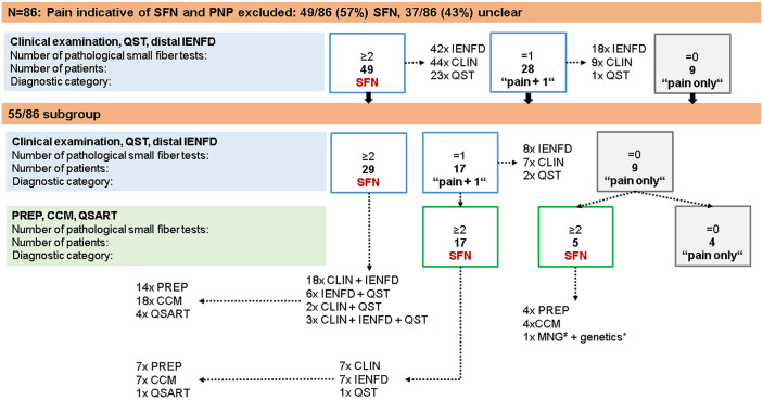
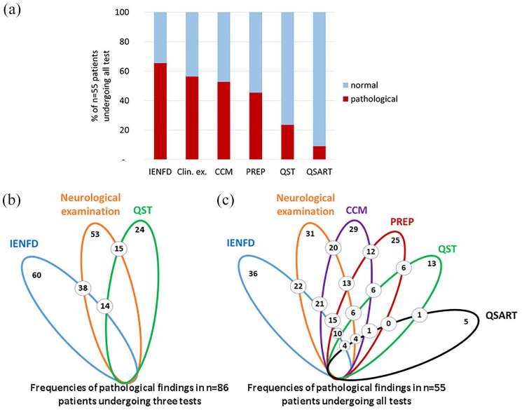
Methods: In this case-control study, we prospectively recruited 86 patients with a medical history and clinical phenotype suggestive of SFN. Patients underwent neurological examination, quantitative sensory testing (QST), and distal and proximal skin punch biopsy, and were tested for pain-associated gene loci. Fifty-five of these patients additionally underwent pain-related evoked potentials (PREP), corneal confocal microscopy (CCM), and a quantitative sudomotor axon reflex test (QSART).

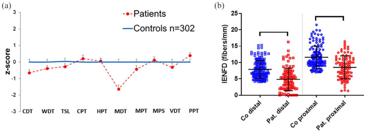
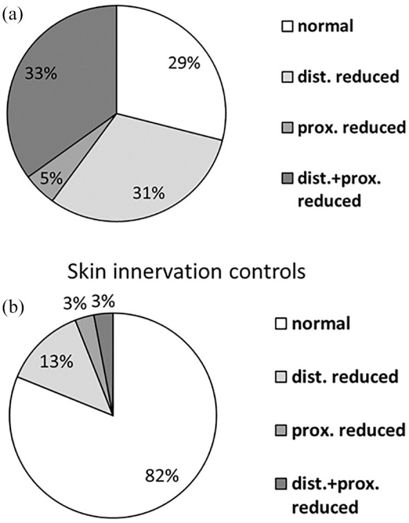
Results: Abnormal distal intraepidermal nerve fiber density (IENFD) (60/86, 70%) and neurological examination (53/86, 62%) most frequently reflected small fiber disease. Adding CCM and/or PREP further increased the number of patients with small fiber impairment to 47/55 (85%). Genetic testing revealed potentially pathogenic gene variants in 14/86 (16%) index patients. QST, QSART, and proximal IENFD were of lower impact.

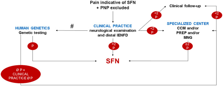
Conclusion: We propose to diagnose SFN primarily based on the results of neurological examination and distal IENFD, with more detailed phenotyping in specialized centers.

Keywords: algorithm; diagnosis; neurological examination; skin punch biopsy; small fiber neuropathy.


Conflict of interest statement

Conflict of interest statement: N.S., C.M.z.A., L.K., F.G., K.E., B.N., T.M., A.K., D.K., I.K. report no conflicts of interest. C.S., R.A.M., N.Ü. have taken part in clinical studies associated with small fiber neuropathy (Biogen, Vertex).

Figures

Figure 1.
Figure 1.
Pain distribution in study cohort. The drawings illustrate the frequency of pain reported in different body areas in the study cohort.
Figure 2.
Figure 2.
QST and IENFD results in patients with suspected SFN compared with healthy controls. (a) The z-score shows elevated MDT in patients with suspected SFN compared with healthy controls (p < 0.01). Data of 86 patients are compared with those of 302 healthy controls all investigated at the dorsal right foot. (b) The scatter plots illustrate reduced distal and proximal IENFD (p < 0.001) in patients compared with healthy controls. CDT, cold detection threshold; Co, controls; CPT, cold pain threshold; HPT, heat pain threshold; IENFD, intraepidermal nerve fiber density; MDT, mechanical detection threshold; MPS, mechanical pain sensitivity; MPT, mechanical pain threshold; Pat., patients; PPT, pressure pain threshold; QST, quantitative sensory testing; SFN, small fiber neuropathy, TSL, thermal sensory limen; VDT, vibration detection threshold; WDT, warm detection threshold.
Figure 3.
Figure 3.
Synopsis of study cohort and results of small fiber test results. Eighty-six patients were included and, upon exclusion of PNP, underwent neurological examination, skin punch biopsy, and QST. Depending on the number of pathological test results, patients were stratified in the subgroups of “SFN”, “pain+1” (i.e. only one small fiber test was pathological), and “pain only” (i.e. small fiber tests were normal). Fifty-five patients agreed to additionally undergo CCM, PREP, and QSART. #One patient agreed to MNG. *Genetic testing including 26 pain-associated genes (see Methods) was performed in all 86 study participants. CCM, corneal confocal microscopy; CLIN, clinical examination; IENFD, intraepidermal nerve fiber density; MNG, microneurography; PNP, polyneuropathy; PREP, pain-related evoked potentials; QSART, quantitative sudomotor axon reflex test; QST, quantitative sensory testing; SFN, small fiber neuropathy.
Figure 4.
Figure 4.
Skin innervation patterns in healthy controls and patients with suspected SFN. (a) Skin innervation was reduced at the distal leg, proximal thigh, or both sites in 71% of patients with suspected SFN. (b) Skin innervation was reduced at the distal leg, proximal thigh, or both sites in 18% of healthy controls. Dist., distal; Prox., proximal; SFN, small fiber neuropathy.
Figure 5.
Figure 5.
Contribution of individual and combined tests of small fiber pathology in patients with suspected SFN. (a) The bar graphs show the frequency of pathological results in individual small fiber tests of 55 patients with suspected SFN. The lotus-shaped figures illustrate the results of the entire study cohort of 86 patients, who underwent clinical examination, skin punch biopsy, and QST (b) and the 55 patients who additionally underwent CCM, PREP, and QSART (c). Colors code for each of these investigations represented by ellipses. The number of pathological results in two small fiber tests (i.e. pairwise comparison) are given with the numbers located at the respective crossing points of the ellipses. CCM, corneal confocal microscopy; Clin. ex., clinical examination; IENFD, intraepidermal nerve fiber density; PREP, pain-related evoked potentials; QSART, quantitative sudomotor axon reflex test; QST, quantitative sensory testing; SFN, small fiber neuropathy.
Figure 6.
Figure 6.
Suggested algorithm for clinical practice. The flow chart shows an algorithm which may be used in clinical practice when investigating patients with suspected SFN and which includes the most informative small fiber tests in our study. #Genetic testing possible at each diagnostic level, if not performed directly upon clinical examination. CCM, corneal confocal microscopy; IENFD, intraepidermal nerve fiber density; MNG, microneurography; P, pathological; PNP, polyneuropathy; PREP, pain-related evoked potentials; SFN, small fiber neuropathy.

References

    1. Terkelsen AJ, Karlsson P, Lauria G, et al. The diagnostic challenge of small fibre neuropathy: clinical presentations, evaluations, and causes. Lancet Neurol 2017; 16: 934–944. - PubMed
    1. Lacomis D. Small-fiber neuropathy. Muscle Nerve 2002; 26: 173–188. - PubMed
    1. Stewart JD, Low PA, Fealey RD. Distal small fiber neuropathy: results of tests of sweating and autonomic cardiovascular reflexes. Muscle Nerve 1992; 15: 661–665. - PubMed
    1. Devigili G, Tugnoli V, Penza P, et al. The diagnostic criteria for small fibre neuropathy: from symptoms to neuropathology. Brain 2008; 131: 1912–1925. - PMC - PubMed
    1. Devigili G, Rinaldo S, Lombardi R, et al. Diagnostic criteria for small fibre neuropathy in clinical practice and research. Brain 2019; 142: 3728–3736. - PMC - PubMed