. 2021 Jun 22;11(16):7767-7778.
doi: 10.7150/thno.59848. eCollection 2021.

Long-term live-cell lipid droplet-targeted biosensor development for nanoscopic tracking of lipid droplet-mitochondria contact sites

Affiliations

Long-term live-cell lipid droplet-targeted biosensor development for nanoscopic tracking of lipid droplet-mitochondria contact sites

Chengying Zhang et al. Theranostics. .

Abstract

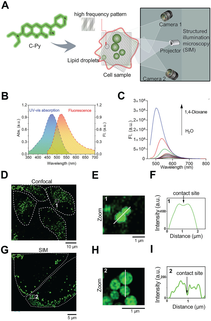
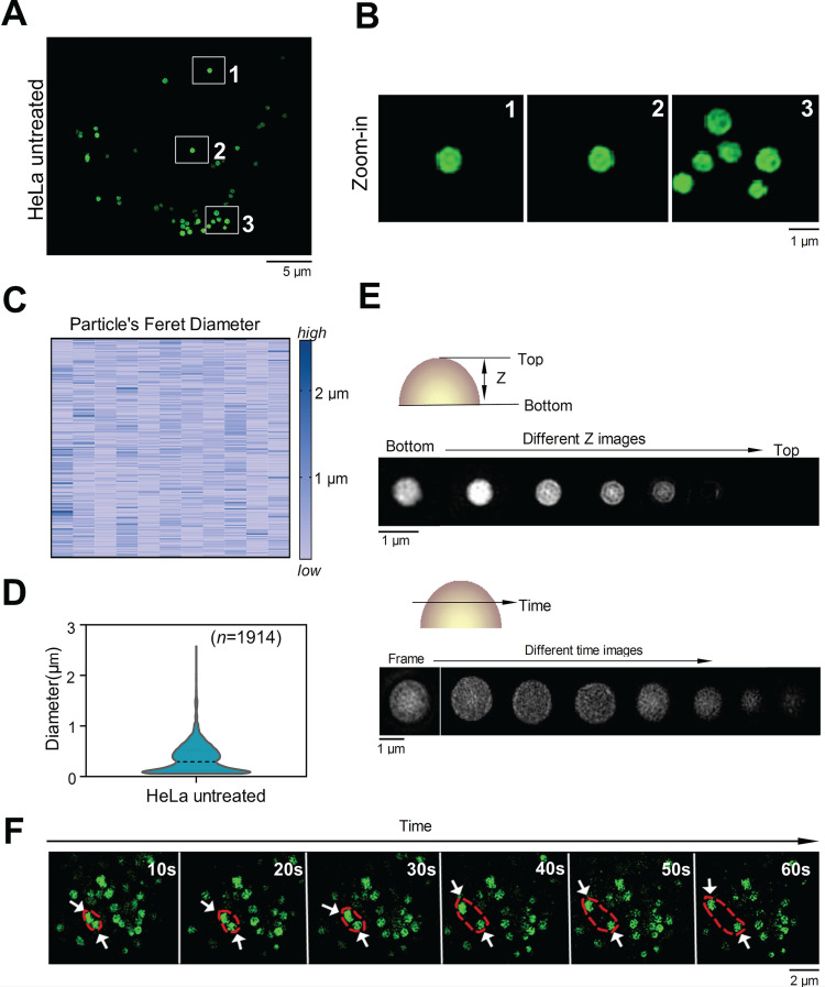
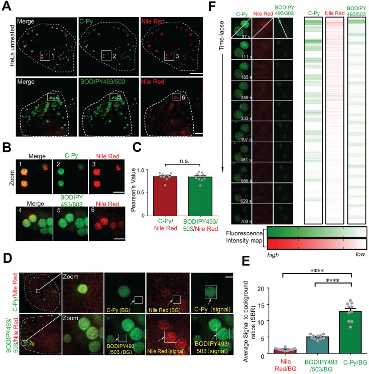
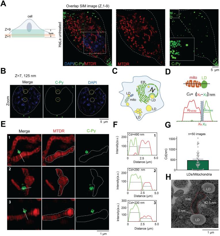
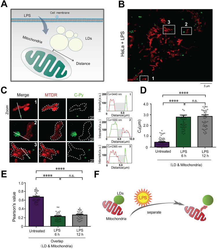
Background: Lipid droplets (LDs) establish a considerable number of contact sites with mitochondria to enable energy transfer and communication. In this study, we developed a fluorescent biosensor to image LD-mitochondria interactions at the nanoscale and further explored the function of LD-mediated matrix transmission in processes involving multi-organelle interactions. Methods: A fluorescent probe called C-Py (C21H19N3O2, 7-(diethylamino) coumarin-3-vinyl-4-pyridine acetonitrile) was designed and synthesized. Colocalization of C-Py and the commercial LD stain Nile Red was analyzed in HeLa cells. The fluorescence stability and signal to background ratio of C-Py under structured illumination microscopy (SIM) were compared to those of the commercial probe BODIPY493/503. The cytotoxicity of C-Py was assessed using CCK-8 assays. The uptake pattern of C-Py in HeLa cells was then observed under various temperatures, metabolic levels, and endocytosis levels. Contact sites between LDs and various organelles, such as mitochondria, nuclei, and cell membrane, were imaged and quantitated using SIM. Physical changes to the contact sites between LDs and mitochondria were monitored after lipopolysaccharide induction. Results: A LD-targeted fluorescent biosensor, C-Py, with good specificity, low background signal, excellent photostability, low cytotoxicity, and high cellular permeability was developed for tracking LD contact sites with multiple organelles using SIM. Using C-Py, the subcellular distribution and dynamic processes of LDs in living cells were observed under SIM. The formation of contact sites between LDs and multiple organelles was visualized at a resolution below ~200 nm. The number of LD-mitochondria contact sites formed was decreased by lipopolysaccharide treatment inducing an inflammatory environment. Conclusions: C-Py provides strategies for the design of ultra-highly selective biosensors and a new tool for investigating the role and regulation of LDs in living cells at the nanoscale.

Keywords: contact sites; extended-resolution imaging; lipid droplets; mitochondria.


Conflict of interest statement

Competing Interests: The authors have declared that no competing interest exists.

Figures

Figure 1
Figure 1
Design and optical characterization of the biosensor C-Py. (A) Schematic representation of SIM of LDs labelled with C-Py. (B) UV-vis absorption and emission spectra of C-Py (10 μM) in buffer solution (10 mM Tris-HCl, 100 mM KCl, pH 7.4). (C) Fluorescence emission spectra of C-Py in mixtures of 1,4-dioxane and water with various proportions (λex = 405 nm). (D) Confocal microscopy image of C-Py-labeled particles. (E) Enlarged image of the indicated region in D. (F) Fluorescence intensity profile along the line drawn in E across a contact site between C-Py-labeled particles. (G) SIM image of C-Py-labeled particles. (H) Enlarged image of the indicated region in G. (I) Fluorescence intensity profile along the line drawn in H across a contact site between C-Py-labeled particles.
Figure 2
Figure 2
SIM images of C-Py puncta in HeLa cells (λex = 488 nm, λem = 505-550 nm). (A) SIM image of cells stained with C-Py (10 µM) for 2 h. (B) Enlarged images of the indicated regions in A. (C and D) Size distribution of the C-Py puncta. Their diameters ranged from 0.1 to 3 µm (n = 1914). (E) Depth- and time-dependent images of cells stained with C-Py. (F) Images of the dynamic C-Py puncta over time. Two interacting puncta are outlined by the red dashed line.
Figure 3
Figure 3
Co-localization, SBR, and photostability of C-Py, BODIPY493/503, and Nile Red in HeLa cells under SIM. (A) Merged SIM images of cells stained with C-Py or BODIPY493/503 and Nile Red. Scale bar, 5 µm. (B) Enlarged images of the indicated regions in A. Scale bar, 1 µm. (C) Quantitative analysis of the colocalization between C-Py or BODIPY493/503 and Nile Red. (D) Merged SIM images of cells stained with C-Py or BODIPY493/503 and Nile Red, with signal and background (BG) regions for each channel indicated. (E) SBRs of C-Py, BODIPY493/503, and Nile Red (****P<0.0001). (F) Photostability of C-Py, BODIPY493/503, and Nile Red during continuous irradiation with the SIM laser. Scale bar, A, 5 µm. B, 1 µm, D, 1 µm.
Figure 4
Figure 4
C-Py nanoscopic tracking of organelle-LD contact sites in living cells. (A) 3D-SIM image collected over 1 μm in the Z-axis of LDs (C-Py), mitochondria (MTDR), and nuclei (DAPI). (B) SIM images collected at 125 nm in the Z-axis of LDs and nuclei. (C) Schematic diagram of the distribution of LDs and other organelles. (D) Schematic diagram of the metric used for estimating the contact distance between LDs and mitochondria. Cd is the absolute distance between the edge of a red mitochondrion (xR) and a green LD (xG) on the x-axis, which are derived from the FWHM of the organelle images. (E) SIM images of contact sites between LDs and mitochondria. (F) Fluorescence intensity profiles along the solid white lines in E and the calculated Cd values. (G) LD-mitochondria contact distances measured using SIM. Data are represented as mean ± SEM (n = 50 images from 20 cells) and three independent experiments were analyzed. (H) TEM image of contact sites between a mitochondrion and LDs.
Figure 5
Figure 5
LPS decreases the number of LD-mitochondria contact sites. (A) Schematic diagram illustrating the predicted changes to the mitochondrial contact sites of LDs during inflammatory processes. (B) SIM image of contact sites established between LDs (C-Py) and mitochondria (MTDR) after induction by LPS. (C) Enlarged images of the indicated regions in B, and quantitative analysis of the fluorescence intensity profiles along the solid white lines. (D) Contact distances between LDs and mitochondria in living cells without LPS stimulation or with LPS stimulation for 6 or 12 h. Data are represented as mean ± SEM (n = 51 from 10 images). Statistical differences between two groups were examined using Mann-Whitney tests (****P < 0.0001). (E) Quantitative analysis of the colocalization between LDs and mitochondria in cells untreated or treated with LPS for 6 or 12 h. Data are represented as mean ± SEM (n = 31 from 31 images) and three independent experiments were analyzed. Statistical differences between two groups were examined using Mann-Whitney tests (****P < 0.0001). (F) Schematic diagram illustrating the relationship between LDs and mitochondria following LPS treatment.

Similar articles

Cited by

References

    1. Thiam AR, Farese RV Jr, Walther TC. The biophysics and cell biology of lipid droplets. Nat Rev Mol Cell Biol. 2013;14:775–86. - PMC - PubMed
    1. Brasaemle DL, Wolins NE. Packaging of fat: an evolving model of lipid droplet assembly and expansion. J Biol Chem. 2012;287:2273–9. - PMC - PubMed
    1. Wu X, Geng F, Cheng X, Guo Q, Zhong Y, Cloughesy TF. et al. Lipid Droplets Maintain Energy Homeostasis and Glioblastoma Growth via Autophagic Release of Stored Fatty Acids. iScience. 2020;23:101569. - PMC - PubMed
    1. Zheng P, Xie Z, Yuan Y, Sui W, Wang C, Gao X. et al. Plin5 alleviates myocardial ischaemia/reperfusion injury by reducing oxidative stress through inhibiting the lipolysis of lipid droplets. Sci Rep. 2017;7:42574. - PMC - PubMed
    1. Boren J, Brindle KM. Apoptosis-induced mitochondrial dysfunction causes cytoplasmic lipid droplet formation. Cell Death Differ. 2012;19:1561–70. - PMC - PubMed

Publication types

MeSH terms

Substances