. 2021 Jul 6;11(16):8092-8111.
doi: 10.7150/thno.58739. eCollection 2021.

N-terminus-independent activation of c-Src via binding to a tetraspan(in) TM4SF5 in hepatocellular carcinoma is abolished by the TM4SF5 C-terminal peptide application

Affiliations

N-terminus-independent activation of c-Src via binding to a tetraspan(in) TM4SF5 in hepatocellular carcinoma is abolished by the TM4SF5 C-terminal peptide application

Haeng Eun Song et al. Theranostics. .

Abstract

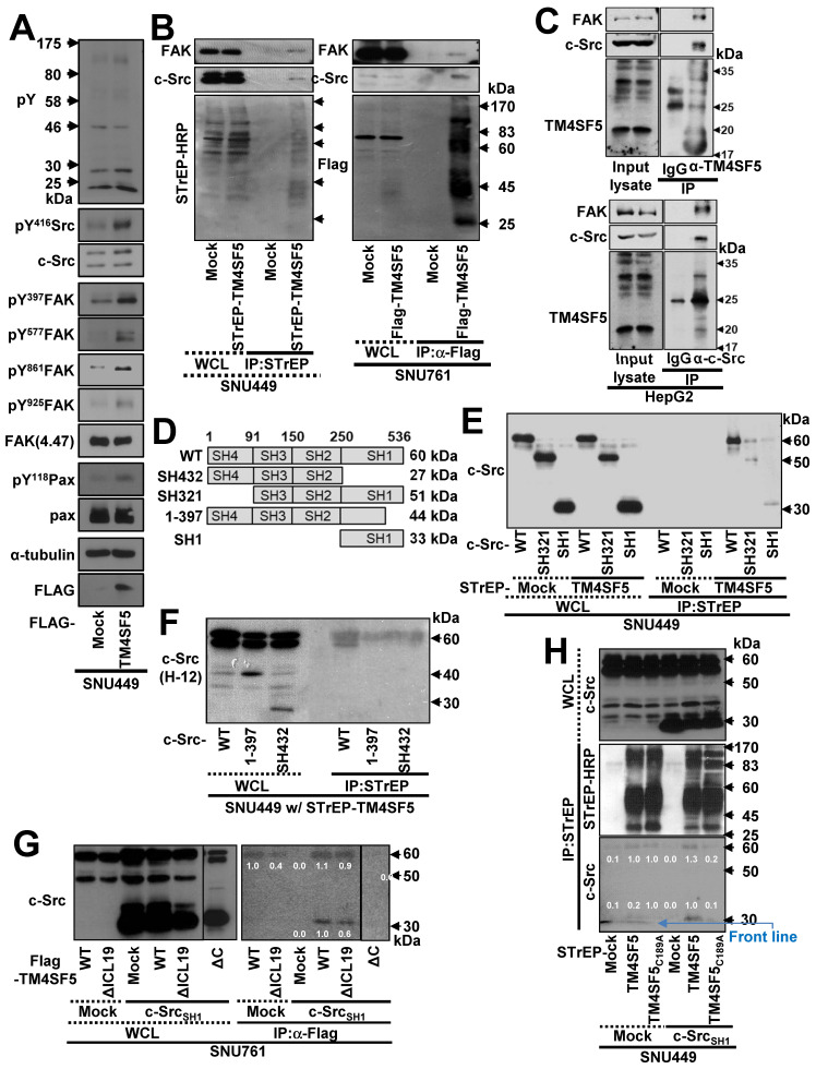
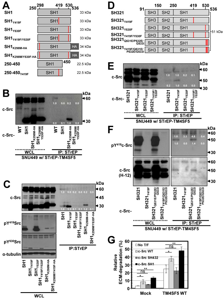
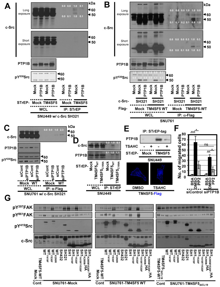
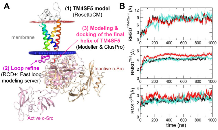
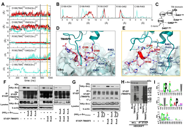
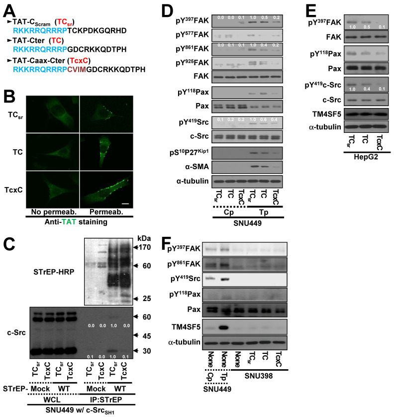
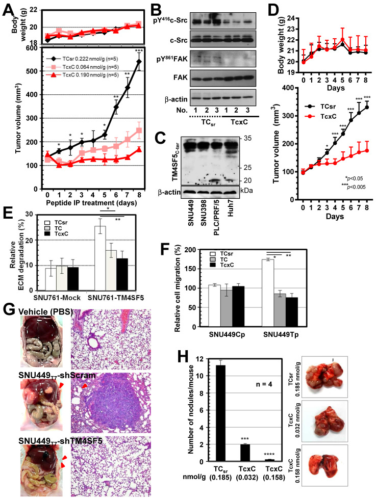
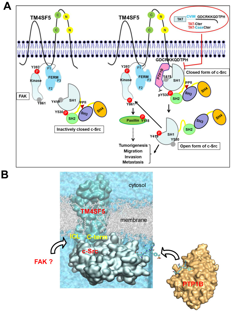
Active c-Src non-receptor tyrosine kinase localizes to the plasma membrane via N-terminal lipid modification. Membranous c-Src causes cancer initiation and progression. Even though transmembrane 4 L six family member 5 (TM4SF5), a tetraspan(in), can be involved in this mechanism, the molecular and structural influence of TM4SF5 on c-Src remains unknown. Methods: Here, we investigated molecular and structural details by which TM4SF5 regulated c-Src devoid of its N-terminus and how cell-penetrating peptides were able to interrupt c-Src activation via interference of c-Src-TM4SF5 interaction in hepatocellular carcinoma models. Results: The TM4SF5 C-terminus efficiently bound the c-Src SH1 kinase domain, efficiently to the inactively-closed form. The complex involved protein tyrosine phosphatase 1B able to dephosphorylate Tyr530. The c-Src SH1 domain alone, even in a closed form, bound TM4SF5 to cause c-Src Tyr419 and FAK Y861 phosphorylation. Homology modeling and molecular dynamics simulation studies predicted the directly interfacing residues, which were further validated by mutational studies. Cell penetration of TM4SF5 C-terminal peptides blocked the interaction of TM4SF5 with c-Src and prevented c-Src-dependent tumor initiation and progression in vivo. Conclusions: Collectively, these data demonstrate that binding of the TM4SF5 C-terminus to the kinase domain of inactive c-Src leads to its activation. Because this binding can be abolished by cell-penetrating peptides containing the TM4SF5 C-terminus, targeting this direct interaction may be an effective strategy for developing therapeutics that block the development and progression of hepatocellular carcinoma.

Keywords: PTPIB; TM4SF5; c-Src; metastasis; protein-protein interaction.


Conflict of interest statement

Competing Interests: The authors have declared that no competing interest exists.

Figures

Figure 1
Figure 1
The c-Src kinase domain binds the TM4SF5 C-terminus. (A) Parental SNU449 cells null for TM4SF5 were transiently transfected with mock or TM4SF5-FLAG for 48 h, prior to whole cell lysate preparation and standard western blots for the indicated molecules. (B) SNU449 cells stably-transfected with STrEP-tagged empty vector (mock) or TM4SF5, and SNU761 cells stably-expressing either FLAG-tagged empty vector (mock) or FLAG-TM4SF5 were harvested for whole cell lysates (WCL). The lysates were pulled-down with streptavidin-beads or with sepharose beads coated with anti-Flag antibody. The proteins pulled-down and WCL were then immunoblotted for anti-FAK, anti-c-Src, anti-STrEP-HRP, or anti-FLAG tag. (C) HepG2 cell lysates were immunoprecipitated with normal IgG or anti-TM4SF5 or anti-c-Src antibody before immunoblots for FAK, c-Src, and TM4SF5. (D) Schematic presentation of the human c-Src constructs. (E-F) SNU449 cells transfected with STrEP-mock, STrEP-TM4SF5 and either pcDNA3-c-Src WT, SH321, or SH1 construct (E) or either pcDNA3-c-Src WT, SH432, or 1-397 construct (F) for 48 h were harvested. The whole cell lysates (WCL) were precipitated with streptavidin beads prior to immunoblotting for c-Src. (G) SNU761 cells stably expressing TM4SF5WT, TM4SF5△ICL19 (△ICL19), or TM4SF5△C (△C) deletion mutant were transiently transfected with either c-Src WT or the SH1 construct, were harvested for WCL. The lysates were immunoprecipitated with anti-FLAG beads prior to immunoblotting for c-Src. (H) SNU449 cells transiently transfected with STrEP-mock, -TM4SF5WT, or -TM4SF5C189A mutant together with either pcDNA3 mock or c-Src SH1 construct were harvested for WCL, and the lysates were precipitated with streptavidin beads, prior to immunoblotting in parallel with the WCL. Quantitative comparison ratios of band intensities were calculated by measurement of band intensities using Image J and their normalization to those of loading controls. The data shown represent three independent experiments.
Figure 2
Figure 2
TM4SF5 preferentially binds inactively-closed c-Src forms. (A) Schematic presentation of the human c-Src SH1 domain constructs. (B and C) SNU449 cells stably expressing STrEP-TM4SF5 transiently transfected with either c-Src construct were harvested for whole cell lysates (WCL), and the lysates were precipitated with streptavidin beads, prior to immunoblotting for c-Src. The various c-Src constructs included c-Src WT, SH1, SH1K298M-HA, and SH1K298MY530F-HA construct in (B), or SH1, SH1Y419F, SH1Y530F, SH1Y419F/Y530F, SH1K298M-HA, and SH1K298M/Y530F-HA constructs in (C). (D) Schematic presentation of the human c-Src SH321 domain constructs. (E and F) SNU449 cells transiently transfected with STrEP-TM4SF5WT construct together with either c-Src SH321 construct were harvested and precipitated with streptavidin agarose beads, prior to immunoblotting for c-Src, or phospho-Y419 c-Src. The various c-Src SH321 constructs included c-Src SH321, SH321Y419F, SH321Y530F, SH321Y419F/Y530F constructs in (E), or SH321, SH321Y419F, SH321Q531E/P532E/G533I, and SH321Y419F/Q531E/P532E/G533I constructs in (F). (G) SNU761 cells stably expressing mock or TM4SF5 WT were transiently transfected without (No T/F) or with c-Src WT, c-Src SH432, or c-Src SH1 expression constructs for 48 h prior to invasive ECM degradation analysis for 4 h. Quantitative comparison ratios were calculated from the blots by measurement of band intensities using Image J and normalization of them to those of loading controls. The data shown represent three independent experiments.
Figure 3
Figure 3
Triple-complex between TM4SF5, inactive c-Src, and PTP1B leads to c-Src activation. (A and B) SNU449 cells transiently transfected with the c-Src SH321 construct together with the indicated constructs were harvested (A). SNU761 cells stably expressing mock or FLAG-TM4SF5 were transiently transfected with the indicated constructs were harvested (B). The whole cell lysates (WCL) were precipitated with streptavidin beads, prior to immunoblotting. (C) SNU761 cells stably expressing mock or FLAG-TM4SF5WT (WT) were transiently transfected with the indicated construct and siRNAs against a scrambled sequence (siCont) or a sequence in PTP1B (siPTP1B). Immunoprecipitation from the WCL using anti-Flag-precoated sepharose beads was performed, before immunoblots. (D) SNU449 cells transfected with the indicated plasmids were processed for precipitation using streptavidin beads, prior to immunoblotting. (E) Stable SNU449 cells were treated with DMSO (-) or TSAHC (20 μM, +) for 24 h, before harvest, precipitation using streptavidin beads, and immunoblotting. Another cell set with TM4SF5-Flag and treated with DMSO or TSAHC as above was processed for immunofluorescence analysis. (F) SNU761 cells stably expressing TM4SF5WT were transiently transfected with the indicated siRNAs for 48 h, prior to transwell migration assay. The mean ± SD were presented in the graph. ANOVA with Tukey's range-test or two-tailed unpaired Student's t-test was done to determine the significance. NS depicts non-significance. *, **, and *** depict p value less than 0.05, 0.01, and 0.005, respectively. (G) SNU761 cells stably expressing the indicated form were transiently transfected with either c-Src expression construct for 48 h were processed to immunoblotting. For comparison of their levels, control (Cont) lysates prepared from mock, TM4SF5WT, or TM4SF5△ICL19 cells without further c-Src construct transfection were also immunoblotted in parallels. Quantitative comparison ratios were calculated from the blots by measurement of band intensities using Image J and normalization of them to those of loading controls. The data shown represent three independent experiments.
Figure 4
Figure 4
Prediction of the complex structure of TM4SF5 and c-Src. (A) Model generation process and the resulting complex structure. The transmembrane region of TM4SF5 is colored by blue-to-red (N-to-C termini). The loop regions are colored in white except the ICL in magenta where the loop refinement was conducted. The inactive c-Src structure in the docked complex is colored in light yellow. The active c-Src (pink) was aligned to the inactive c-Src structure to speculate the possible conformational change after the kinase activation. (B) The Root-Mean-Square-Deviation (RMSD) values for the C-terminal region of TM4SF5 (top), the transmembrane region of TM4SF5 (middle), and c-Src (bottom) during the MD simulation. The results from the three independent trajectories are colored in black, red, and cyan. Note that the C-terminal tail of TM4SF5 showed dramatic increase in their RMSDs, which means this region underwent drastic conformational change.
Figure 5
Figure 5
Direct key interactions and dynamic hydrogen bonding network between the TM4SF5 C-terminus and the c-Src SH1 kinase domain. (A) The time traces of the distances between the key residue pairs. The results from the three independent trajectories are colored in black, red, and cyan. (B) The histogram of the distances in (A). (C) Schematic diagram of the dynamic hydrogen bonding network observed during the simulation. (D and E) Detailed molecular interactions at the time point marked in the gray and yellow boxes in (A). The TM4SF5 transmembrane region is colored in teal, and the C-terminal tail residues are represented in sticks with their carbon atoms in pink. The interacting residues in c-Src are depicted in sticks with their carbon atoms in light blue. The molecular surface of c-Src is also displayed in light blue. The hydrogen bonding or ionic interactions are displayed in black dashed lines. (F and G) SNU449 cells were transiently transfected with indicated plasmids for 48 h, before whole cell lysate preparation and co-immunoprecipitation, before immunoblots. (H) HEK3293FT cells transfected with the indicated expression constructs were processed for co-precipitation and immunoblotting. The data shown represent three independent experiments. (I) Sequence conservation of the key residues. The larger sequence character size corresponds to the higher sequence conservation. The X-axis is for the residue number of human TM4SF5. The highly conserved cysteine residues in the mutational analysis are depicted with red arrows, and the residues involved in direct binding to c-Src are marked with blue arrow.
Figure 6
Figure 6
Cell-penetrating TM4SF5 C-terminal peptides inhibit the TM4SF5 binding-mediated c-Src activation. (A) The sequences of the CPPs containing TAT-conjugated with a control (i.e., scrambled) sequence (TCsr) and the TM4SF5 C-terminal sequence without (TAT-Cter, TC) or with a CVIM motif (TAT-Caax-Cter, TcxC). (B) SNU449 cells were treated with the CPPs at 10 μM, prior to performing indirect immunofluorescence using anti-TAT (green). Incubation of the cells with the primary antibody was performed without (No Permeab.) or with pre-permeabilization (Permeab.). (C) SNU449 cells transiently transfected with STrEP-mock or TM4SF5WT (WT) together with pcDNA3 c-Src SH1 construct were treated with the CPPs (10 μM) for 24 h and then harvested for the WCL. The lysates were precipitated with streptavidin beads prior to immunoblotting for c-Src and Streptavidin (STrEP-HRP). (D to F) Control SNU449Cp (Cp) or ectopically TM4SF5-expressing SNU449Tp (Tp) cells (D), HepG2 cells endogenously expressing TM4SF5 (e), or TM4SF5-null SNU398 cells (F) were treated with the CPPs (10 μM), before harvests of WCL and immunoblotting for the indicated molecules. The lysates from SNU449Cp (Cp) and SNU449Tp (Tp) cells were used as a negative or a positive control of TM4SF5 expression. Quantitative comparison ratios were calculated from the blots by measurement of band intensities using Image J and normalization of them to those of loading controls. The data shown represent three independent experiments.
Figure 7
Figure 7
TM4SF5 C-terminal peptides suppress TM4SF5-mediated tumorigenesis and metastasis. (A and B) SNU449T7 cells stably expressing TM4SF5 were injected subcutaneously into nude mice (5 x 10⁶ cells/mouse, n =5). The cell-penetrating scramble or Caax peptides were administrated via intraperitoneal injections (at 0.222 nmol/g for TCsr and 0.064 or 0.190 nmol/g for TcxC) every day for 8 days, before tumor xenograft analysis, as explained in Materials and Methods. (B) Tissue extracts prepared from the xenograft tumors were processed to immunoblotting. (C and D) Different hepatoma cells including PLC/PRF/5 cells endogenously expressing TM4SF5, were immunoblotted for TM4SF5 expression (C). PLC/PRF/5 cells were injected subcutaneously into nude mice (1 x 10⁷ cells/mouse, n =6). The control TCsr or TcxC peptides were administrated via intraperitoneal injections (at 0.074 nmol/g for TCsr and 0.064 nmol/g for TcxC) every day for 8 days, before tumor xenograft analysis. (E) Degradation of Oregon Green® 488-conjugated gelatin were analyzed for SNU761 cells stably expressing mock (mock) or TM4SF5WT with the 10 μM CPPs treatment for 24 h (mean ± SD values). (F) TM4SF5-null SNU449Cp (Cp) and stably TM4SF5-expressing SNU449Tp (Tp) cells were treated with the 10 μM CPPs for 24 h and then analyzed for transwell migration for 4 h (mean ± SD values in the graph). (G) Vehicle (PBS), SBU449T7 cells with infected with shRNA against either control sequence (shScram) or TM4SF5 (shTM4SF5) were once orthotopically injected to livers (n≥5) and 4 weeks later lung tissues were analyzed for the metastatic cell masses. (H) SNU449T7 cells were injected into the tail veins of mice (5 x 10⁶ cells/100 μl/mice, n=4). After 2 weeks, the CPPs were administrated at 0.037 or 0.185 nmol/g for TCsr and 0.032 or 0.158 nmol/g for TcxC every other day for 2 weeks. Two additional weeks later, animals were sacrificed for examination of in vivo lung metastasis. Representative lung images in each experimental condition are shown. Surface nodules were counted and the mean ± SD values are presented (graph). ANOVA with Tukey's range-test or two-tailed unpaired Student's t-test was done to determine the significance. NS depicts non-significance. *, **, ***, and **** depict p value less than 0.05, 0.01, 0.005, and 0.0001, respectively. The data represent three independent experiments.
Figure 8
Figure 8
The working model. (A) TM4SF5, whose ICL interacts with the F1 lobe of FAK, prefers to directly bind to inactively-closed c-Src that is phosphorylated at the Y530 residue. After binding, TM4SF5 mediates dephosphorylation of phospho-Y530 by further recruiting PTP1B (i.e., unlatching). In addition, the TM4SF5 C-terminus-mediated interaction with the c-Src SH1 kinase domain may open its conformation (unclamping). Then the opened c-Src can be autophosphorylated at the Y419 residue (i.e., switching), leading to its full activation. Activated c-Src can phosphorylate Y861 of neighboring FAK. Activated c-Src and FAK then transduce downstream signaling pathways for TM4SF5-medated tumorigenesis and tumor progression. The CPPs containing the TM4SF5 C-terminal sequence interrupt the interaction between TM4SF5 and c-Src, suppress TM4SF5-mediated c-Src activation, and consequently inhibit TM4SF5-dependent tumorigenesis and metastatic potential. (B) Structural model of the membrane protein TM4SF5 complexed with c-Src. The plausible directions of the PTP1B and FAK bindings are depicted with black open arrows.

Similar articles

Cited by

References

    1. Yeatman TJ. A renaissance for SRC. Nat Rev Cancer. 2004;4:470–80. - PubMed
    1. Roskoski R Jr. Src protein-tyrosine kinase structure, mechanism, and small molecule inhibitors. Pharmacol Res. 2015;94:9–25. - PubMed
    1. Gargalionis AN, Karamouzis MV, Papavassiliou AG. The molecular rationale of Src inhibition in colorectal carcinomas. Intern J Cancer. 2014;134:2019–29. - PubMed
    1. Roskoski R Jr. Src protein-tyrosine kinase structure and regulation. Biochem Biophys Res Commun. 2004;324:1155–64. - PubMed
    1. Moarefi I, LaFevre-Bernt M, Sicheri F, Huse M, Lee CH, Kuriyan J. et al. Activation of the Src-family tyrosine kinase Hck by SH3 domain displacement. Nature. 1997;385:650–3. - PubMed

Publication types

MeSH terms