Review
. 2021 Jul 19:2021:9945424.
doi: 10.1155/2021/9945424. eCollection 2021.

Ischemia-Modified Albumin: Origins and Clinical Implications

Affiliations
Review

Ischemia-Modified Albumin: Origins and Clinical Implications

Alla Shevtsova et al. Dis Markers. .

Abstract

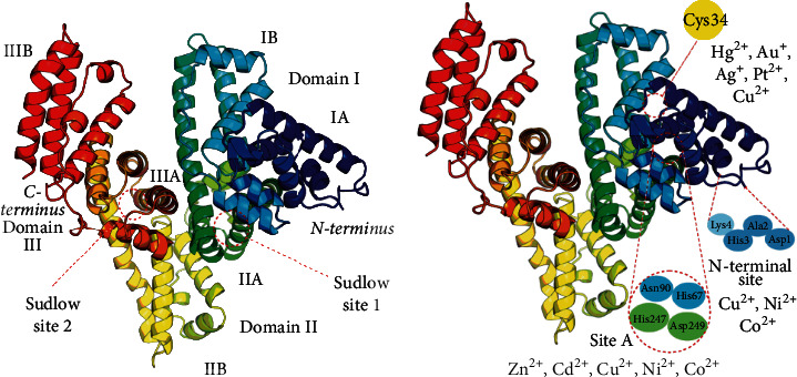
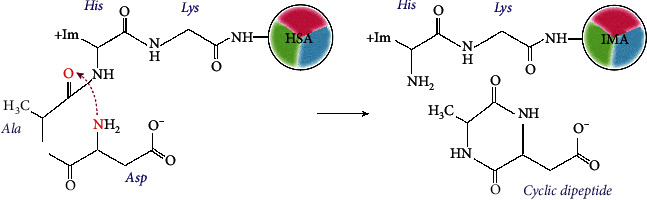
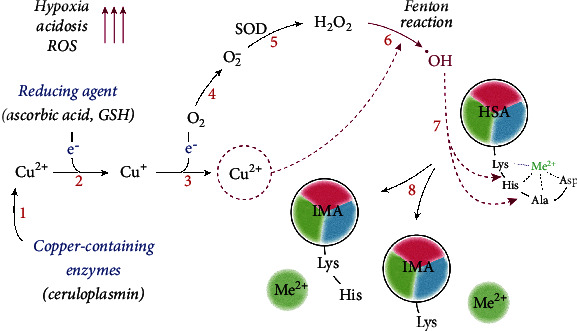
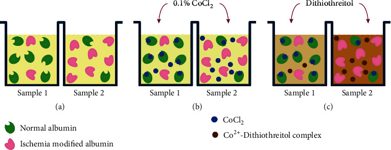
Albumin is one of the most abundant proteins in the body of mammals: about 40% of its pool is located in the intravascular space and the remainder is found in the interstitial space. The content of this multifunctional protein in blood is about 60-65% of total plasma proteins. A decrease in its synthesis or changes of functional activity can destabilize oncotic blood pressure, cause a violation of transporting hormones, fatty acids, metals, and drugs. Albumin properties change under ischemic attacks associated with oxidative stress, production of reactive oxygen species, and acidosis. Under these conditions, ischemia-modified albumin (IMA) is generated that has a reduced metal-binding capacity, especially for transition metals, such as copper, nickel, and cobalt. The method of determining the cobalt-binding capability of HSA was initially proposed to evaluate IMA level and then licensed as an ACB test for routine clinical analysis for myocardial ischemia. Subsequent studies have shown the viability of the ACB test in diagnosing other diseases associated with the development of oxidative stress. This review examines recent data on IMA generation mechanisms, describes principles, advantages, and limitations of methods for evaluation of IMA levels, and provides detailed analysis of its use in diagnostic and monitoring therapeutic efficacy in different diseases.


Conflict of interest statement

The authors declare that they have no conflicts of interest.

Figures

Figure 1
Figure 1
Structure of human serum albumin. (a) The molecule consists of a single polypeptide chain; about half of its length is an α-helix. The albumin structure comprises three homologous domains: I, marked in blue and cyan; II, green and yellow; III, orange and red. Each domain contains two subdomains, A and B, and two sites to bind hydrophobic molecules (Sudlow sites 1 and 2). (b) Sites for binding transition metal ions: N-terminal site, Cys34, and site А (multimetal binding site). Site B is not shown because its exact position is unknown.
Figure 2
Figure 2
ІМА formation through dipeptide cleavage. A nucleophilic attack by the α-amino nitrogen on the carbonyl of Ala2-His3 peptide bond cleaves and releases the cyclic dipeptide. The truncated NTS cannot bind transition metal ions [20].
Figure 3
Figure 3
The mechanism of ІМА formation driven by oxidative stress. Tissue hypoxia and activation of anaerobic glycolysis induce acidosis and release Cu2+ ions from copper-containing proteins, such as ceruloplasmin (1). In the presence of reducing agents, e.g., ascorbic acid, Cu2+ is reduced to Cu+ (2), followed by the formation of superoxide anion O2 (3-4). Superoxide dismutase (SOD) catalyzes the dismutation of superoxide O2 to hydrogen peroxide H2O2 (5), which, in the presence of Cu2+, undergoes the Fenton reaction with the formation of hydroxyl radicals ·OH (6). These radicals contribute to the degradation of NTS (7) and IMA formation (8), which cannot bind Cu2+ and other metal ions.
Figure 4
Figure 4
The scheme of the ACB test. Serum samples (100 μL) are added in the wells of the microplate (a), then add 25 μL of CoCl2 (b), incubate for 10 min, and then add dithiothreitol (c), which binds to free cobalt, staining the medium brown. The color intensity is proportional to the amount of free cobalt and the amount of IMA.

Similar articles

Cited by

References

    1. Ahn S. M., Simpson R. J. Body fluid proteomics: prospects for biomarker discovery. Proteomics – Clinical Applications. 2007;1(9):1004–1015. doi: 10.1002/prca.200700217. - DOI - PubMed
    1. Ellmerer M., Schaupp L., Brunner G. A., et al. Measurement of interstitial albumin in human skeletal muscle and adipose tissue by open-flow microperfusion. American Journal of Physiology-Endocrinology and Metabolism. 2000;278(2):E352–E356. doi: 10.1152/ajpendo.2000.278.2.E352. - DOI - PubMed
    1. Peters T. “All about Albumin,” Biochemistry, Genetics, and Medical Applications. San Diego, California: Academic Press, Inc.; 1996.
    1. Nahon J. L., Tratner I., Poliard A., et al. Albumin and alpha-fetoprotein gene expression in various nonhepatic rat tissues. Journal of Biological Chemistry. 1988;263(23):11436–11442. doi: 10.1016/S0021-9258(18)37976-6. - DOI - PubMed
    1. Shamay A., Homans R., Fuerman Y., et al. Expression of albumin in nonhepatic tissues and its synthesis by the bovine mammary gland. Journal of Dairy Science. 2005;88(2):569–576. doi: 10.3168/jds.S0022-0302(05)72719-3. - DOI - PubMed

LinkOut - more resources