

OCT Biomarkers in Neovascular Age-Related Macular Degeneration: A Narrative Review
- PMID: 34336265
- PMCID: PMC8313359
- DOI: 10.1155/2021/9994098
OCT Biomarkers in Neovascular Age-Related Macular Degeneration: A Narrative Review
Abstract
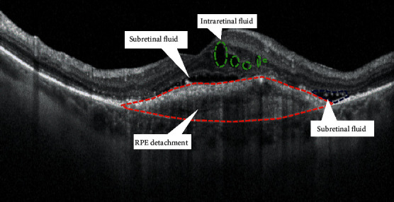
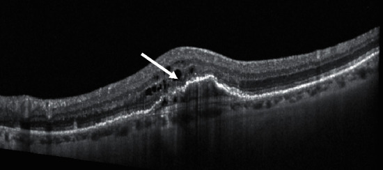
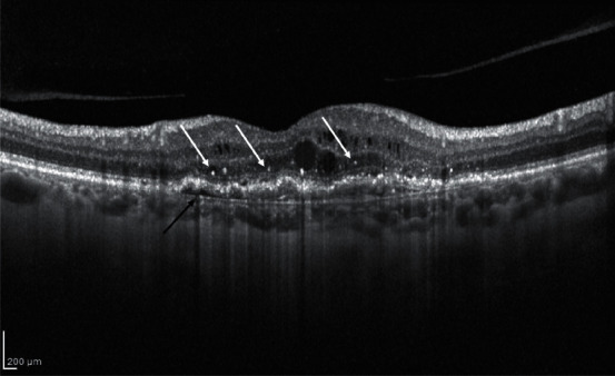
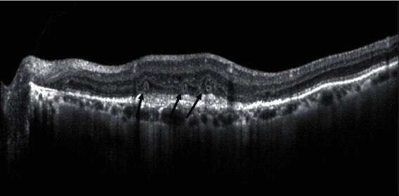
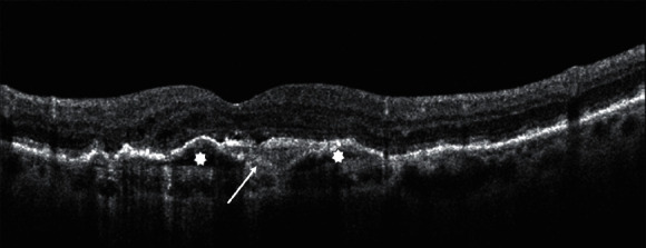
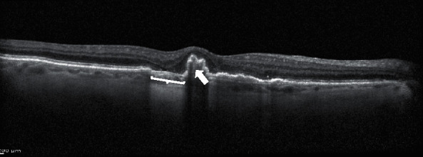
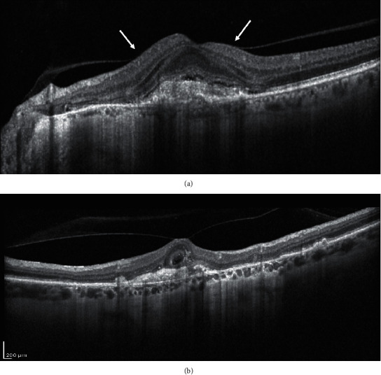
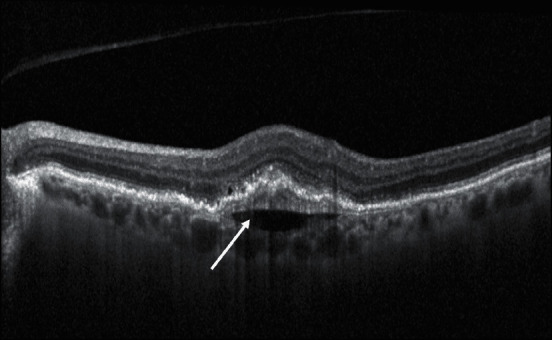
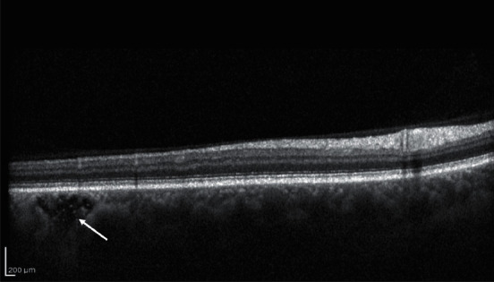
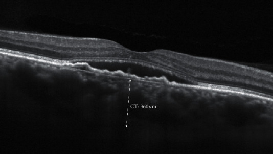
Age-related macular degeneration (AMD) is the leading cause of legal blindness in elderly people. Neovascular AMD (nAMD) is responsible for the majority of cases of severe visual loss in eyes with AMD. Optical coherence tomography (OCT) is the most widely used technology for the diagnosis and follow-up of nAMD patients, which is widely used to study and guide the clinical approach, as well as to predict and evaluate treatment response. The aim of this review is to describe and analyze various structural OCT-based biomarkers, which have practical value during both initial assessment and treatment follow-up of nAMD patients. While central retinal thickness has been the most common and one of the first OCT identified biomarkers, today, other qualitative and quantitative biomarkers provide novel insight into disease activity and offer superior prognostic value and better guidance for tailored therapeutic management. The key importance of retinal fluid compartmentalization (intraretinal fluid, subretinal fluid, and subretinal pigment epithelium (RPE) fluid) will be discussed firstly. In the second part, the structural alterations of different retinal layers in various stages of the disease (photoreceptors layer integrity, hyperreflective dots, outer retinal tubulations, subretinal hyperreflective material, and retinal pigment epithelial tears) will be analyzed in detail. The last part of the review will focus on how alterations of the vitreoretinal interface (vitreomacular adhesion and traction) and of the choroid (sub-RPE hyperreflective columns, prechoroidal clefts, choroidal caverns, choroidal thickness and choroidal volume, and choroidal vascular index) interact with nAMD progression. OCT technology is evolving very quickly, and new retinal biomarkers are continuously described. This up-to-date review article provides a comprehensive description on how structural OCT-based biomarkers provide a valuable tool to monitor the progression of the disease and the treatment response in nAMD patients. Thus, in this perspective, clinicians will be able to allocate hospital resources in the best possible way and tailor treatment to the individual patient's needs.
Copyright © 2021 Cristian Metrangolo et al.
Conflict of interest statement
Authors declare no conflicts of interest.
Figures
References
-
- Branisteanu D., Branisteanu D., Feraru C., et al. Influence of unilateral intravitreal bevacizumab injection on the incidence of symptomatic choroidal neovascularization in the fellow eye in patients with neovascular age‑related macular degeneration (review) Experimental and Therapeutic Medicine. 2020;20(6):p. 182. doi: 10.3892/etm.2020.9312. - DOI - PMC - PubMed
Publication types
LinkOut - more resources
Full Text Sources
