Case Reports
. 2021 Jul 17:2021:3626276.
doi: 10.1155/2021/3626276. eCollection 2021.

Modified Arthroscopic Suture Fixation of Posterior Cruciate Ligament Tibial Avulsion Fracture in the Setting of Multiligament Knee Injury in Teenager

Affiliations
Case Reports

Modified Arthroscopic Suture Fixation of Posterior Cruciate Ligament Tibial Avulsion Fracture in the Setting of Multiligament Knee Injury in Teenager

Miguel Quesado et al. Case Rep Orthop. .

Abstract

The posterior cruciate ligament (PCL) avulsion fracture is a rare injury and occurs mainly in young patients. The development of arthroscopic techniques and fixation methods has improved the treatment of this entity. This report describes a modified arthroscopic suture fixation of a small tibial avulsion fracture of the PCL. A 17-year-old male, injured in a motorcycle crash, was admitted to the Emergency Department and diagnosed with left knee PCL tibial avulsion fracture, lateral collateral ligament (LCL) femoral avulsion fracture, and patella fracture. The PCL was fixed arthroscopically using a Knee Scorpion and two SutureTapes (Arthrex, Munich-Germany) through of an interlaced configuration at the base of the fragment using a transseptal approach and fixed distally over a cortical button on the anterior cortex. The LCL was repaired with two cannulated screws by a percutaneous approach. At 1 year of follow-up, the fragment was healed with tibiofemoral congruence, and the knee was stable with complete range of motion. The Tegner Lysholm Knee Scoring Scale (TLKSS) was 92.


Conflict of interest statement

The authors have no conflict of interests to declare.

Figures

Figure 1
Figure 1
Coronal (a) and sagittal (b) X-ray on admission to the Emergency Department.
Figure 2
Figure 2
Coronal (a, d), axial (b), and sagittal (c) CT scan on admission to the Emergency Department.
Figure 3
Figure 3
MRI (fat-saturated T2-weighted) lateral view showing a displaced avulsion fracture of PCL at tibial attachment.
Figure 4
Figure 4
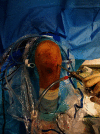
Arthroscopic portals used during the procedure (two standard anterior and two accessory posterior).
Figure 5
Figure 5
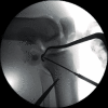
Insertion of K-wires to the PCL tibial footprint controlled by fluoroscopy.
Figure 6
Figure 6
MRI (T2-weighted) axial view - schematic representation of each suture (yellow and blue lines) crossing the posterior part of the base of the PCL and exiting the contralateral tunnel (a). Arthroscopic posteromedial view - interlaced suture configuration for PCL avulsion fracture (b).
Figure 7
Figure 7
Coronal (a) and sagittal (b) intraoperative X-ray showing the final reduction and fixation of PCL and LCL avulsion fractures.
Figure 8
Figure 8
Coronal (a, b), sagittal (c), and axial (d) control CT scan after 3 months postoperative with evidence of fracture healing.

References

    1. Lamoria R., Goyal D., Bansal M., Kaler S., Upadhyay R. Clinical experience with arthroscopic suture pull technique in isolated PCL avulsion injuries. Journal of Clinical Orthopaedics and Trauma. 2020;11(Suppl 3):S362–S367. doi: 10.1016/j.jcot.2019.06.020. - DOI - PMC - PubMed
    1. Zhu W., Lu W., Cui J., et al. Treatment of tibia avulsion fracture of posterior cruciate ligament with high-strength suture fixation under arthroscopy. European Journal of Trauma and Emergency Surgery. 2017;43(1):137–143. doi: 10.1007/s00068-015-0606-9. - DOI - PMC - PubMed
    1. Chen L. B., Wang H., Tie K., Mohammed A., Qi Y. J. Arthroscopic fixation of an avulsion fracture of the tibia involving the posterior cruciate ligament. The Bone & Joint Journal. 2015;97-B(9):1220–1225. doi: 10.1302/0301-620X.97B9.35765. - DOI - PubMed
    1. Song J.-G., Nha K.-W., Lee S.-W. Open posterior approach versus arthroscopic suture fixation for displaced posterior cruciate ligament avulsion fractures: systematic review. Knee Surgery and Related Research. 2018;30(4):275–283. doi: 10.5792/ksrr.17.073. - DOI - PMC - PubMed
    1. Nakagawa S., Arai Y., Hara K., Inoue H., Hino M., Kubo T. Arthroscopic pullout fixation for a small and comminuted avulsion fracture of the posterior cruciate ligament from the tibia. Knee Surgery & Related Research. 2017;29(4):316–320. doi: 10.5792/ksrr.16.025. - DOI - PMC - PubMed

Publication types

LinkOut - more resources