. 2015 Sep;1(3):292-308.
doi: 10.15302/j-eng-2015078. Epub 2016 Mar 16.

Systems Neuroengineering: Understanding and Interacting with the Brain

Affiliations

Systems Neuroengineering: Understanding and Interacting with the Brain

Bradley J Edelman et al. Engineering (Beijing). 2015 Sep.

Abstract

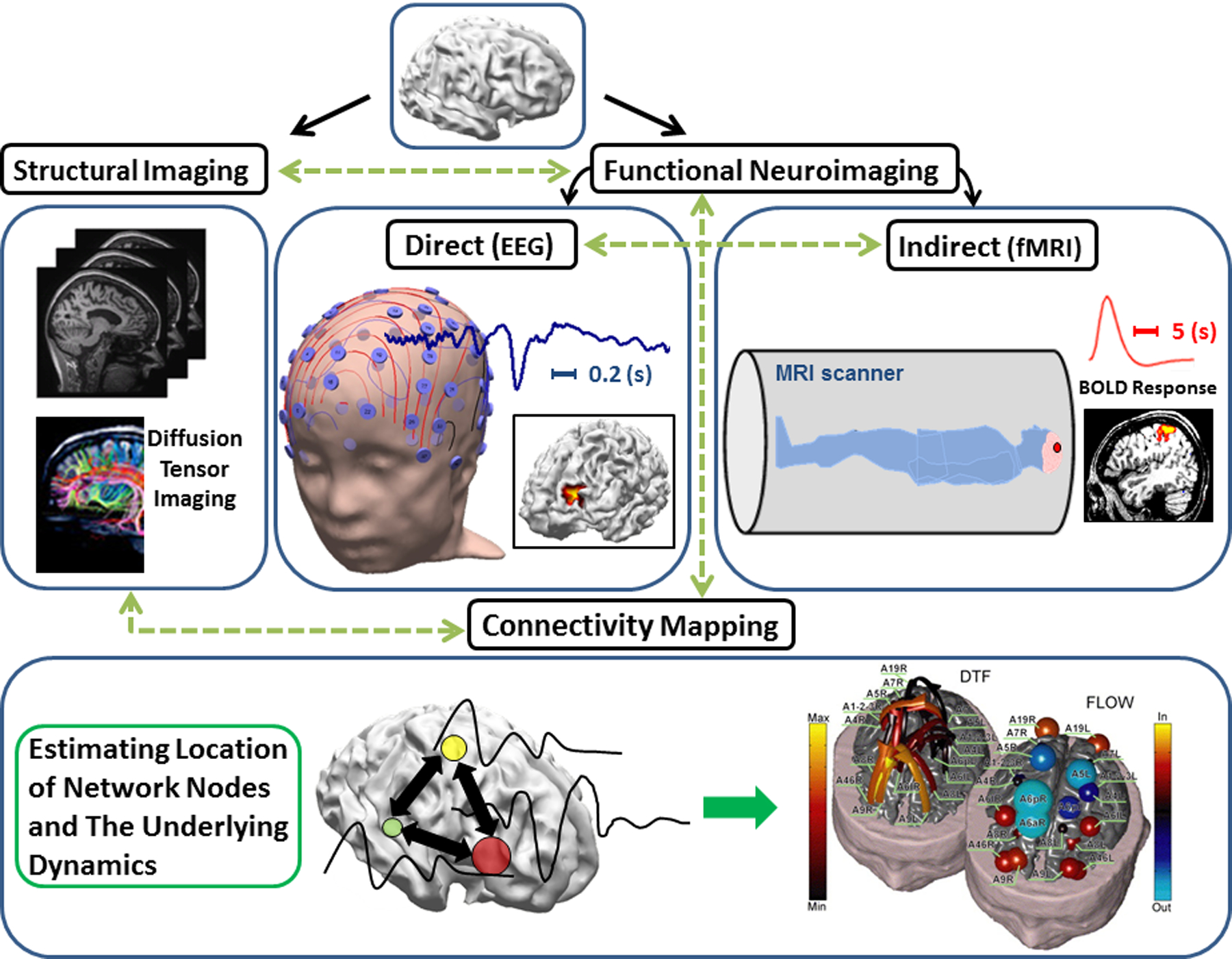
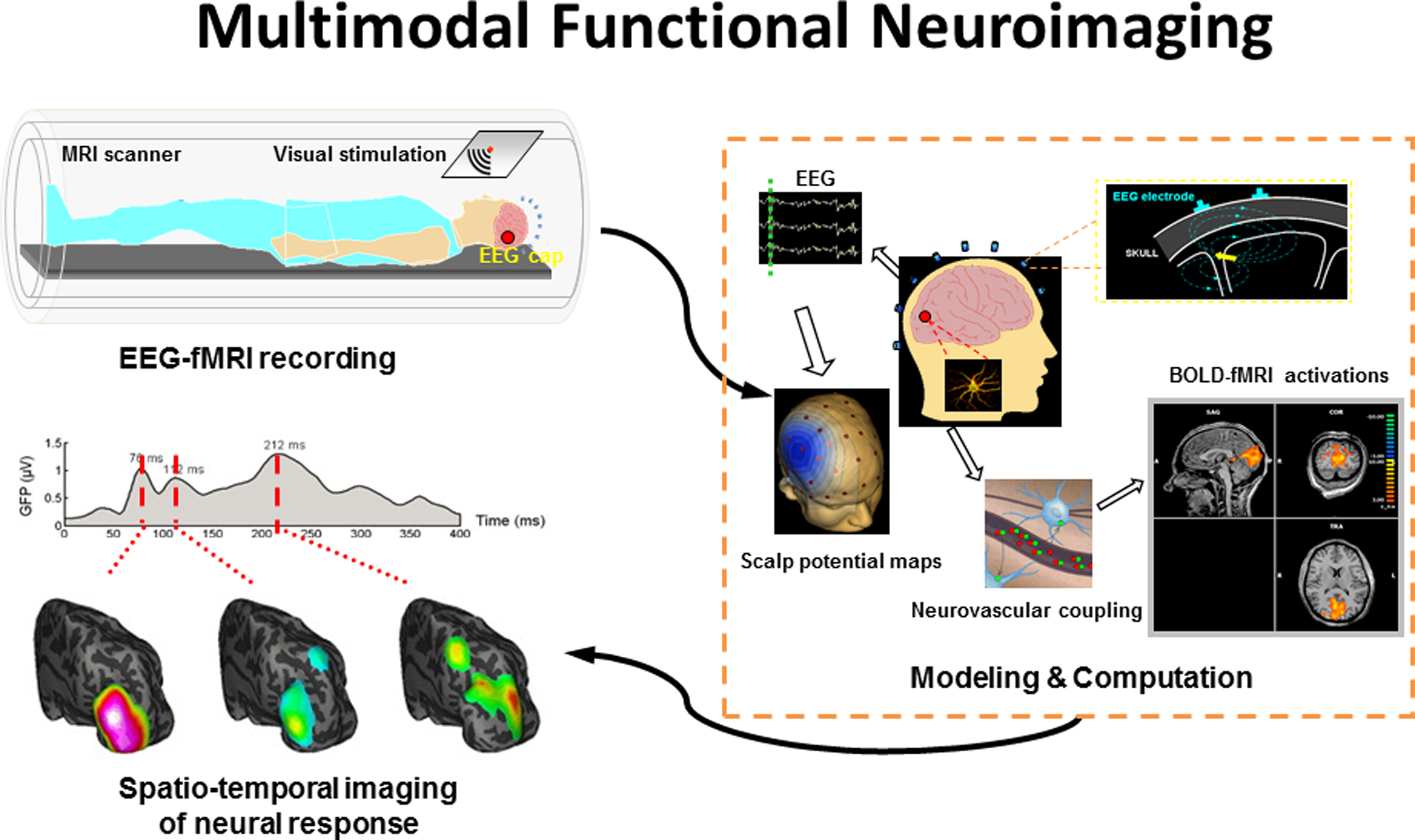
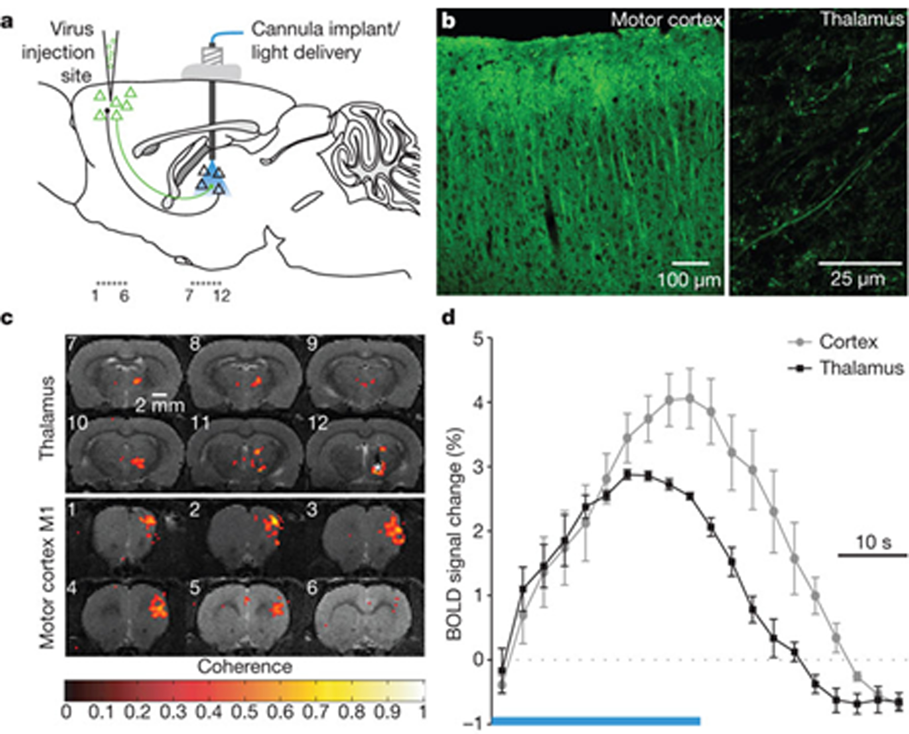
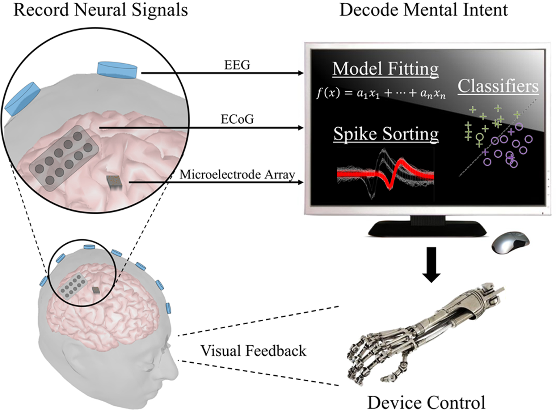
In this paper, we review the current state-of-the-art techniques used for understanding the inner workings of the brain at a systems level. The neural activity that governs our everyday lives involves an intricate coordination of many processes that can be attributed to a variety of brain regions. On the surface, many of these functions can appear to be controlled by specific anatomical structures; however, in reality, numerous dynamic networks within the brain contribute to its function through an interconnected web of neuronal and synaptic pathways. The brain, in its healthy or pathological state, can therefore be best understood by taking a systems-level approach. While numerous neuroengineering technologies exist, we focus here on three major thrusts in the field of systems neuroengineering: neuroimaging, neural interfacing, and neuromodulation. Neuroimaging enables us to delineate the structural and functional organization of the brain, which is key in understanding how the neural system functions in both normal and disease states. Based on such knowledge, devices can be used either to communicate with the neural system, as in neural interface systems, or to modulate brain activity, as in neuromodulation systems. The consideration of these three fields is key to the development and application of neuro-devices. Feedback-based neuro-devices require the ability to sense neural activity (via a neuroimaging modality) through a neural interface (invasive or noninvasive) and ultimately to select a set of stimulation parameters in order to alter neural function via a neuromodulation modality. Systems neuroengineering refers to the use of engineering tools and technologies to image, decode, and modulate the brain in order to comprehend its functions and to repair its dysfunction. Interactions between these fields will help to shape the future of systems neuroengineering-to develop neurotechniques for enhancing the understanding of whole-brain function and dysfunction, and the management of neurological and mental disorders.

Keywords: brain-computer interface; brain-machine interface; neural interface; neural stimulation; neuroimaging; neuromodulation; neurotechnology; systems neuroengineering.


Conflict of interest statement

Brad Edelman, Nessa Johnson, Abbas Sohrabpour, Shanbao Tong, Nitish Thakor, and Bin He declare that they have no conflict of interest or financial conflicts to disclose.

Figures

Figure 1.
Figure 1.. Neuroimaging at a glance: Different neuroimaging modalities interact with each other to delineate underlying brain networks (not inclusive of all modalities).
Adapted from Ref. [33, 41] with permissions.
Figure 2.
Figure 2.. Principles of EEG-fMRI.
Indirect functional neuroimaging modalities such as fMRI are related to direct electrophysiological activities via some sort of coupling (neurovascular coupling in case of fMRI). GFP: Global Field Power. Adapted from Ref. [33] with permission.
Figure 3.
Figure 3.. Optogenetics functional MRI (ofMRI) [69].
This technique inherits the high spatial resolution and wide field-of-view of fMRI along with the specificity of optogenetics, making it ideal for imaging selectively brain-wide large-scale networks. (a) Injecting the virus to encode specific cells to be responsive to light; (b) confirming the induction of the desired pattern; (c) the BOLD of MRI data and (d) of MRI-HRF, for BOLD signals elicited by optical stimulation. Adapted from Ref. [69] with permission.
Figure 4.
Figure 4.. Schematic of a brain-computer/machine interface system.
Signals are acquired from the brain through the use of internal or external stimuli. A computer then decodes these signals to interpret the user’s goal and translates the result into an action of the output device. Subjects can often observe such effects and modulate their brain signals to accomplish the desired task.
Figure 5.
Figure 5.. Concept diagram of using a noninvasive BMI to control a wireless quadcopter in three dimensions [77].
A camera mounted on the quadcopter allows users to view their environment. The user can then continuously navigate the quadcopter using motor imagery tasks. Adapted from Ref. [77] with permission. See Ref. [78] for a video clip demonstrating how the mind-controlled quadcopter works.
Figure 6.
Figure 6.. Schematic of a bi-directional BMI [116].
Motor intent is decoded from signals collected in the primary motor cortex. As the controlled device interacts with the environment, sensory cues are translated into pulse trains and used to stimulate the primary sensory cortex. In this approach, both efferent and afferent signals contribute to BMI control. Adapted from Ref. [116] with permission.
Figure 7.
Figure 7.. A summary of invasive and noninvasive neuromodulation technologies.
Invasive techniques include DBS, in which a lead is implanted into a deep brain structure, and cortical stimulation, in which electrodes are placed on the brain surface. Noninvasive techniques include transcranial magnetic stimulation (TMS), typically applied with a figure-eight coil, transcranial direct-current stimulation (tDCS) via scalp sponge electrodes, or transcranial focused ultrasound stimulation (tFUS) using pulsed ultrasound from a transducer on the scalp.
Figure 8.
Figure 8.. Deep brain stimulation for Parkinson’s disease [129].
The stimulation lead is implanted to a deep brain structure, and connected to the pulse generator in the chest via a lead tunneled through the neck (left panel). For Parkinson’s disease, the stimulation lead is targeted to either the internal segment of the globus pallidus (middle right panel) or to the sub-thalamic nucleus (lower right panel). Adapted from Ref. [129] with permission.

Similar articles

Cited by

References

    1. Herman GT. Fundamentals of Computerized Tomography: Image Reconstruction from Projections. London: Springer Science & Business Media, 2009
    1. Bogdahn U, Becker G, Winkler J, Greiner K, Perez J, Meurers B. Transcranial color-coded real-time sonography in adults. Stroke, 1990, 21(12): 1680–1688doi:10.1161/01.STR.21.12.1680. - DOI - PubMed
    1. Basser PJ, Mattiello J, LeBihan D. MR diffusion tensor spectroscopy and imaging. Biophys. J, 1994, 66(1): 259–267doi:10.1016/S0006-3495(94)80775-1. - DOI - PMC - PubMed
    1. Uğurbil K, et al.; WU-Minn HCP Consortium. Pushing spatial and temporal resolution for functional and diffusion MRI in the Human Connectome Project. Neuroimage, 2013, 80: 80–104doi:10.1016/j.neuroimage.2013.05.012. - DOI - PMC - PubMed
    1. Setsompop K, et al. Pushing the limits of in vivo diffusion MRI for the Human Connectome Project. Neuroimage, 2013, 80: 220–233doi:10.1016/j.neuroimage.2013.05.078. - DOI - PMC - PubMed

LinkOut - more resources