

doi: 10.1016/j.eats.2021.04.010.
eCollection 2021 Jul.
Lateral Patellar Retinaculum Z-Lengthening
Affiliations
- PMID: 34336590
- PMCID: PMC8322702
- DOI: 10.1016/j.eats.2021.04.010
Item in Clipboard
Lateral Patellar Retinaculum Z-Lengthening
Arthrosc Tech.
.
Abstract
The lateral retinaculum is a 2-layered structure. The plane between the superficial oblique fibers and the deep transverse fibers allows for coronal plane Z-lengthening of the lateral retinaculum. The lengthening procedure can be used for treatment of lateral patellar hypercompression syndrome or as an adjunct to surgical procedures undertaken to address patellar instability. This article describes the surgical technique for lateral retinacular lengthening.
Level 1: Knee.
Level 2: Malalignment, patellofemoral, other.
© 2021 by the Arthroscopy Association of North America. Published by Elsevier.
Figures

Illustration of excessive lateral patellar tilt greater than 20˚ with reference to the posterior femoral condyles.
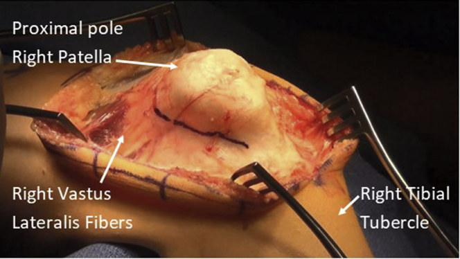
Midline approach to the right anterior knee with patient in supine position and planned lateral retinacular incision, with emphasis on avoiding proximal extension into the vastus lateralis tendon.
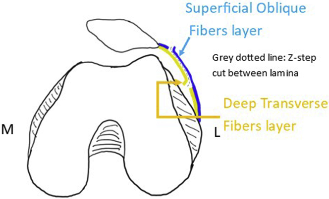
Illustration of 30˚ lateral patellar tilt superficial oblique fibers (blue) and deep transverse fibers (yellow) for coronal Z-lengthening step-cut (dotted black).

Patient's right knee, in supine position. Development of the fascial plane between incised superficial oblique (blue) and intact deep transverse fibers (yellow).

Patient's right knee, in supine position. Longitudinal incision through the deep transverse fibers (yellow) and capsular layer (white) completing the Z-incision. The lateral femoral condyle is visible in this example.

Illustration of neutral patellar position after lengthening the lateral retinaculum with provisional suturing.

Illustration of 30˚ (maximum) medial patellar tilt after repair, verifying the adequacy of lengthening and provisional repair.
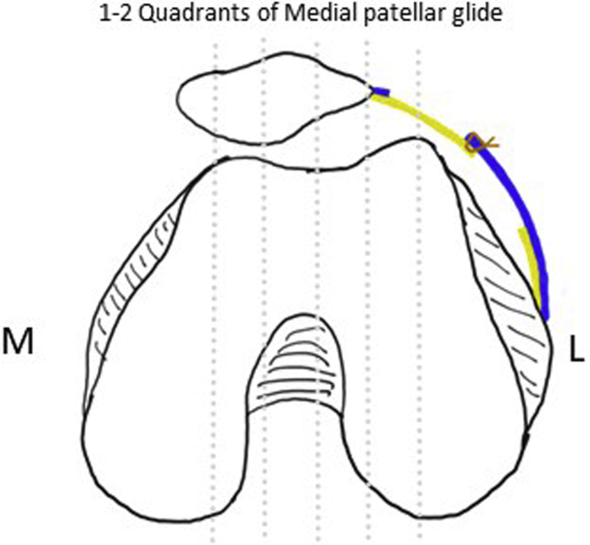
Illustration of acceptable 1-2 quadrants of medial patellar subluxation after retinacular lengthening.
References
-
- Ceder L.C., Larson R.L. Z-plasty lateral retinacular release for the treatment of patellar compression syndrome. Clin Orthop Relat Res. 1979;144:110–113. - PubMed
-
- Pagenstert G., Wolf N., Bachmann M., et al. Open lateral patellar retinacular lengthening versus open retinacular release in lateral patellar hypercompression syndrome: A prospective double-blinded comparative study on complications and outcome. Arthroscopy. 2012;28:788–797. doi: 10.1016/j.arthro.2011.11.004. - DOI - PubMed
LinkOut - more resources
Full Text Sources
