Review
. 2021 Jul 16:9:702404.
doi: 10.3389/fcell.2021.702404. eCollection 2021.

Caspases in the Developing Central Nervous System: Apoptosis and Beyond

Affiliations
Review

Caspases in the Developing Central Nervous System: Apoptosis and Beyond

Trang Thi Minh Nguyen et al. Front Cell Dev Biol. .

Abstract

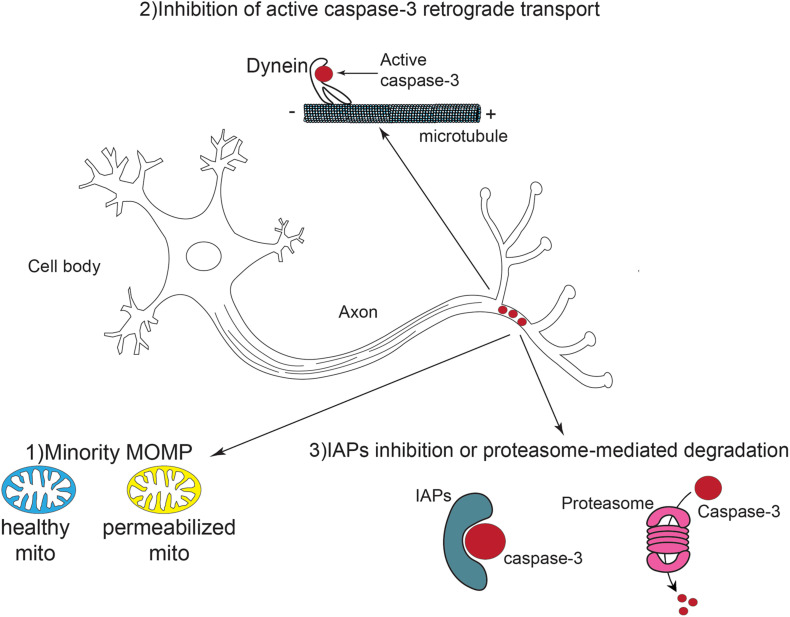
The caspase family of cysteine proteases represents the executioners of programmed cell death (PCD) type I or apoptosis. For years, caspases have been known for their critical roles in shaping embryonic structures, including the development of the central nervous system (CNS). Interestingly, recent findings have suggested that aside from their roles in eliminating unnecessary neural cells, caspases are also implicated in other neurodevelopmental processes such as axon guidance, synapse formation, axon pruning, and synaptic functions. These results raise the question as to how neurons regulate this decision-making, leading either to cell death or to proper development and differentiation. This review highlights current knowledge on apoptotic and non-apoptotic functions of caspases in the developing CNS. We also discuss the molecular factors involved in the regulation of caspase-mediated roles, emphasizing the mitochondrial pathway of cell death.

Keywords: apoptosis; caspases; central nervous system; embryonic development; mitochondria.


Conflict of interest statement

The authors declare that the research was conducted in the absence of any commercial or financial relationships that could be construed as a potential conflict of interest.

Figures

FIGURE 1
FIGURE 1
The caspase family of proteases. (A) Members of the caspase family, are synthesized as inactive precursors called zymogens composed of a prodomain, a p20 large subunit and a p10 small subunit. The p20 subunit contains the active site of the enzyme harboring a “QACXG” pentapeptide motif. Initiator Caspases are characterized by the presence of a long N-terminal prodomain whereas the N-terminus domain of effector caspases is shorter. (B) Caspase activation is achieved by proteolytic cleavage between the large and small subunits and removal of the N-terminus prodomain. This post translational modification leads to new conformational state in which caspase homodimers are fully active.
FIGURE 2
FIGURE 2
Schematic representation of caspase activation and regulation during MOMP. During apoptosis, the MOM is permeabilized, leading to the release of pro-apoptotic molecules into the cytosol. When released in the cytosol, cytochrome c interacts with the adaptor protein Apaf-1 in presence of ATP. A multiprotein complex called the apoptosome, comprising cytochrome c, Apaf-1 and caspase-9 activates caspase-3, leading to cell death. The IAPs prevent caspase activation and apoptosis whereas Smac/Diablo and HtrA2/Omi exert the opposite effect by reversible direct binding and/or proteolytic cleavage. Crosstalk between extrinsic and intrinsic apoptosis pathways takes place through caspase-8-mediated Bid cleavage which activates Bax and Bak and subsequent MOMP.
FIGURE 3
FIGURE 3
Schematic representation of local caspase activation and potential regulation pathways in neural development. Caspase-3 is activated locally and transiently in the axons of developing and mature neurons. This activation might result from a phenomenon called “minority MOMP” in which only a subpopulation of mitochondria is permeabilized while the others remain healthy. The retrograde transport of active caspases via dynein is also inhibited. Finally, IAPs can suppress caspase-3 activity through direct inhibition or proteasome mediated degradation.

References

    1. Adolf A., Leondaritis G., Rohrbeck A., Eickholt B. J., Just I., Ahnert-Hilger G., et al. (2016). The intermediate filament protein vimentin is essential for axonotrophic effects of Clostridium botulinum C3 exoenzyme. J. Neurochem. 139 234–244. 10.1111/jnc.13739 - DOI - PubMed
    1. Allan L. A., Clarke P. R. (2007). Phosphorylation of caspase-9 by CDK1/cyclin B1 protects mitotic cells against apoptosis. Mol. Cell 26 301–310. 10.1016/j.molcel.2007.03.019 - DOI - PubMed
    1. Alvarado-Kristensson M., Andersson T. (2005). Protein phosphatase 2A regulates apoptosis in neutrophils by dephosphorylating both p38 MAPK and its substrate caspase 3∗. J. Biol. Chem. 280 6238–6244. 10.1074/jbc.M409718200 - DOI - PubMed
    1. Alvarado-Kristensson M., Melander F., Leandersson K., Rönnstrand L., Wernstedt C., Andersson T. (2004). p38-MAPK signals survival by phosphorylation of caspase-8 and caspase-3 in human neutrophils. J. Exp. Med. 199 449–458. 10.1084/jem.20031771 - DOI - PMC - PubMed
    1. Ameisen J. C. (2002). On the origin, evolution, and nature of programmed cell death: a timeline of four billion years. Cell Death Differ. 9 367–393. 10.1038/sj.cdd.4400950 - DOI - PubMed

LinkOut - more resources