Case Reports
. 2021 Jun 23;8(7):ofab329.
doi: 10.1093/ofid/ofab329. eCollection 2021 Jul.

Critical Presentation of a Severe Acute Respiratory Syndrome Coronavirus 2 Reinfection: A Case Report

Affiliations
Case Reports

Critical Presentation of a Severe Acute Respiratory Syndrome Coronavirus 2 Reinfection: A Case Report

Marta Massanella et al. Open Forum Infect Dis. .

Abstract

Background: Severe acute respiratory syndrome coronavirus 2 (SARS-CoV-2) reinfections have been reported; however, most cases are milder than the primary infection. We report the first case of a life-threatening critical presentation of a SARS-CoV-2 reinfection.

Methods: A 62-year-old man from Palamós (Spain) suffered a first mild coronavirus disease 2019 (COVID-19) episode in March 2020, confirmed by 2 independent SARS-CoV-2 nasopharyngeal polymerase chain reaction (PCR) assays and a normal radiograph. He recovered completely and tested negative on 2 consecutive PCRs. In August 2020, the patient developed a second SARS-CoV-2 infection with life-threatening bilateral pneumonia and Acute respiratory distress syndrome criteria, requiring COVID-19-specific treatment (remdesivir + dexamethasone) plus high-flow oxygen therapy. Nasopharyngeal swabs from the second episode were obtained for virus quantification by real-time PCR, for virus outgrowth and sequencing. In addition, plasma and peripheral blood mononuclear cells during the hospitalization period were used to determine SARS-CoV-2-specific humoral and T-cell responses.

Results: Genomic analysis of SARS-CoV-2 showed that the virus had probably originated shortly before symptom onset. When the reinfection occurred, the subject showed a weak immune response, with marginal humoral and specific T-cell responses against SARS-CoV-2. All antibody isotypes tested as well as SARS-CoV-2 neutralizing antibodies increased sharply after day 8 postsymptoms. A slight increase of T-cell responses was observed at day 19 after symptom onset.

Conclusions: The reinfection was firmly documented and occurred in the absence of robust preexisting humoral and cellular immunity. SARS-CoV-2 immunity in some subjects is unprotective and/or short-lived; therefore, SARS-CoV-2 vaccine schedules inducing long-term immunity will be required to bring the pandemic under control.

Keywords: 19; 2; CoV; SARS; immune responses; life; reinfection; secondary infection; threatening COVID.


Figures

Figure 1.
Figure 1.
Clinical characterization of the reinfected patient. A, First coronavirus disease 2019 (COVID-19) episode, 30 May 2020, with no radiologic infiltrates. Panels B–D correspond to 3 consecutive days of the second COVID-19 episode in September 2020, that is, 2 September (day of hospital admission, showing bilateral lung infiltrates, B), 3 September (lung angiogram that ruled out a lung thromboembolism but showed extensive bilateral lung infiltrates with left pneumonic consolidation, C) and 4 September (with clear radiologic progression in 48 hours, D).
Figure 2.
Figure 2.
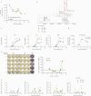
Virological and immunological characterization of the second episode of severe acute respiratory syndrome coronavirus 2 (SARS-CoV-2) reinfection. A, Viral load of SARS-CoV-2 was determined in nasopharyngeal swabs (green circles) and saliva (orange squares) by Real-Time Quantitative Reverse Transcription polymerase chain reaction up to 21 days post–symptom onset. Viral load values were log10 transformed. B, Phylogenetic tree reconstruction of SARS-CoV-2 including European samples from April–May (blue squares) and August–September 2020 (red circles). The sample obtained from our patient from 31 August 2020 is indicated in green. C, The presence of anti–SARS-CoV-2 antibodies, including immunoglobulin G (IgG, left panel), immunoglobulin A (IgA, middle panel), and immunoglobulin M (IgM, right panel), against receptor-binding domain (RBD, blue squares), nucleocapsid (NP, orange triangles), and spike (green circles) antigens (Ag) was quantified by enzyme-linked immunosorbent assay in serum or plasma samples up to 16 days postsymptoms. The signal was analyzed as the optical density (OD) with noise correction. The specific signal for each Ag was calculated after subtracting the background signal obtained for each sample in Ag-free wells. Dotted lines indicate the limit of positivity. D, The presence of neutralizing capacity was determined incubating a serial dilution of serum (light blue squares) or plasma (dark blue circles) with HIV reporter pseudoviruses expressing SARS-CoV-2 S protein and then infecting ACE2-overexpressing HEK293T cells up to 21 days postsymptoms. Neutralization titer was calculated. E, T-cell responses were evaluated using the interferon gamma (IFN-γ) enzyme-linked immunosorbent spot (ELISPOT) assay against NP and spike recombinant proteins in peripheral blood mononuclear cells (PBMCs) up to 21 days postsymptoms. Triplicate wells at each time point are depicted as well as the phytohemagglutinin (PHA)–stimulated positive control (left panel). Longitudinal evolution of spot-forming cells (SFCs) per million PBMCs are shown (right panel). F, Intracellular cytokine production (IFN-γ, interleukin 2 [IL-2], and tumor necrosis factor alpha [TNF-α]) was evaluated in CD4 and CD8 T cells by flow cytometry after stimulation of PBMCs with NP and spike recombinant proteins. Activation-induced markers (CD25 and OX40) after stimulation were also evaluated. Samples below the limit of the detection of the assay are indicated as open symbols.

References

    1. To KK-W, Hung IF-N, IpJD, et al. . COVID-19 re-infection by a phylogenetically distinct SARS-coronavirus-2 strain confirmed by whole genome sequencing [manuscript published online ahead of print 25 August 2020]. Clin Infect Dis 2020. doi:10.1093/cid/ciaa1275. - PMC - PubMed
    1. Van Elslande J, Vermeersch P, Vandervoort K, et al. . Symptomatic SARS-CoV-2 reinfection by a phylogenetically distinct strain [manuscript published online ahead of print 5 September 2020]. Clin Infect Dis 2020. doi:10.1093/cid/ciaa1330. - PMC - PubMed
    1. Gupta V, Bhoyar RC, Jain A, et al. . Asymptomatic reinfection in two healthcare workers from India with genetically distinct SARS-CoV-2 [manuscript published online ahead of print 23 September 2020]. Clin Infect Dis 2020. doi:10.1093/cid/ciaa1451. - PMC - PubMed
    1. Duggan NM, Ludy SM, Shannon BC, et al. . Is novel coronavirus 2019 reinfection possible? Interpreting dynamic SARS-CoV-2 test results through a case report. Am J Emerg Med 2021; et al.:256.e1–3. - PMC - PubMed
    1. Prado-Vivar B, Becerra-Wong M, Guadalupe JJ, et al. . COVID-19 re-infection by a phylogenetically distinct SARS-CoV-2 variant, first confirmed event in South America. SSRN [Preprint]. Posted online 8 September 2020. doi:10.2139/ssrn/3686174.

Publication types