

Relationships Between 24-hour LH and Testosterone Concentrations and With Other Pituitary Hormones in Healthy Older Men
- PMID: 34337275
- PMCID: PMC8315483
- DOI: 10.1210/jendso/bvab075
Relationships Between 24-hour LH and Testosterone Concentrations and With Other Pituitary Hormones in Healthy Older Men
Abstract
Objective: To investigate the relationship between LH and testosterone (T), which characteristics associate with the strength of this relationship, and their interrelationships with GH, TSH, cortisol, and ACTH.
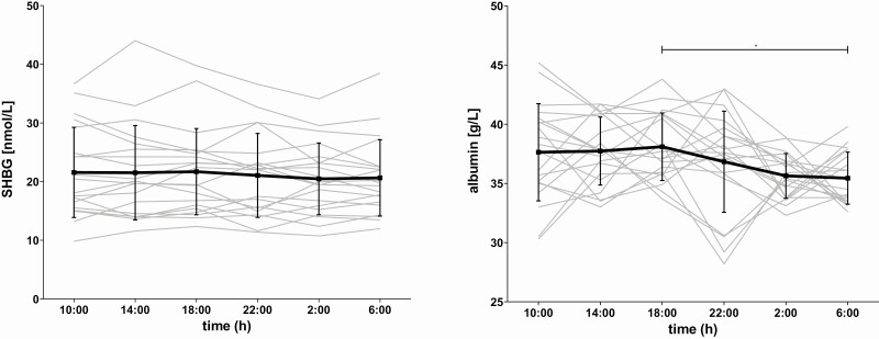
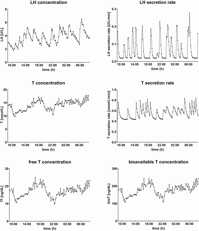
Design: Hormones were measured in serum samples collected every 10 minutes during 24 hours from 20 healthy men, comprising 10 offspring of long-lived families and 10 control subjects, with a mean (SD) age of 65.6 (5.3) years. We performed cross-correlation analyses to assess the relative strength between 2 timeseries for all possible time shifts.
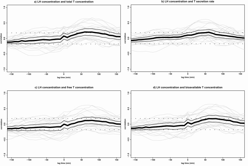
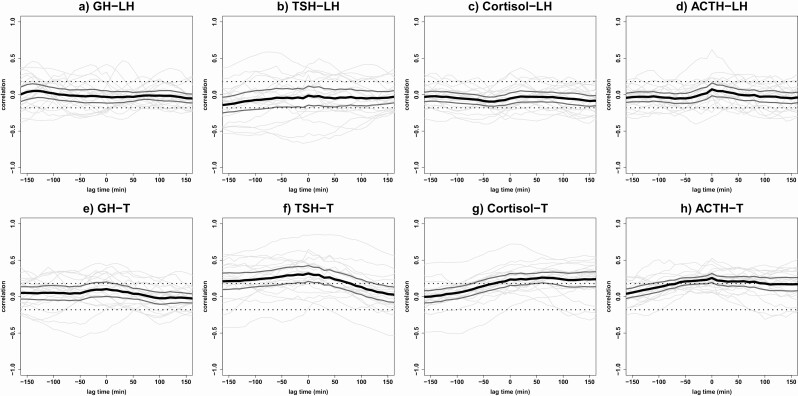
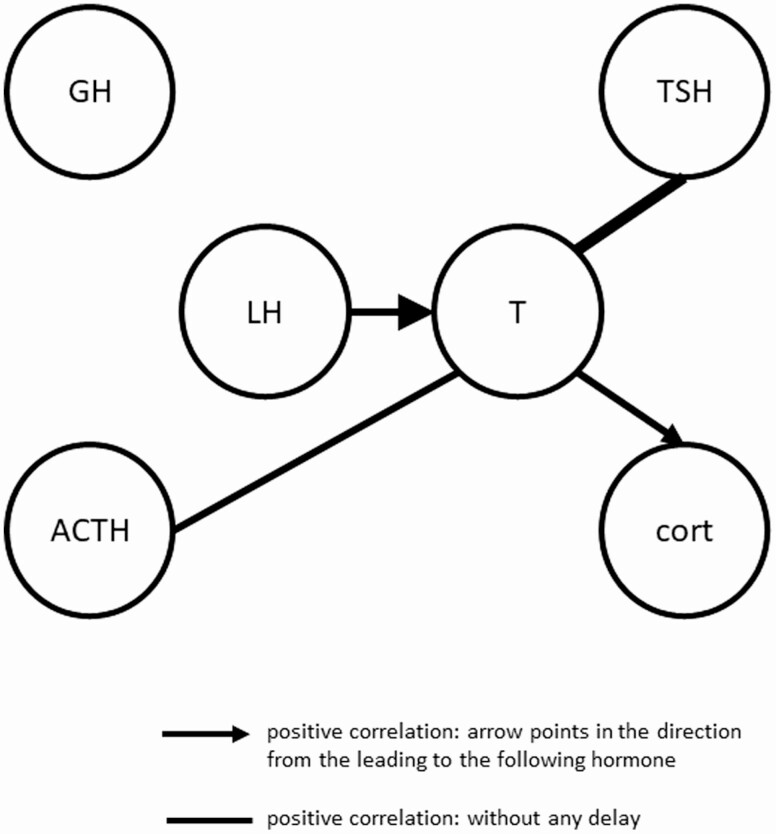
Results: Mean (95% CI) maximal correlation was 0.21 (0.10-0.31) at lag time of 60 minutes between LH and total T concentrations. Results were comparable for calculated free, bioavailable, or secretion rates of T. Men with strong LH-T cross-correlations had, compared with men with no cross-correlation, lower fat mass (18.5 [14.9-19.7] vs. 22.3 [18.4-29.4] kg), waist circumference (93.6 [5.7] vs. 103.1 [12.0] cm), high-sensitivity C-reactive protein (0.7 [0.4-1.3] vs. 1.8 [0.8-12.3] mg/L), IL-6 (0.8 [0.6-1.0] vs. 1.2 [0.9-3.0] pg/mL), and 24-hour mean LH (4.3 [2.0] vs. 6.1 [1.5] U/L), and stronger LH-T feedforward synchrony (1.5 [0.3] vs. 1.9 [0.2]). Furthermore, T was positively cross-correlated with TSH (0.32 [0.21-0.43]), cortisol (0.26 [0.19-0.33]), and ACTH (0.26 [0.19-0.32]).
Conclusions: LH is followed by T with a delay of 60 minutes in healthy older men. Men with a strong LH-T relationship had more favorable body composition, inflammatory markers, LH levels, and LH-T feedforward synchrony. We observed positive correlations between T and TSH, cortisol, and ACTH.
Keywords: ageing; cross-correlation; luteinizing hormone; men; pituitary hormones; testosterone.
© The Author(s) 2021. Published by Oxford University Press on behalf of the Endocrine Society.
Figures
Similar articles
-
Effects of megestrol acetate on pituitary function and end-organ hormone secretion: a post hoc analysis of serum samples from a 12-week study in healthy older men.Am J Geriatr Pharmacother. 2005 Sep;3(3):160-7. doi: 10.1016/s1543-5946(05)80022-4. Am J Geriatr Pharmacother. 2005. PMID: 16257818
-
Interrelationships Between Pituitary Hormones as Assessed From 24-hour Serum Concentrations in Healthy Older Subjects.J Clin Endocrinol Metab. 2020 Apr 1;105(4):e1201-14. doi: 10.1210/clinem/dgz253. J Clin Endocrinol Metab. 2020. PMID: 31853555 Free PMC article.
-
Fasting suppresses pulsatile luteinizing hormone (LH) secretion and enhances orderliness of LH release in young but not older men.J Clin Endocrinol Metab. 1998 Jun;83(6):1967-75. doi: 10.1210/jcem.83.6.4856. J Clin Endocrinol Metab. 1998. PMID: 9626127
-
Five-day pulsatile gonadotropin-releasing hormone administration unveils combined hypothalamic-pituitary-gonadal defects underlying profound hypoandrogenism in men with prolonged critical illness.J Clin Endocrinol Metab. 2001 Jul;86(7):3217-26. doi: 10.1210/jcem.86.7.7680. J Clin Endocrinol Metab. 2001. PMID: 11443192 Clinical Trial.
-
Nature of altered pulsatile hormone release and neuroendocrine network signalling in human ageing: clinical studies of the somatotropic, gonadotropic, corticotropic and insulin axes.Novartis Found Symp. 2000;227:163-85; discussion 185-9. doi: 10.1002/0470870796.ch10. Novartis Found Symp. 2000. PMID: 10752070 Review.
Cited by
-
Salivary testosterone and cortisol responses to seven weeks of practical blood flow restriction training in collegiate American football players.Front Physiol. 2025 Jan 8;15:1507445. doi: 10.3389/fphys.2024.1507445. eCollection 2024. Front Physiol. 2025. PMID: 39844895 Free PMC article.
-
Neuroendocrine and Developmental Impacts of Early Life Exposure to EDCs.J Endocr Soc. 2024 Dec 10;9(1):bvae195. doi: 10.1210/jendso/bvae195. eCollection 2024 Nov 26. J Endocr Soc. 2024. PMID: 39659541 Free PMC article.
-
The influence of light and heavy training weeks on the cortisol and testosterone awakening responses of elite male judokas: is skeletal muscle damage a mediating factor?Biol Sport. 2024 Oct;41(4):187-195. doi: 10.5114/biolsport.2024.135415. Epub 2024 Apr 25. Biol Sport. 2024. PMID: 39416512 Free PMC article.
-
Effects of a 12-week training programme on selected hormonal and psychological parameters and their interrelationships in highly-trained male and female swimmers.Biol Sport. 2025 Apr;42(2):279-287. doi: 10.5114/biolsport.2025.145910. Epub 2024 Dec 13. Biol Sport. 2025. PMID: 40182718 Free PMC article.
-
Pituitary cells in man during aging: An immunohistological and morphometric study.J Med Biochem. 2025 Mar 21;44(2):203-210. doi: 10.5937/jomb0-54605. J Med Biochem. 2025. PMID: 40386521 Free PMC article.
References
-
- Kaufman JM, Lapauw B, Mahmoud A, T’Sjoen G, Huhtaniemi IT. Aging and the male reproductive system. Endocr Rev. 2019;40(4):906-972. - PubMed
-
- Wu FC, Tajar A, Pye SR, et al. ; European Male Aging Study Group . Hypothalamic-pituitary-testicular axis disruptions in older men are differentially linked to age and modifiable risk factors: the European Male Aging Study. J Clin Endocrinol Metab. 2008;93(7):2737-2745. - PubMed
-
- Camacho EM, Huhtaniemi IT, O’Neill TW, et al. ; EMAS Group . Age-associated changes in hypothalamic-pituitary-testicular function in middle-aged and older men are modified by weight change and lifestyle factors: longitudinal results from the European Male Ageing Study. Eur J Endocrinol. 2013;168(3):445-455. - PubMed
-
- Travison TG, Araujo AB, Kupelian V, O’Donnell AB, McKinlay JB. The relative contributions of aging, health, and lifestyle factors to serum testosterone decline in men. J Clin Endocrinol Metab. 2007;92(2):549-555. - PubMed
LinkOut - more resources
Full Text Sources
Research Materials