

Evaluation of Cystic and Solid Renal Lesions with Contrast-Enhanced Ultrasound: A Retrospective Study
- PMID: 34337312
- PMCID: PMC8315990
- DOI: 10.1055/a-1522-8969
Evaluation of Cystic and Solid Renal Lesions with Contrast-Enhanced Ultrasound: A Retrospective Study
Abstract
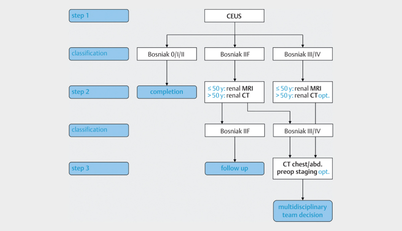
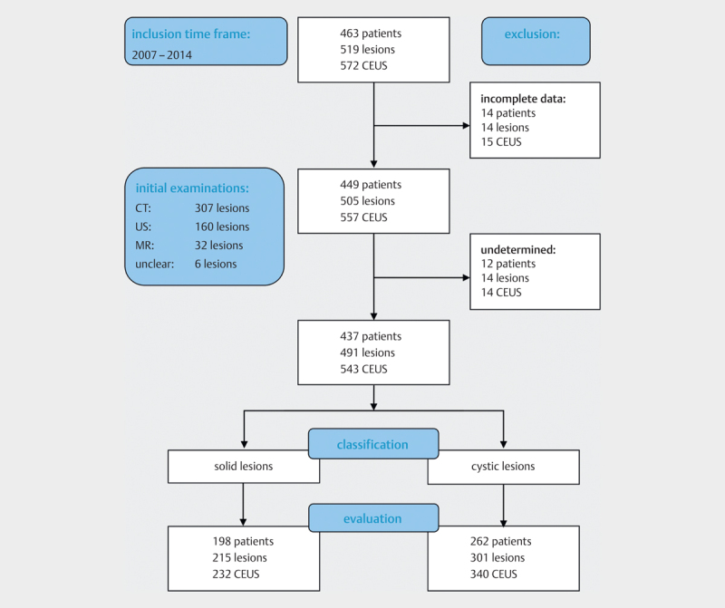
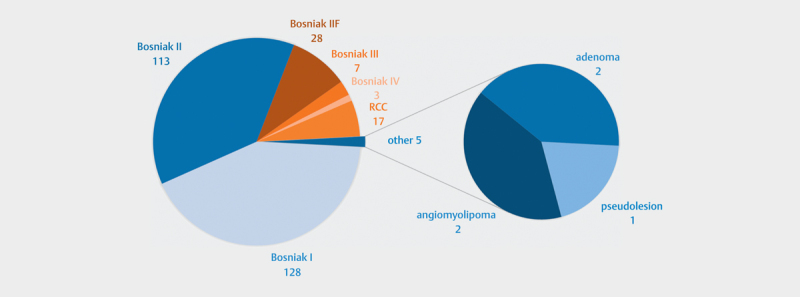
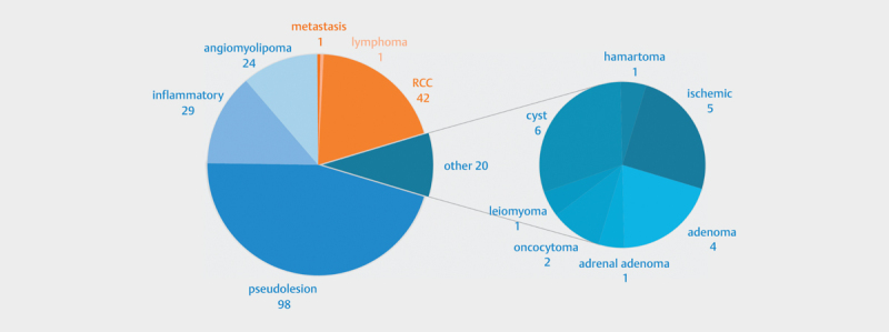
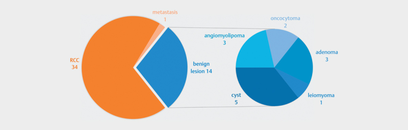
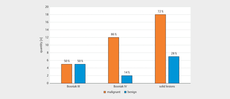
Purpose Renal lesions are frequent random findings on CT, MRI, and conventional ultrasound. Since they are usually found accidentally, the respective examinations have not been performed optimally to provide a conclusive diagnosis, making additional multiphase contrast-enhanced examinations necessary. The aim of the study is to correlate CEUS findings with the final diagnosis and to determine whether it is a suitable method for the conclusive characterization of undetermined renal lesions. Materials and Methods All CEUS examinations of focal renal lesions performed at our institute between 2007 and 2014 were retrospectively examined. 437 patients with a total of 491 lesions and 543 examinations were included. 54 patients had bilateral lesions. One patient had three lesions in one kidney. Histology was available in 49 cases and follow-ups in 124 cases. The sensitivity, specificity, positive and negative predictive value as well as positive and negative likelihood ratios were calculated. Results There were 54 malignant and 437 benign lesions. The sensitivity and specificity were 0.981/0.954 overall, 1.000/0.956 for cystic lesions, 0.977/0.906 for solid lesions, and 0.971/0.071 for the histologically confirmed lesions. Bosniak classification was consistent in 289 of 301 lesions (96%). Only 12 lesions (3.9%) were falsely assessed as malignant. Conclusion CEUS is an appropriate method for the clarification of undetermined renal lesions. The characterization of cystic lesions according to Bosniak is adequately possible, especially for potentially malignant lesions (types III and IV).
Keywords: Bosniak; CEUS; Contrast-Enhanced Ultrasound; RCC; Renal.
The Author(s). This is an open access article published by Thieme under the terms of the Creative Commons Attribution-NonDerivative-NonCommercial-License, permitting copying and reproduction so long as the original work is given appropriate credit. Contents may not be used for commercial purposes, or adapted, remixed, transformed or built upon. (https://creativecommons.org/licenses/by-nc-nd/4.0/).
Conflict of interest statement
Conflict of Interest The authors declare that they have no conflict of interest.
Figures
Similar articles
-
The added value of contrast-enhanced ultrasound in evaluation of indeterminate small solid renal masses and risk stratification of cystic renal lesions.Eur Radiol. 2021 Nov;31(11):8468-8477. doi: 10.1007/s00330-021-07964-0. Epub 2021 Apr 29. Eur Radiol. 2021. PMID: 33912992
-
Contrast-Enhanced Ultrasound (CEUS) for the Evaluation of Bosniak III Complex Renal Cystic Lesions-A 10-Year Specialized European Single-Center Experience with Histopathological Validation.Medicina (Kaunas). 2020 Dec 12;56(12):692. doi: 10.3390/medicina56120692. Medicina (Kaunas). 2020. PMID: 33322683 Free PMC article.
-
Multislice computed tomography versus contrast-enhanced ultrasound in evaluation of complex cystic renal masses using the Bosniak classification system.Clin Hemorheol Microcirc. 2008;39(1-4):171-8. Clin Hemorheol Microcirc. 2008. PMID: 18503122
-
[Reliable diagnosis of cystic renal lesions].Radiologe. 2018 Oct;58(10):887-893. doi: 10.1007/s00117-018-0444-y. Radiologe. 2018. PMID: 30159584 Review. German.
-
[CEUS-diagnostic workup of cystic renal lesions].Radiologe. 2018 Jun;58(6):545-552. doi: 10.1007/s00117-018-0394-4. Radiologe. 2018. PMID: 29728745 Review. German.
Cited by
-
Contrast-enhanced ultrasound in renal cystic lesions: an update.J Med Ultrason (2001). 2024 Oct;51(4):635-647. doi: 10.1007/s10396-024-01489-x. Epub 2024 Aug 20. J Med Ultrason (2001). 2024. PMID: 39164480 Free PMC article. Review.
-
Case report: Ultrasonographic findings of retroperitoneum and abdominal wall metastases of renal cell carcinoma with FH gene deletion.Front Oncol. 2022 Oct 18;12:896477. doi: 10.3389/fonc.2022.896477. eCollection 2022. Front Oncol. 2022. PMID: 36330469 Free PMC article.
-
Contrast-Enhanced Ultrasound in the Diagnosis of Solid Renal Lesions.J Clin Med. 2024 Jun 29;13(13):3821. doi: 10.3390/jcm13133821. J Clin Med. 2024. PMID: 38999387 Free PMC article. Review.
-
DNA Methylation and Epigenetic Events Underlying Renal Cell Carcinomas.Cureus. 2022 Oct 27;14(10):e30743. doi: 10.7759/cureus.30743. eCollection 2022 Oct. Cureus. 2022. PMID: 36447689 Free PMC article. Review.
-
Contrast-enhanced ultrasound of renal masses in the pre-transplant setting: literature review with case highlights.Abdom Radiol (NY). 2024 Dec;49(12):4521-4530. doi: 10.1007/s00261-024-04366-w. Epub 2024 Jun 20. Abdom Radiol (NY). 2024. PMID: 38900316 Free PMC article. Review.
References
-
- Bosniak M A. The current radiological approach to renal cysts. Radiology. 1986;158:1–10. - PubMed
-
- Israel G M, Bosniak M A. An update of the Bosniak renal cyst classification system. Urology. 2005;66:484–488. - PubMed
-
- Huber J, Hallscheidt P, Wagener N.Raumforderungen der Niere: CT vs. MRT. Zwei fast gleichwertige Alternativen mit feinen Unterschieden [Tumours of the Kidney: CT vs. MRI. Nearly equal alternatives with minor differences] Urologe A 201049345–350.German - PubMed
LinkOut - more resources
Full Text Sources