. 2021;4(2):244-263.
doi: 10.20517/cdr.2020.89. Epub 2021 Jun 19.

Exploiting DNA repair pathways for tumor sensitization, mitigation of resistance, and normal tissue protection in radiotherapy

Affiliations

Exploiting DNA repair pathways for tumor sensitization, mitigation of resistance, and normal tissue protection in radiotherapy

Jac A Nickoloff et al. Cancer Drug Resist. 2021.

Abstract

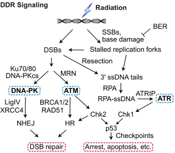
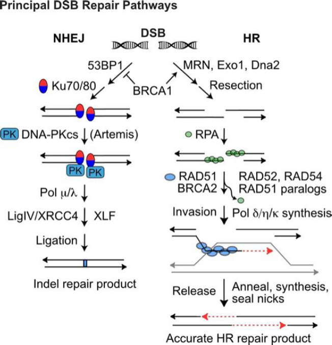
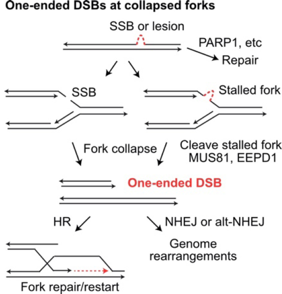
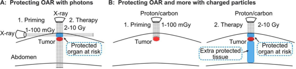
More than half of cancer patients are treated with radiotherapy, which kills tumor cells by directly and indirectly inducing DNA damage, including cytotoxic DNA double-strand breaks (DSBs). Tumor cells respond to these threats by activating a complex signaling network termed the DNA damage response (DDR). The DDR arrests the cell cycle, upregulates DNA repair, and triggers apoptosis when damage is excessive. The DDR signaling and DNA repair pathways are fertile terrain for therapeutic intervention. This review highlights strategies to improve therapeutic gain by targeting DDR and DNA repair pathways to radiosensitize tumor cells, overcome intrinsic and acquired tumor radioresistance, and protect normal tissue. Many biological and environmental factors determine tumor and normal cell responses to ionizing radiation and genotoxic chemotherapeutics. These include cell type and cell cycle phase distribution; tissue/tumor microenvironment and oxygen levels; DNA damage load and quality; DNA repair capacity; and susceptibility to apoptosis or other active or passive cell death pathways. We provide an overview of radiobiological parameters associated with X-ray, proton, and carbon ion radiotherapy; DNA repair and DNA damage signaling pathways; and other factors that regulate tumor and normal cell responses to radiation. We then focus on recent studies exploiting DSB repair pathways to enhance radiotherapy therapeutic gain.

Keywords: DNA double-strand break repair; DNA repair; cancer therapy; homologous recombination; non-homologous end-joining; radioprotection; radiosensitization.


Conflict of interest statement

Conflicts of interest All authors declared that there are no conflicts of interest.

Figures

Figure 1
Figure 1
DDR signaling. Ionizing radiation and genotoxic chemotherapy create single- and double-strand DNA damage including DSBs that activate three PIKKs: DNA-PK, ATM, and ATR. Single-strand breaks and base damage, if not repaired by base excision repair (BER), block replication, which produces ssDNA when the replisome decouples from the MCM helicase or stalled forks are cleaved to produce DSBs, which, along with frank DSBs, are resected to 3’ single-stranded tails that are coated by RPA. This activates ATR to signal checkpoint responses through Chk1 and p53. Non-resected DSB ends are bound by the Ku70/Ku80 heterodimer, which recruits and activates DNA-PKcs in the DNA-PK holoenzyme, LigIV/XRCC4 ligates DNA ends to effect NHEJ. The competing HR pathway initiates with limited DSB end resection by MRE11/RAD50/NBS1 (MRN), more extensive resection by Exo1 and Dna2, and RAD51 binding to ssDNA (mediated by BRCA1, BRCA2, and other proteins) to yield the RAD51-ssDNA nucleoprotein filament that effects HR. DDR: DNA damage response; DSBs: DNA double-strand breaks
Figure 2
Figure 2
DSB repair by NHEJ and HR. Pathway choice is determined by end-resection, prevented by 53BP1, or promoted by BRCA1/MRN and Exo1/Dna2 exonucleases. (Left) NHEJ of unresected DSB ends initiates with Ku70/80 binding to ends and recruitment of DNA-PKcs to form the activated DNA-PK holoenzyme. Artemis is required to trim certain types of end-structures, and small gaps may be filled with polymerases m and l prior to LigIV/XRCC4/XLF-mediated ligation. NHEJ repair usually produces small indels (1-20-bp deletions, few-bp insertions). (Right) Resected 3’ single-strand ends are coated with RPA, which is then exchanged with RAD51, mediated by BRCA2, RAD52, RAD54, and RAD51 paralogs. The RAD51 nucleoprotein filament seeks and invades a homologous donor duplex (grey). RAD51 dissociates before repair synthesis; the newly synthesized strand (red dash) is released from the donor duplex and anneals with the complementary strand on the opposite side of the DSB. A second round of repair synthesis and nick sealing completes repair. DDR: DNA damage response; DSB: DNA double-strand break; NHEJ: non-homologous end-joining; HR: homologous recombination
Figure 3
Figure 3
Repair and mis-repair of one-ended DSBs at collapsed replication forks. Single-strand breaks (SSB) and single-strand DNA lesions may be repaired prior to replication. If encountered by a replication fork, SSBs and single-strand lesions cause fork collapse and fork stalling, respectively. Stalled forks may collapse when cleaved by MUS81 or EEPD1. Collapsed forks create one-ended DSBs that can initiate fork restart by HR, or they may be joined to other DSB ends (at collapsed forks or frank DSBs) by NHEJ or alt-NHEJ, creating large-scale genome rearrangements. DSBs: DNA double-strand breaks; HR: homologous recombination; NHEJ: non-homologous end-joining
Figure 4
Figure 4
Proposed approach to protect normal tissue by stimulating radioadaptive responses. Horizontal X-ray beam delivers a priming dose to protect OAR (blue), but not the tumor (red) from subsequent, high doses delivered with a vertical beam(A); low priming dose of charged particles (left) protect OAR (blue) from subsequent high doses (right) (B). With charged particles, priming and therapeutic doses can be delivered along the same beamline since particles stop at predetermined depths. Charged particles also protect normal tissue distal to the tumor (larger blue section). OAR: organs at risk

References

    1. Otter SJ, Stewart AJ, Devlin PM. Modern brachytherapy. Hematol Oncol Clin North Am. 2019;33:1011–25. doi: 10.1016/j.hoc.2019.08.011. - DOI - PubMed
    1. Daguenet E, Louati S, Wozny AS, et al. Radiation-induced bystander and abscopal effects: important lessons from preclinical models. Br J Cancer. 2020;123:339–48. doi: 10.1038/s41416-020-0942-3. - DOI - PMC - PubMed
    1. Halperin EC, Wazer DE, Perez CA, Brady LW. Perez & Brady’s Principles and Practice of Radiation Oncology. Lippincott Williams & Wilkins; 2018. p. 24.
    1. Mounier NM, Abdel-Maged AE, Wahdan SA, Gad AM, Azab SS. Chemotherapy-induced cognitive impairment (CICI): An overview of etiology and pathogenesis. Life Sci. 2020;258:118071. doi: 10.1016/j.lfs.2020.118071. - DOI - PubMed
    1. Delou JMA, Souza ASO, Souza LCM, Borges HL. Highlights in resistance mechanism pathways for combination therapy. Cells. 2019;8:1013. doi: 10.3390/cells8091013. - DOI - PMC - PubMed

LinkOut - more resources