. 2021 Aug 12;64(15):11379-11394.
doi: 10.1021/acs.jmedchem.1c00717. Epub 2021 Jul 31.

High-Throughput Crystallography Reveals Boron-Containing Inhibitors of a Penicillin-Binding Protein with Di- and Tricovalent Binding Modes

Affiliations

High-Throughput Crystallography Reveals Boron-Containing Inhibitors of a Penicillin-Binding Protein with Di- and Tricovalent Binding Modes

Hector Newman et al. J Med Chem. .

Abstract

The effectiveness of β-lactam antibiotics is increasingly compromised by β-lactamases. Boron-containing inhibitors are potent serine-β-lactamase inhibitors, but the interactions of boron-based compounds with the penicillin-binding protein (PBP) β-lactam targets have not been extensively studied. We used high-throughput X-ray crystallography to explore reactions of a boron-containing fragment set with the Pseudomonas aeruginosa PBP3 (PaPBP3). Multiple crystal structures reveal that boronic acids react with PBPs to give tricovalently linked complexes bonded to Ser294, Ser349, and Lys484 of PaPBP3; benzoxaboroles react with PaPBP3 via reaction with two nucleophilic serines (Ser294 and Ser349) to give dicovalently linked complexes; and vaborbactam reacts to give a monocovalently linked complex. Modifications of the benzoxaborole scaffold resulted in a moderately potent inhibition of PaPBP3, though no antibacterial activity was observed. Overall, the results further evidence the potential for the development of new classes of boron-based antibiotics, which are not compromised by β-lactamase-driven resistance.


Conflict of interest statement

The authors declare no competing financial interest.

Figures

Figure 1
Figure 1
BCIs are proposed to act as mimics of “tetrahedral” transition states arising from enzyme-catalyzed hydrolysis pathways of natural substrates and β-lactams with PBPs. Outline of general mechanisms of (a) transpeptidase reactions catalyzed by a class B PBP, for example, PaPBP3 and (b) the reaction of β-lactam with PBP3, exemplified with a penicillin. Hydrolysis of the acyl–enzyme complex is typically slow in PBPs but is rapid in serine-BL (SBL) catalysis. (c) sp3 form of BCIs may mimic the proposed “tetrahedral” transition states and thus bind tightly to the PBP active site. The ability of boron to “morph” between sp2 and sp3 hybridization states is important in the context of SBL and likely MBL inhibition.
Figure 2
Figure 2
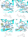
Boronates 1 and 2 react with PaPBP3 in a tricovalent manner. Boronates (a) 1 and (b) 2 react with Ser294, Ser349, and Lys484 (PDB: 7ATM and 7ATO respectively). A dimethyl sulfoxide molecule at the active site in both structures is not shown for clarity (see Figure S10). Hydrogen bonds are shown as black dashed lines. Unbiased omit Fo-Fc maps are shown (light blue mesh) for the ligand and covalently attached side chains (contoured at 1σ), as calculated by “comit” in the ccp4 suite.
Figure 3
Figure 3
Benzoxaboroles react with PaPBP3 in a dicovalent manner. (a) 3 and (b) 4 react covalently with Ser294 and Ser349 and hydrogen bond with Lys484 (PDB: 7ATW and 7ATX, respectively). Hydrogen bonds are shown as black dashed lines. Unbiased omit Fo-Fc maps (light blue mesh) are shown for the ligand and covalently attached residue side chains (contoured at 1σ), as calculated by “comit” in the ccp4 suite.
Figure 4
Figure 4
Binding mode of PaPBP3 inhibited by piperacillin compared with those for benzoxaboroles 7 and 11. (a) PaPBP3 inhibited by piperacillin (PDB: 6R3X); (b) predicted binding mode of 11 as determined by docking, showing how groups 1 and 2 may engage in the same manner as analogous groups in piperacillin; (c) binding mode of 7 complexed with PaPBP3 (PDB: 7AU0). Hydrogen bonds are shown as black dashed lines. An unbiased omit Fo-Fc map is shown (light blue mesh) of the ligand and covalently attached residue side chains (contoured at 1σ), as calculated by “comit” in the ccp4 suite. (d) Structure of piperacillin colored according to its functional groups: β-lactam (orange), C-3 carboxylate (pink), group 1 (blue), and group 2 (green); (e) benzoxaborole 11 was designed to mimic the piperacillin binding mode. Colors match analogous groups within piperacillin which were hoped would engage the same parts of the protein in the case of 11.
Scheme 1
Scheme 1. Synthesis of Benzoxaborole Derivatives (A) 5–10 and 14, (B) 11, and (C) 15
Reagents and conditions: (a) 1,1′-carbonyldiimidazole, N,N-dimethylformamide (DMF), 40 °C, 4–16 h; (b) LiOH·H2O, 1,4-dioxane/H2O (3:1), 40 °C, 60 min; (c) HCl (4 M soln. in 1,4-dioxane), CH2Cl2, rt,16 h; (d) Br2, CH2Cl2, rt, 18 h; (e) benzyl bromide, K2CO3, DMF, rt, 4 h; (f) bis(pinacolato)diboron, Pd(dppf)Cl2, KOAc, dioxane, 80 °C, 12 h; (g) EtOAc, LDA (1 M solution in THF), −78 to −20 °C, 2 h; (h) LiOH·H2O, THF/H2O (1:1), rt, 2 h. Note that low isolation yields for substituted benzoxaboroles (e.g., 8–10) in part reflect significant losses during purification on the silica gel and provide scope for further optimization. For B1, R1 = H and R2 = CH(R3)CO2Me. The complete structures of 57, 14, and 810 are shown in Table 1. Dppf: 1,1′-bis (diphenylphosphino)ferrocene; Boc, tert-butoxycarbonyl; Bn, benzyl.
Figure 5
Figure 5
Structures of benzoxaboroles with a C-3 acid group (12, 13, and 15) and 14 complexed with PaPBP3. (a) PaPBP3/12 complex (PDB: 7AU1); (b) PaPBP3/13 complex (PDB: 7AU8); (c) PaPBP3/14 (PDB: 7AU9); and (d) PaPBP3/15 complex (PDB: 7AUB). For clarity, only one of the two refined conformations of Tyr409 is shown in (a). Hydrogen bonds: dashed black lines. Unbiased omit Fo-Fc maps are shown (light blue mesh) of the ligand and covalently attached residue side chains (contoured at 1σ), as calculated by “comit” in the CCP4 suite.
Figure 6
Figure 6
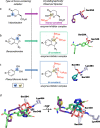
Boron-containing compounds react with a PBP in three distinct modes. The formation of different complexes with exemplary crystallographically observed complexes are shown. (a) Vaborbactam reacts monocovalently with Ser294; (b) Benzoxaboroles (315) bind dicovalently with Ser294 and Ser349; (c) phenylboronic acids (e.g., 1, 2, and alkyl boronic acids) bind tricovalently to Ser294, Ser349, and Lys484. The order of nucleophilic residue reactions is unknown; (d) overlay of crystallographically observed states. The position of the boron can be described by rotations of the chi angles of the Ser294 side chain. In the monocovalently and dicovalently bound crystal structures [and in the piperacillin-reacted structure (PDB: 6R3X), the chi1 angle of Ser294 is gauche- (∼−60°) relative to the serine amine. In contrast, in tricovalent mode (e.g., 1), the chi1 angle of Ser294 is trans (∼−161°) relative to the amine. There is an 80° difference between the chi2 angles of dicovalent mode and monocovalent mode (Figure S9).
Figure 7
Figure 7
Binding of 2 causes changes in the PaPBP3 active-site conformation. Views of benzoxaborole PaPBP3/2 (orange, PDB: 7ATO) and piperacillin-reacted PaPBP3 (black, PDB: 6R3X) (protein backbone colored blue and gray, respectively) are shown. A clear unprecedented difference in the α10-β3 loop conformation (residues 466–473) is observed. See also Figure S14.
Figure 8
Figure 8
Binding of vaborbactam to PaPBP3. Hydrogen bonds: black dashed lines. An unbiased omit Fo-Fc map is shown (light blue mesh) of the ligand and covalently attached residue side chains (contoured at 1 σ), as calculated by “comit” in the ccp4 suite. PDB code 6AUH.

References

    1. Spratt B. G. Distinct Penicillin Binding Proteins Involved in the Division, Elongation, and Shape of Escherichia Coli K12. Proc. Natl. Acad. Sci. U.S.A. 1975, 72, 2999–3003. 10.1073/pnas.72.8.2999. - DOI - PMC - PubMed
    1. Spratt B. G. Temperature-Sensitive Cell Division Mutants of Escherichia Coli with Thermolabile Penicillin-Binding Proteins. J. Bacteriol. 1977, 131, 293–305. 10.1128/jb.131.1.293-305.1977. - DOI - PMC - PubMed
    1. Sauvage E.; Kerff F.; Terrak M.; Ayala J. A.; Charlier P. The Penicillin-Binding Proteins: Structure and Role in Peptidoglycan Biosynthesis. FEMS Microbiol. Rev. 2008, 32, 234–258. 10.1111/j.1574-6976.2008.00105.x. - DOI - PubMed
    1. Adam M.; Fraipont C.; Rhazi N.; Nguyen-Distèche M.; Lakaye B.; Frère J. M.; Devreese B.; Van Beeumen J.; van Heijenoort Y.; van Heijenoort J.; Ghuysen J. M. The Bimodular G57-V577 Polypeptide Chain of the Class B Penicillin-Binding Protein 3 of Escherichia Coli Catalyzes Peptide Bond Formation from Thiolesters and Does Not Catalyze Glycan Chain Polymerization from the Lipid II Intermediate. J. Bacteriol. 1997, 179, 6005–6009. 10.1128/jb.179.19.6005-6009.1997. - DOI - PMC - PubMed
    1. Tipper D. J.; Strominger J. L. Mechanism of Action of Penicillins: A Proposal Based on Their Structural Similarity to Acyl-D-Alanyl-D-Alanine. Proc. Natl. Acad. Sci. U.S.A. 1965, 54, 1133–1141. 10.1073/pnas.54.4.1133. - DOI - PMC - PubMed

Publication types

MeSH terms