Review
. 2022 Jan 1;102(1):379-410.
doi: 10.1152/physrev.00043.2020. Epub 2021 Aug 2.

The osteocyte as a signaling cell

Affiliations
Review

The osteocyte as a signaling cell

Jesus Delgado-Calle et al. Physiol Rev. .

Abstract

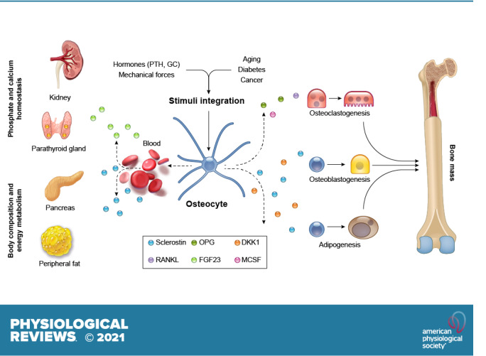
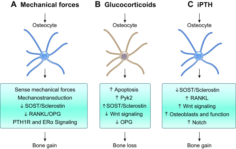
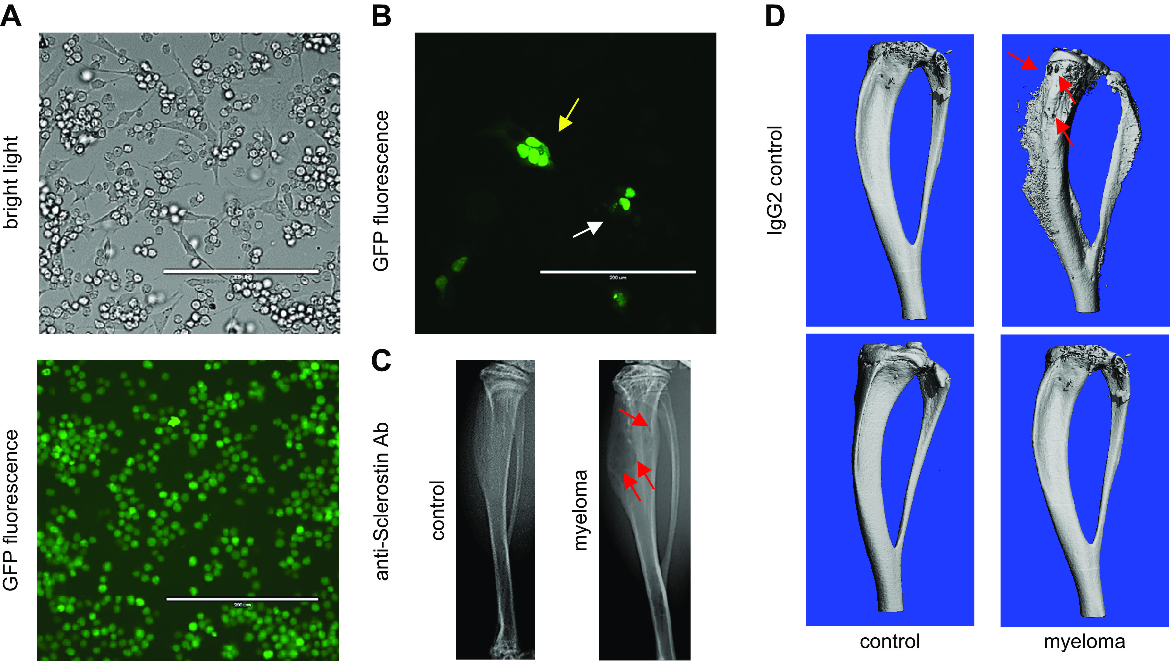
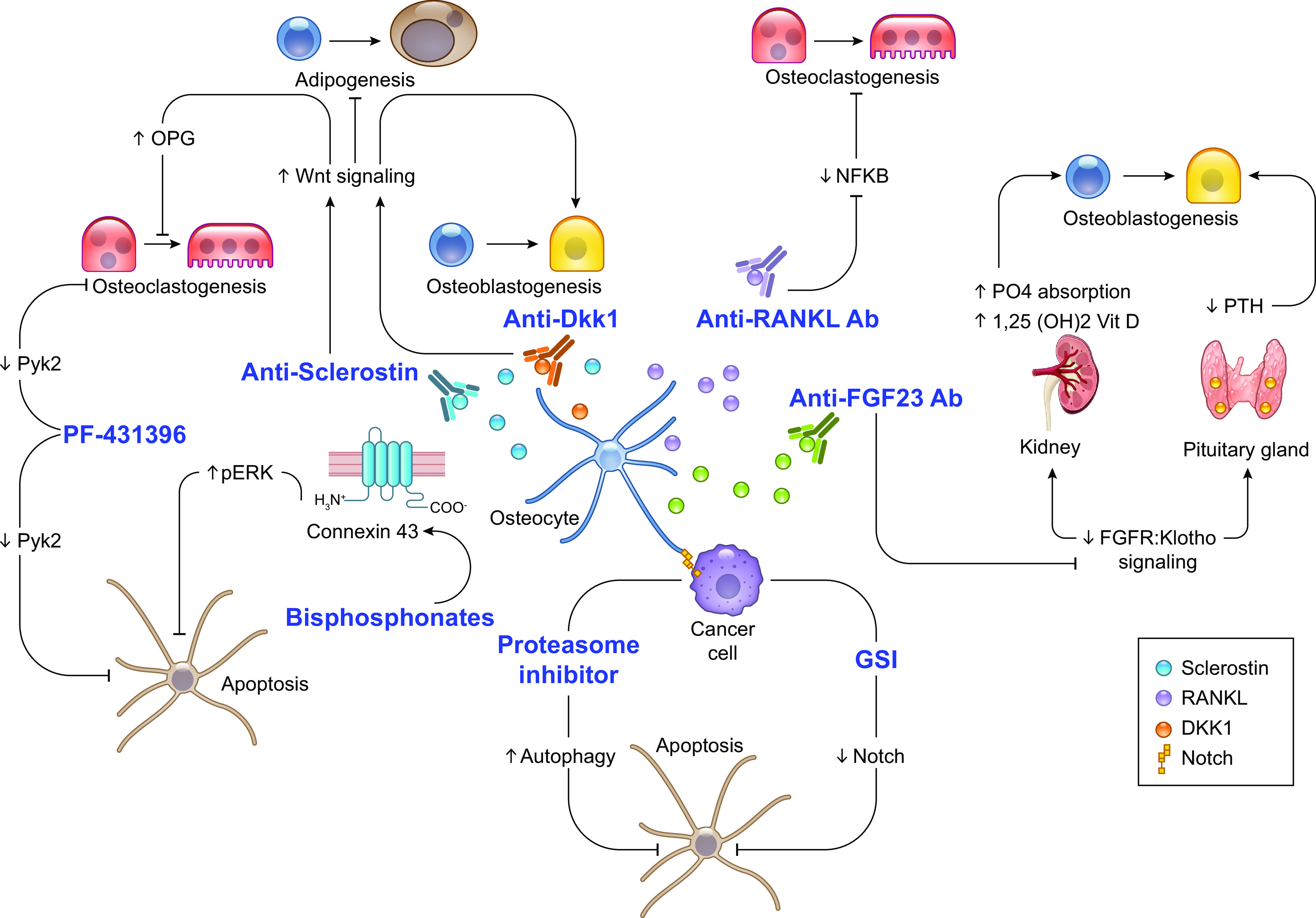
Osteocytes, former osteoblasts encapsulated by mineralized bone matrix, are far from being passive and metabolically inactive bone cells. Instead, osteocytes are multifunctional and dynamic cells capable of integrating hormonal and mechanical signals and transmitting them to effector cells in bone and in distant tissues. Osteocytes are a major source of molecules that regulate bone homeostasis by integrating both mechanical cues and hormonal signals that coordinate the differentiation and function of osteoclasts and osteoblasts. Osteocyte function is altered in both rare and common bone diseases, suggesting that osteocyte dysfunction is directly involved in the pathophysiology of several disorders affecting the skeleton. Advances in osteocyte biology initiated the development of novel therapeutics interfering with osteocyte-secreted molecules. Moreover, osteocytes are targets and key distributors of biological signals mediating the beneficial effects of several bone therapeutics used in the clinic. Here we review the most recent discoveries in osteocyte biology demonstrating that osteocytes regulate bone homeostasis and bone marrow fat via paracrine signaling, influence body composition and energy metabolism via endocrine signaling, and contribute to the damaging effects of diabetes mellitus and hematologic and metastatic cancers in the skeleton.

Keywords: Sclerostin; bone; cancer; hormonal signals; mechanical signals.


Conflict of interest statement

No conflicts of interest, financial or otherwise, are declared by the authors.

Figures

None
Graphical abstract
FIGURE 1.
FIGURE 1.
Osteocyte morphology and lacuno-canalicular network. A: osteocytes are highly interconnected to each other and to cells on the bone surface. Canaliculi connect osteocytes to the endocortical bone surface and the bone marrow compartment. Electron microscopy imaging of an acid etched bone. B: osteocyte-derived factors circulate through the canalicular system to reach other osteocytes and bone cells on the bone surface or in bone marrow. Immunostaining of Sclerostin in paraffin-embedded bone tissue. C: MLO-Y4 osteocyte-like cells display a stellate morphology with dendritic processes resembling those found in primary osteocytes. White arrows point to individual osteocytes; yellow arrows point to canaliculi.
FIGURE 2.
FIGURE 2.
Osteocyte apoptosis: key molecular mechanisms. A: scheme showing that proapoptotic stimuli activate death receptors in osteocytes and initiate caspase-3-mediated programmed cell death. Osteocytes adjacent to apoptotic osteocytes are thought to release proinflammatory and proosteoclastogenic cytokines that attract osteoclasts and promote local bone resorption. B: image showing TUNEL assay to detect degraded DNA in apoptotic osteocytes in paraffin-embedded bones infiltrated with myeloma cancer cells. C: image showing active caspase-3 immunostaining of paraffin-embedded human bone from an osteoporotic patient. Black arrows point to apoptotic osteocytes; red arrows point to healthy osteocytes. AGE, advanced glycation end-products; HMGB1, high mobility group box 1; ROS, reactive oxygen species; TNF, tumor necrosis factor-α; TRAIL, tumor necrosis factor-related apoptosis inducing ligand.
FIGURE 3.
FIGURE 3.
Osteocyte paracrine signaling in bone. Osteocytes exert paracrine actions on osteoblasts and osteoclasts via cell-to-cell physical communication and/or secretion of molecules that stimulate or inhibit osteoblast or osteoclast differentiation. A: osteocytes produce the proosteoclastogenic cytokines receptor activator of nuclear factor-κΒ ligand (RANKL) and macrophage colony-stimulating factor (M-CSF) in both membrane-bound and soluble forms and the anti-osteoclastogenic factor osteoprotegerin (OPG). B: osteocyte-derived Sclerostin acts on mesenchymal stem cell precursors and promotes bone marrow adipogenesis. C: Wnt antagonists Dkk-1 and Sclerostin produced by osteocytes inhibit osteoblast differentiation and bone forming function. Furthermore, osteocytes express Notch receptors that can communicate physically with osteoblasts and potentially with osteoclasts.
FIGURE 4.
FIGURE 4.
Osteocyte regulation of bone formation via Sclerostin production. A: Sclerostin immunostaining in paraffin-embedded human bone. Black arrow points to Sclerostin-positive osteocytes; red arrow points to Sclerostin-negative osteocytes. B: micro-computed tomography (μCT) image of vertebral cancellous bone from wild-type mice (WT) and mice with global deletion of SOST (SOST KO). C: toluidine blue-tartrate-resistant acid phosphatase (TRAP)-stained histological image of tibial cancellous bone from WT and SOST KO mice. D: dynamic histomorphometry in plastic-embedded bones from WT mice and mice overexpressing human SOST under the control of the DMP1-8kb promoter (Dmp1-8kb-hSOST). Green calcein labels and red alizarin labels are shown. E: toluidine blue-TRAP stained histological image of bones from C57BL/6 mice treated with IgG (control) or neutralizing anti-Sclerostin antibody (anti-Sclerostin-Ab). Yellow arrows point to osteoblasts on the bone surface.
FIGURE 5.
FIGURE 5.
The osteocyte as the anabolic Wnt signaling cell in bone. Mice with genetic constitutive activation of canonical Wnt/β-catenin signaling in osteocytes (daβcatOt) have been used to determine the cell responsible for the bone anabolic effects of canonical Wnt signaling. A: daβcatOt mice are smaller in size compared with wild-type (WT) control littermates and exhibit denser bones. B: daβcatOt mice exhibit exuberant bone formation that expands into the bone marrow cavity of the femoral midshaft. C: dynamic histomorphometry in a plastic-embedded bone from a daβcatOt mouse. Green calcein labels and red alizarin labels are shown.
FIGURE 6.
FIGURE 6.
Osteocytes as mediators of mechanical and hormonal signals in bone. A: osteocytes sense mechanical forces in bone and transduce them into biological cues to effector cells. Mechanical forces decrease Sclerostin, increase Wnt signaling, and decrease osteoprotegerin (OPG) expression in osteocytes to enable bone gain. ER, estrogen receptor; PTH1R, parathyroid hormone (PTH) receptor 1. B: glucocorticoid direct actions on osteocytes increase apoptosis (via Pyk2 activation) and Sclerostin expression, leading to decreased Wnt signaling and OPG expression and overall contributing to the bone loss induced by glucocorticoids. C: intermittent administration of PTH (iPTH) requires osteocytic PTH1R signaling to induce full bone anabolism. Mechanistically, PTH decreases SOST expression, activates Wnt signaling, and increases osteoblast number and function. Additionally, iPTH increases receptor activator of nuclear factor-κΒ ligand (RANKL) expression and promotes Notch signaling to regulate PTH-induced bone resorption.
FIGURE 7.
FIGURE 7.
Osteocytes generate a microenvironment supportive for the growth of cancer cells and exacerbated bone resorption. A: cocultures of osteocytes and myeloma-green fluorescent protein (GFP) cancer cells enable the study of communication by cell-to-cell direct cell contact and by the exchange of soluble factors. Under these conditions, myeloma cells establish physical contact with the body and dendritic processes of osteocytes. B: osteocytes transfected with nuclear GFP cocultured with myeloma cancer cells. Fragmented (yellow arrow) and normal (white arrow) nuclei in MLO-Y4 osteocytes cocultured with MM1.S myeloma cells are displayed. C: murine intratibial injection of myeloma cells engrafts and produces multiple myeloma (MM) tumors in the bone marrow tumors that induce osteolytic lesions (red arrows) similar to those seen in myeloma patients. D: pharmacological blockade of the osteocyte-derived factor Sclerostin with a neutralizing antibody (anti-Sclerostin Ab) prevents the progression of the myeloma-induced bone disease in mice bearing myeloma tumors. Three-dimensional micro-computed tomography (μCT) reconstructions of C57BL/KaLwRijHsd bones bearing 5TGM1 myeloma cells are shown.
FIGURE 8.
FIGURE 8.
Osteocytes and osteocyte-derived factors as therapeutic targets. A better understanding of osteocyte biology prompted the generation of therapeutics targeting osteocytes and osteocyte-secreted signaling molecules. Antibodies designed to neutralize Sclerostin (clinically tested and FDA approved) and Dkk1 (clinically tested but not FDA approved) function were developed to boost bone formation in osteoporotic patients. Similarly, a neutralizing antibody against receptor activator of nuclear factor-κΒ ligand (RANKL) (clinically tested and FDA approved) was generated to stop bone loss in osteoporotic and cancer patients. More recently, a neutralizing antibody against fibroblast growth factor (FGF)23 (clinically tested and FDA approved) has been approved for the treatment of X-linked hypophosphatemia and tumor-induced osteomalacia. Furthermore, we know now that bisphosphonates (clinically tested and FDA approved), proteasome inhibitors (clinically tested and FDA approved), and pan-inhibitors of the Notch pathway (clinically tested but not FDA approved; bone-targeting molecule (BT)-gamma secretase inhibitor (GSI) experimental] preserve osteocyte viability. Finally, pharmacological inhibition of Pyk2 signaling with PF-431396 (experimental, not FDA approved) decreases glucocorticoid induced osteocyte apoptosis and promotes osteoclast cell death. NFKB, nuclear factor-κΒ; OPG, osteoprotegerin; PTH, parathyroid hormone.

References

    1. Parfitt AM. Life history of osteocytes: relationship to bone age, bone remodeling, and bone fragility. J Musculoskelet Neuronal Interact 2: 499–500, 2002. - PubMed
    1. Plotkin LI, Bellido T. Osteocytic signalling pathways as therapeutic targets for bone fragility. Nat Rev Endocrinol 12: 593–605, 2016. doi: 10.1038/nrendo.2016.71. - DOI - PMC - PubMed
    1. Paic F, Igwe JC, Nori R, Kronenberg MS, Franceschetti T, Harrington P, Kuo L, Shin DG, Rowe DW, Harris SE, Kalajzic I. Identification of differentially expressed genes between osteoblasts and osteocytes. Bone 45: 682–692, 2009. doi: 10.1016/j.bone.2009.06.010. - DOI - PMC - PubMed
    1. Piemontese M, Onal M, Xiong J, Han L, Thostenson JD, Almeida M, O’Brien CA. Low bone mass and changes in the osteocyte network in mice lacking autophagy in the osteoblast lineage. Sci Rep 6: 24262, 2016. doi: 10.1038/srep24262. - DOI - PMC - PubMed
    1. Palumbo C, Palazzini S, Zaffe D, Marotti G. Osteocyte differentiation in the tibia of newborn rabbit: an ultrastructural study of the formation of cytoplasmic processes. Acta Anat (Basel) 137: 350–358, 1990. doi: 10.1159/000146907. - DOI - PubMed

Publication types

LinkOut - more resources