Review
. 2021 Sep 20;50(18):10451-10485.
doi: 10.1039/d0cs01275k.

Advances in chemical probing of protein O-GlcNAc glycosylation: structural role and molecular mechanisms

Affiliations
Review

Advances in chemical probing of protein O-GlcNAc glycosylation: structural role and molecular mechanisms

Abhijit Saha et al. Chem Soc Rev. .

Abstract

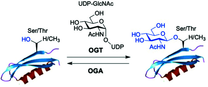
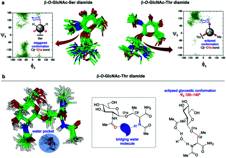
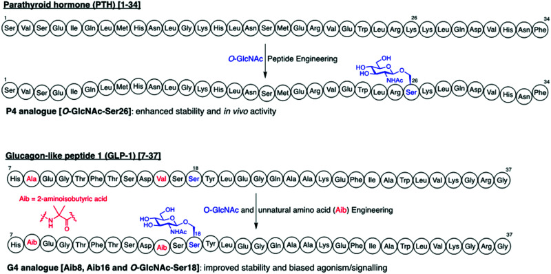
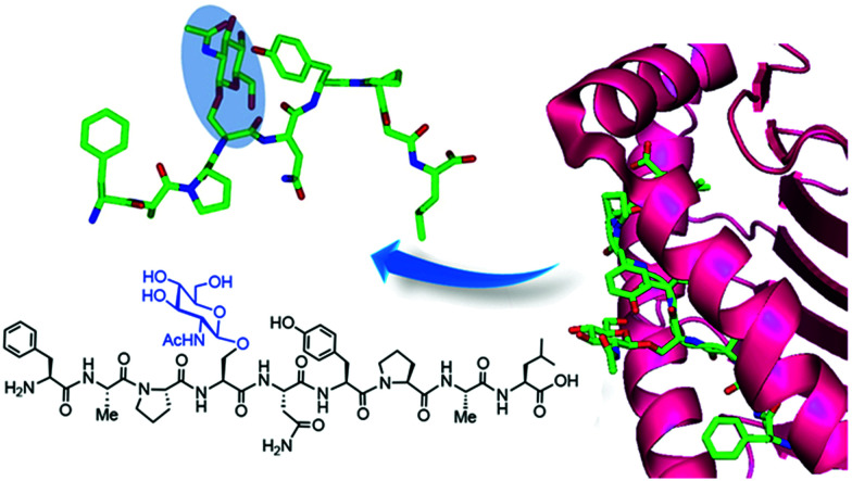
The addition of O-linked-β-D-N-acetylglucosamine (O-GlcNAc) onto serine and threonine residues of nuclear and cytoplasmic proteins is an abundant, unique post-translational modification governing important biological processes. O-GlcNAc dysregulation underlies several metabolic disorders leading to human diseases, including cancer, neurodegeneration and diabetes. This review provides an extensive summary of the recent progress in probing O-GlcNAcylation using mainly chemical methods, with a special focus on discussing mechanistic insights and the structural role of O-GlcNAc at the molecular level. We highlight key aspects of the O-GlcNAc enzymes, including development of OGT and OGA small-molecule inhibitors, and describe a variety of chemoenzymatic and chemical biology approaches for the study of O-GlcNAcylation. Special emphasis is placed on the power of chemistry in the form of synthetic glycopeptide and glycoprotein tools for investigating the site-specific functional consequences of the modification. Finally, we discuss in detail the conformational effects of O-GlcNAc glycosylation on protein structure and stability, relevant O-GlcNAc-mediated protein interactions and its molecular recognition features by biological receptors. Future research in this field will provide novel, more effective chemical strategies and probes for the molecular interrogation of O-GlcNAcylation, elucidating new mechanisms and functional roles of O-GlcNAc with potential therapeutic applications in human health.


Conflict of interest statement

There are no conflicts to declare.

Figures

Fig. 1
Fig. 1. The enzyme O-GlcNAc transferase (OGT) catalyses the addition of O-GlcNAc to nuclear and cytoplasmic proteins, whereas the enzyme O-GlcNAcase (OGA) catalyses the removal of the sugar.
Fig. 2
Fig. 2. (a) Active site view of truncated OGT in complex with UDP-5S-GlcNAc and CKII peptide (PDB: 4GYY) showing key interacting residues and polar contacts (black dashed lines) (left panel); mechanism of O-GlcNAcylation by OGT highlighting important active site enzyme residues and interactions (right panel). (b and c) Chemical structures of some OGT inhibitors and their reported potencies: (b) BZX1, BZX2, OSMI-1, OSMI-4 identified via high-throughput screening, (c) Ac4-5S-GlcNAc, 5S-GlcNHex, goblin1 and its S-linked analogue, designed based on the structures of the UDP-GlcNAc donor substrate and the UDP reaction product.
Fig. 3
Fig. 3. (a) Catalytic mechanism of human O-GlcNAcase (hOGA) deglycosylation (hydrolysis) proceeds through an oxazoline intermediate with the assistance of Asp174 and Asp175 as catalytic acid–base residues. (b) Active site view of OGA in complex with thiamEt-G (depicted as an overlay of three different PDB structures (5UN9, 5M7S, 5UHL)) showing key interacting residues and contacts (black dashed lines). (c) Chemical structure of some OGA inhibitors with their reported potencies: PUGNAc, GlcNAcstatin G, NAG-thiazoline, NButGT, thiamEt-G, its methyl analogue thiamMe-G, its difluoro congener MK-8719, and iminocyclitol derivative VV347.
Fig. 4
Fig. 4. (a) Schemes of the GlcNAc de novo hexosamine biosynthetic pathway (HBP) and salvage pathway from exogenous GlcNAc, showing a range of synthetic metabolic chemical reporters (MCRs). The engineered GlcNAc analogues enter the salvage pathway and are converted to the corresponding UDP-GlcNAc derivatives in the cell; subsequent OGT-catalysed O-GlcNAcylation enables labelling of the relevant O-GlcNAc modified proteins. (b) General workflow showing how the probes are applied for the identification of the O-GlcNAcylated proteins.
Fig. 5
Fig. 5. Identification of O-GlcNAc-mediated protein interactions and binding partners using a metabolic labelling photocrosslinking approach. Cells are cultured with a synthetic diazirine-functionalised, cell-permeable precursor [Ac3GlcNDAz-1-P(Ac-SATE)2], which is deprotected inside the cell by esterases and converted to UDP-GlcNDAz by an AGX1 mutant. OGT (or an engineered OGT with enhanced substrate preference) transfers GlcNDAz to its native substrates, labelling O-GlcNAcylated proteins with the unnatural modification. Short UV irradiation (365 nm) activates the diazirine for carbene-mediated crosslinking with the neighbouring binding partners. After cell lysis, immunoprecipitation of the covalent protein complex with an anti-O-GlcNAc antibody, followed by SDS-PAGE separation, in-gel tryptic digestion and subsequent proteomics analysis by LC-MS/MS, enables the identification of O-GlcNAc interacting proteins.
Fig. 6
Fig. 6. Chemo-enzymatic labelling of endogenous O-GlcNAcylated proteins by enzymatic modification with unnatural UDP-Gal analogues ((a) keto or (b) azide-functionalised) using a mutant GalT(Y289L). Subsequent chemical probing with (cleavable) biotin, fluorescent or PEG mass tags using (a) oxime or (b) click-chemistry for identification of the O-GlcNAcylated proteins.
Fig. 7
Fig. 7. Bioorthogonal ligation reactions employed for probing O-GlcNAcylation: oxime ligation, Staudinger ligation, “click” chemistry (CuAAC and SPAAC), iEDDA reaction, and isonitrile–tetrazine ligation.
Fig. 8
Fig. 8. Chemical methods for site-specific installation of O-GlcNAcylation on target proteins: (a) chemical protein modification (post-translational mutagenesis) via the tag-and-modify approach using dehydroalanine (Dha) and GlcNAc mimics. (b) Synthetic glycopeptide and protein chemistry for NCL/EPL-enabled O-GlcNAc glycoprotein (semi)synthesis.
Fig. 9
Fig. 9. (a) Distribution of the ϕs/ψs torsion angles (glycosidic linkage) and major conformations (calculated ensembles) in solution for β-O-GlcNAc-Ser (left) and β-O-GlcNAc-Thr (right) derived from NMR-guided MD simulations. The Newman projections for the Cβ-O1s bond are also included, showing the staggered conformation (Ser derivative) and eclipsed conformation (Thr derivative). (b) Key water pocket (bridging water molecule) between the GlcNAc (N-acetyl group) and the peptide backbone (Thr-nitrogen) derived from NMR-guided MD simulations for β-O-GlcNAc-Thr due to its distinct conformational behaviour in solution.
Fig. 10
Fig. 10. Synthetic engineering of therapeutic peptides PTH and GLP-1 via artificial O-GlcNAc installation.
Fig. 11
Fig. 11. (a) Model of WGA lectin derived from the X-ray crystal structure in complex with GlcNAc (PDB: 2UVO). (b) High affinity tetravalent neoglycopeptide (IC50 = 0.9 μM) and divalent GlcNAc ligands display potent multivalent binding to WGA. (c) Selenium-based GlcNAc affinity ligands applied for detection of GlcNAc–WGA interactions by 77Se NMR. (d) Fluoroacetamide-containing chitobiose and chitotriose mimics used as NMR probes for molecular recognition studies between WGA and GlcNAc glycans.
Fig. 12
Fig. 12. Chemical structure of synthetic lectins ((a) first-generation design, (b) second-generation “eclipsed” receptor) for β-O-GlcNAc, showing intermolecular CH-aromatic contacts (purple dashed lines) and polar hydrogen-bond interactions (green dashed lines).
Fig. 13
Fig. 13. Crystal structure of the FAPS(β-O-GlcNAc)NYPAL glycopeptide (termed K3G) in complex with the MHC class I H-2Db molecule, showing one of the two major conformers for the O-GlcNAc residue.
None
Abhijit Saha
None
Davide Bello
None
Alberto Fernández-Tejada

References

    1. Torres C. R. Hart G. W. J. Biol. Chem. 1984;259:3308–3317. doi: 10.1016/S0021-9258(17)43295-9. - DOI - PubMed
    1. Holt G. D. Hart G. W. J. Biol. Chem. 1986;261:8049–8057. doi: 10.1016/S0021-9258(19)57510-X. - DOI - PubMed
    1. van der Laarse S. A. M. Leney A. C. Heck A. J. R. FEBS J. 2018;285:3152–3167. doi: 10.1111/febs.14491. - DOI - PubMed
    1. Lubas W. A. Frank D. W. Krause M. Hanover J. A. J. Biol. Chem. 1997;272:9316–9324. doi: 10.1074/jbc.272.14.9316. - DOI - PubMed
    1. Kreppel L. K. Blomberg M. A. Hart G. W. J. Biol. Chem. 1997;272:9308–9315. doi: 10.1074/jbc.272.14.9308. - DOI - PubMed

MeSH terms