Review
. 2022 Jan 17;26(1):105-122.
doi: 10.5935/1518-0557.20210026.

Oocyte quality and aging

Affiliations
Review

Oocyte quality and aging

Ali Reza Eftekhari Moghadam et al. JBRA Assist Reprod. .

Abstract

It is well known that female reproduction ability decreases during the forth decade of life due to age-related changes in oocyte quality and quantity; although the number of women trying to conceive has today increased remarkably between the ages of 36 to 44. The causes of reproductive aging and physiological aspects of this phenomenon are still elusive. With increase in the women's age, during Assisted Reproductive Technologies (ART) we have perceived a significant decline in the number and quality of retrieved oocytes, as well as in ovarian follicle reserves. This is because of increased aneuploidy due to factors such as spindle apparatus disruption; oxidative stress and mitochondrial damage. The aim of this review paper is to study data on the potential role of the aging process impacting oocyte quality and female reproductive ability. We present the current evidence that show the decreased oocyte quality with age, related to reductions in female reproductive outcome. The aging process is complicated and it is caused by many factors that control cellular and organism life span. Although the factors responsible for reduced oocyte quality remain unknown, the present review focuses on the potential role of ovarian follicle environment, oocyte structure and its organelles. To find a way to optimize oocyte quality and ameliorate clinical outcomes for women with aging-related causes of infertility.

Keywords: aging; female infertility; oocyte quality; ovary.


Conflict of interest statement

CONFLICT OF INTERESTS

The authors declare that there is no conflict of interests.

Figures
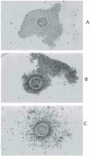
Figure 1
Figure 1
Different classes of COCs (de Wit et al., 2000). Class A. Oocyte is surrounded by a compact cumulus with a homogeneous cytoplasm. Class B. Oocyte is surrounded by a less compact and darker cumulus. The cytoplasm is dark and moderately granular. Class C. Oocyte is surrounded by dispersed cumulus cells. Oocyte has a granular cytoplasm. Reproduced from Lasiene et al. (2009) with permission.
Figure 2
Figure 2
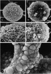
Conventional SEM. (a): Unfertilized human oocyte. Porousnet appearance of ZP (X 2,000). (b): Unfertilized human oocyte. Compact and smooth surfaced ZP (X 2,000). (c): Higher magnification of (a). The spongy ZP structure is evident (X 4,000). (d): Higher magnification of (b). It shows a dense and compact ZP structure (X 4,000). (e): Unfertilized mouse oocyte with very high magnification showing a branch of the spongy structure of the ZP (X 350,000). Familiari et al. (2006) with permission.
Figure 3
Figure 3
(a): The outer surface of the ZP of a human mature oocyte. Many fenestrations are present in which the filaments form a large meshed network (X 9,000). (b): The outer surface of the ZP of a human atretic oocyte. The filaments form a tight meshed network (X 9,000). Familiari et al. (2006) with permission.
Figure 4
Figure 4
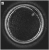
An ideal oocyte which displaying good spindle retardance and length, trilaminar structure of zona with good retardance of inner layer. Magnification ×200. Reproduced from Rama Raju et al. (2007) with permission.
Figure 5
Figure 5
Calculation method for measuring the size of each part of the oocyte. ZP: Zona pellucida. PS: perivitelline space. PB: 1st polar body. Diameter of cytoplasm (A) (A1+A2) / 2. Inner diameter of zona pellucida (B) = (B1+B2) / 2. Outer diameter of zona pellucida (C) = (C1+C2) / 2. Thickness of zona pellucida = (C B) / 2. Size of perivitelline space = (B A) / 2. Yoshida & Niimura (2010) with permission.
Figure 6
Figure 6
Young oocytes (A) expanded against the zona pellucida with a minimal perivitelline space (PVS) and a clear indentation at the polar body, whereas old oocytes (B) had wide PVS with no indentation from the polar body. Reproduced from Li et al. (2017) with permission.
Figure 7
Figure 7
MII oocytes after denudation of cumulus cells before microinjection. (A) normal PBI and (B) fragmented PBI. Halvaei et al. (2011) with permission.
Figure 8
Figure 8
(A) Mature oocyte. Tubules (SER) and vesicles (V) of smooth endoplasmic reticulum associated with mitochondria (M) to form M-SER aggregates and MV complexes. TEM, bar is 0,5 µm. (B) Early degenerating mature oocyte. Numerous vacuoles (Va) associated with lysosomes (Ly) are found in the ooplasm. TEM bar is 1,2 µm. Nottola et al. (2007) with permission.
Figure 9
Figure 9
Confocal micrographs of fresh and aged mice oocytes. Configuration of meiotic spindle and chromosomes in (A) Normal spindle morphology of fresh oocytes, microtubules traversing between both poles and chromosomes aligned in a compact group on the equatorial plane (B) and (c) abnormal spindle morphologies of aged oocytes. Miao et al. (2009) with permission.
Figure 10
Figure 10
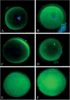
Confocal microscopy images demonstrating mitochondria distribution during maturation of human oocytes. (A) GV oocyte displays the distribution of mitochondria in the peripheral zone. (B) GV oocyte with mitochondria diffused fairly in the cytoplasm. (C) MI oocyte with mitochondria displaying a peripheral type of distribution. (D) In vitro matured oocyte indicating the semiperipheral distribution of mitochondria in the oocyte. (E) In vitro matured oocyte showing the distribution of mitochondria throughout the cytoplasm. (F) In vivo matured oocyte: mitochondria are disseminated throughout the cytoplasm, and there are mitochondria in the central region. Scale bar represents 50 µm. Liu et al. (2010) with permission.
Figure 11
Figure 11
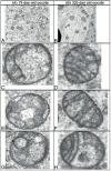
TEM images of mitochondria from young and old mice oocytes. MII oocytes from hyperstimulated young and old mice were evaluated and the mitochondrial structures were compared. Representative electron micrographs of ooplasm at 11,000x magnification are shown; higher magnification views of individual mitochondria are also presented. (A) abundant mitochondria per field, also notice different size mitochondria compared to B. (B) relatively few mitochondria of uniform size per field in ooplasm of an aged animal. (C & E) undifferentiated round mitochondria with an electron dense matrix vs D, F & H. More differentiated mitochondria with an elongated shape and distinct cristae G. arrow indicating vacuoles within mitochondria, Kushnir et al. (2012) with permission.

References

    1. Absalan F, Saremy S, Mansori E, Taheri Moghadam M, Eftekhari Moghadam AR, Ghanavati R. Effects of Mono-(2-Ethylhexyl) Phthalate and Di-(2-Ethylhexyl) Phthalate Administrations on Oocyte Meiotic Maturation, Apoptosis and Gene Quantification in Mouse Model. Cell J. 2017;18:503–513. doi: 10.22074/cellj.2016.4717. - DOI - PMC - PubMed
    1. Akiyama T, Kim JM, Nagata M, Aoki F. Regulation of histone acetylation during meiotic maturation in mouse oocytes. Mol Reprod Dev. 2004;69:222–227. doi: 10.1002/mrd.20121. - DOI - PubMed
    1. Al-Edani T, Assou S, Ferrières A, Bringer Deutsch S, Gala A, Lecellier CH, Aït-Ahmed O, Hamamah S. Female aging alters expression of human cumulus cells genes that are essential for oocyte quality. Biomed Res Int. 2014;2014:964614–964614. doi: 10.1155/2014/964614. - DOI - PMC - PubMed
    1. Baird DT, Collins J, Egozcue J, Evers LH, Gianaroli L, Leridon H, Sunde A, Templeton A, Van Steirteghem A, Cohen J, Crosignani PG, Devroey P, Diedrich K, Fauser BC, Fraser L, Glasier A, Liebaers I, Mautone G, Penney G, Tarlatzis B, ESHRE Capri Workshop Group Fertility and ageing. Hum Reprod Update. 2005;11:261–276. doi: 10.1093/humupd/dmi006. - DOI - PubMed
    1. Balasch J. Ageing and infertility: an overview. Gynecol Endocrinol. 2010;26:855–860. doi: 10.3109/09513590.2010.501889. - DOI - PubMed