. 2021 Aug 2;17(8):e1009329.
doi: 10.1371/journal.ppat.1009329. eCollection 2021 Aug.

Structural and functional studies of the first tripartite protein complex at the Trypanosoma brucei flagellar pocket collar

Affiliations

Structural and functional studies of the first tripartite protein complex at the Trypanosoma brucei flagellar pocket collar

Charlotte Isch et al. PLoS Pathog. .

Abstract

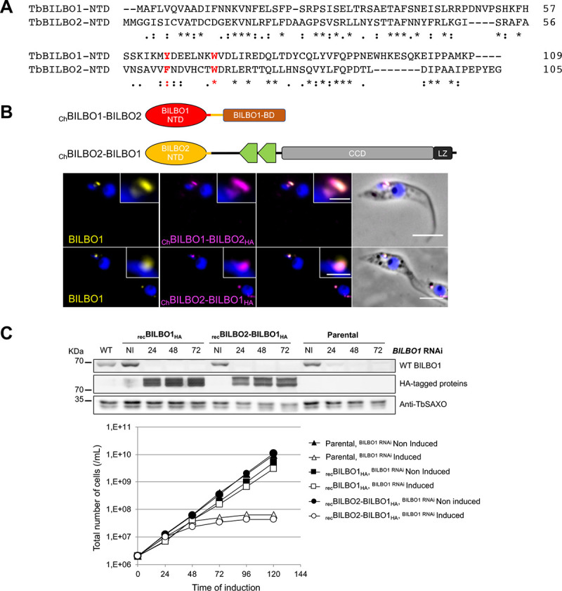
The flagellar pocket (FP) is the only endo- and exocytic organelle in most trypanosomes and, as such, is essential throughout the life cycle of the parasite. The neck of the FP is maintained enclosed around the flagellum via the flagellar pocket collar (FPC). The FPC is a macromolecular cytoskeletal structure and is essential for the formation of the FP and cytokinesis. FPC biogenesis and structure are poorly understood, mainly due to the lack of information on FPC composition. To date, only two FPC proteins, BILBO1 and FPC4, have been characterized. BILBO1 forms a molecular skeleton upon which other FPC proteins can, theoretically, dock onto. We previously identified FPC4 as the first BILBO1 interacting partner and demonstrated that its C-terminal domain interacts with the BILBO1 N-terminal domain (NTD). Here, we report by yeast two-hybrid, bioinformatics, functional and structural studies the characterization of a new FPC component and BILBO1 partner protein, BILBO2 (Tb927.6.3240). Further, we demonstrate that BILBO1 and BILBO2 share a homologous NTD and that both domains interact with FPC4. We have determined a 1.9 Å resolution crystal structure of the BILBO2 NTD in complex with the FPC4 BILBO1-binding domain. Together with mutational analyses, our studies reveal key residues for the function of the BILBO2 NTD and its interaction with FPC4 and evidenced a tripartite interaction between BILBO1, BILBO2, and FPC4. Our work sheds light on the first atomic structure of an FPC protein complex and represents a significant step in deciphering the FPC function in Trypanosoma brucei and other pathogenic kinetoplastids.


Conflict of interest statement

The authors have declared that no competing interests exist.

Figures

Fig 1
Fig 1. Schematic representation of the BILBO1, FPC4, and BILBO2 secondary structures and Y2H interaction tests.
A. The BILBO1 domains T3 (aa 171–587) and T4 (aa 251–587) were previously described in [9]. BILBO2 is presented as three domains: T1 (aa 1–103), T2 (aa 1–150), and the BILBO1-binding domain (B1BD, aa 151–271). FPC4 is presented as three domains: the microtubule binding domain (MT-BD), the coiled-coil domain (CCD), and the BILBO1 binding domain (B1BD). B. Y2H assay with full-length or domains of BILBO2 as bait and BILBO1 as prey, and of BILBO2 as prey and bait tested on minus histidine medium (-HIS). Loading control was on medium plus histidine. The positive control involved the previously demonstrated interaction between the p53 and T-antigen proteins, whereas the negative control involved Lamin and T-antigen proteins that do not interact.
Fig 2
Fig 2. Analysis of BILBO1 and BILBO2 interaction in a heterologous system.
A. U-2 OS whole cells expressing BILBO1 (a), HABILBO2 (b), or co-expressing BILBO1 with HABILBO2 (c) or with HABILBO2-T1 (d), T2 (e), BILBO2-B1BD (f) domains, and processed for immunofluorescence. B. Immunolabeling on detergent-extracted U-2 OS cells co-expressing HABILBO2 and BILBO1GFP (a), BILBO1-T3GFP (b), BILBO1-T4GFP (c), and BILBO1-T3GFP with HABILBO2-B1BD (d). C. Immunolabeling on detergent-extracted U-2 OS cells expressing BILBO1 mutated on the EF-hands (mEFh1, mEFh2, mEFH1+2, a-c), deleted of the Efh1+2 domain (d), and co-expressed with HABILBO2 (e-h). Scale bars, 10 μm.
Fig 3
Fig 3. Cellular localization of BILBO2.
A. Immunolabelling on detergent-extracted PCF cells using anti-BILBO1 and anti-BILBO2 antibodies. B. Immunolabelling on detergent-extracted PCF cells expressing endogenously tagged TY1BILBO2 using anti-BILBO1 and anti-TY1 antibodies. C. Ectopic expression of BILBO2HA and domains was induced for 24H with 1 μg/mL of tetracycline followed by immunofluorescence on whole cells and detergent-extracted cells (Cytoskeleton). Unlike BILBO2-T1HA, faint but consistent labelling of BILBO2-T2HA the FPC on cytoskeleton is indicated by the asterisk and are to be compared in the insets with increased contrast. Scale bars in A, B and D represent 5μm, and 1 μm (insets). D. Sum intensity per collar quantification of BILBO1 and of TY1BILBO2 labelling at the 1K1, 2K1N and 2K2N stages of the PCF cell cycle. Error bars represent the standard error (n = 200).
Fig 4
Fig 4. Depletion of BILBO1 induces cytosolic localization of BILBO2.
A. Comparative growth curves between WT cells and cells expressing TY1BILBO2 and non-induced (NI) or induced (I) for BILBO1 RNAi. B. Immunolabelling of BILBO1 and TY1BILBO2 on BILBO1 RNAi non-induced (NI) or induced 24H and 48H whole cells. C. Representative western-blot analysis of the fate of BILBO2 during BILBO1 RNAi in whole-cell (WC) and detergent-extracted samples (CSK). Anti-enolase and anti-tubulin were used as detergent extraction and loading controls, respectively. D. Quantification of the Western-blot in C. Error bars represent the standard error from two independent experiments. Scale bars in B represent 5 μm.
Fig 5
Fig 5. BILBO1 and BILBO2 share a conserved N-terminal domain with conserved residues.
A. Alignment of the BILBO1 and BILBO2 NTD domains. Asterisks indicate identical residues; colons indicate conserved substitution; periods indicate semi-conserved substitutions. B. Immunolocalization of chimeric BILBO1-BILBO2 proteins. Anti-BILBO1 labels both BILBO1 and ChBILBO1-BILBO2HA; anti-HA labels ChBILBO1-BILBO2HA or ChBILBO2-BILBO1HA. Cells were induced 18h with 1 μg.mL-1 tetracycline and detergent-extracted for immuno-labelling. Scale bars represent 5 μm. C. Cells inducible for BILBO1 RNAi and for the expression of recoded recBILBO1HA or recBILBO2-BILBO1HA were induced with 2 μg.mL-1 of tetracycline. The parental cells are inducible for BILBO1 RNAi only. Top panel: Western blot analysis of whole cells non induced or induced at different time points. Anti-TbSAXO was used as loading control. Bottom panel: Growth curves of non-induced and induced cells showing that recBILBO1HA can rescue the RNAi growth defect, contrary to recBILBO2-BILBO1HA.
Fig 6
Fig 6. Interaction between BILBO2 and FPC4 is similar to that of BILBO1 and FPC4.
A. Y2H interaction assay between BILBO2 and FPC4 and domains. B. Immuno-colocalisation of TY1FPC4 and BILBO2 in detergent-extracted PCF cells. Scale bars 5 μm. C. Immunolocalization of BILBO1, mycBILBO2 and TY1FPC4 using U-ExM in detergent-extracted PCF cells. Scale bars 10 μm and 5 μm in enlarged inset. D. Expression in U-2 OS cell and immunolocalization of FPC4GFP and FPC4 deleted of its B1BD (FPC4-ΔB1BDGFP) and HABILBO2 and domains demonstrating that the BILBO2-T1 (aa 1–103) domain is not sufficient for a stable interaction between BILBO2 and FPC4 whereas a longer domain (BILBO2-T2) is stabilizing the interaction. Cells were detergent-extracted before the IF to reduce the FPC4 and BILBO2 cytosolic labelling. Scale bars 10 μm. E. A tripartite interaction is demonstrated in U-2 OS cells by the co-labelling of FPC4 and BILBO2 and domains onto the BILBO1 polymers. Scale bars 10 μm. F. Schematic representation of the interactions between BILBO1, BILBO2 and FPC4.
Fig 7
Fig 7. Crystal structure the BILBO2-NTD/FPC4-B1BD complex.
A. Superposition of the two copies of BILBO2-NTD in the asymmetric unit cell of the crystal lattice (blue and light blue), together with BILBO1-NTD (pink, 6SJQ.pdb). Shown in yellow and orange are polypeptides of FPC4 bound to BILBO2-NTD. B. Ribbon diagram of the structure of the BILBO2-NTD/FPC4 complex in two orthogonal views. The structure of BILBO2-NTD is colour-ramped from blue to red at the N- and C-termini, respectively. FPC4 is shown as a yellow tube. C. Crystal structure of the complex with FPC4 residues depicted as sticks and BILBO2-NTD as an electrostatic surface plot. D. Stereo view of the 2Fo-Fc map (purple) around the binding interface between FPC4 and BILBO2 contoured at 1.5 σ level. BILBO2 and FPC4 are coloured in green and yellow, respectively. All visible residues of FPC4 (aa 432–438) and two interface aromatic residues of BILBO2 (F63, W70) are labelled. Plots in (A) and (B) were generated using PyMOL (The PyMOL Molecular Graphics System, Version 1.2r3pre, Schrödinger, LLC.), the one in (C) was done by CCP4mg Presenting your structures: the CCP4mg molecular-graphics software [43], and that in (D) by COOT [39].
Fig 8
Fig 8. Identification of key residues involved in the BILBO2-FPC4 interaction.
A. Interaction between FPC4-B1BD and BILBO2-NTD. FPC4 residues are depicted as sticks in yellow, whereas BILBO2-NTD is shown as an electrostatic surface plot. B. ITC experiments using purified BILBO2-NTD and wild-type (WT) and mutants of FPC4-B1BD. C. Co-immunolocalization of HABILBO2 (magenta) and of mutated forms of FPC4GFP (yellow) in U-2 OS cells (a). Co-immunolocalization of FPC4GFP (yellow) and mutated forms of HABILBO2 (magenta) (b). Scale bars, 10 μm.

References

    1. Gull K. The cytoskeleton of trypanosomatid parasites. Annu Rev Microbiol. 1999;53: 629–55. doi: 10.1146/annurev.micro.53.1.629 - DOI - PubMed
    1. Field MC, Carrington M. The trypanosome flagellar pocket. Nat Rev Microbiol. 2009;7: 775–786. doi: 10.1038/nrmicro2221 - DOI - PubMed
    1. Robinson DR, Sherwin T, Ploubidou A, Byard EH, Gull K. Microtubule polarity and dynamics in the control of organelle positioning, segregation, and cytokinesis in the trypanosome cell cycle. J Cell Biol. 1995;128: 1163–1172. doi: 10.1083/jcb.128.6.1163 - DOI - PMC - PubMed
    1. Bonhivers M, Nowacki S, Landrein N, Robinson DR. Biogenesis of the Trypanosome Endo-Exocytotic Organelle Is Cytoskeleton Mediated. PLOS Biol. 2008;6: e105. doi: 10.1371/journal.pbio.0060105 - DOI - PMC - PubMed
    1. Lacomble S, Vaughan S, Gadelha C, Morphew MK, Shaw MK, McIntosh JR, et al.. Three-dimensional cellular architecture of the flagellar pocket and associated cytoskeleton in trypanosomes revealed by electron microscope tomography. J Cell Sci. 2009;122: 1081–90. doi: 10.1242/jcs.045740 - DOI - PMC - PubMed

Publication types

Substances