Macular Thickness and Microvasculature Loss in Glaucoma Suspect Eyes
- PMID: 34339877
- PMCID: PMC9988288
- DOI: 10.1016/j.ogla.2021.07.009
Macular Thickness and Microvasculature Loss in Glaucoma Suspect Eyes
Abstract
Purpose: To characterize the change of ganglion cell complex (GCC) thickness and macular vessel density in glaucoma suspect eyes with ocular hypertension (OHT) or glaucomatous optic neuropathy (GON).
Design: Prospective, longitudinal study.
Participants: Eight-three eyes (24 healthy, 30 OHT, and 29 GON) of 65 patients who underwent at least 3 visits were included from the Diagnostic Innovations in Glaucoma Study. The mean follow-up was at least 3 years.
Methods: OCT angiography (OCTA)-based vessel density and OCT-based structural thickness of the 3 × 3-mm1 GCC scan slab were evaluated at each visit. The rates of vessel density and thickness change were compared across diagnostic groups using a linear mixed-effects model.
Main outcome measures: Change rates of macula GCC thickness and superficial vessel density.
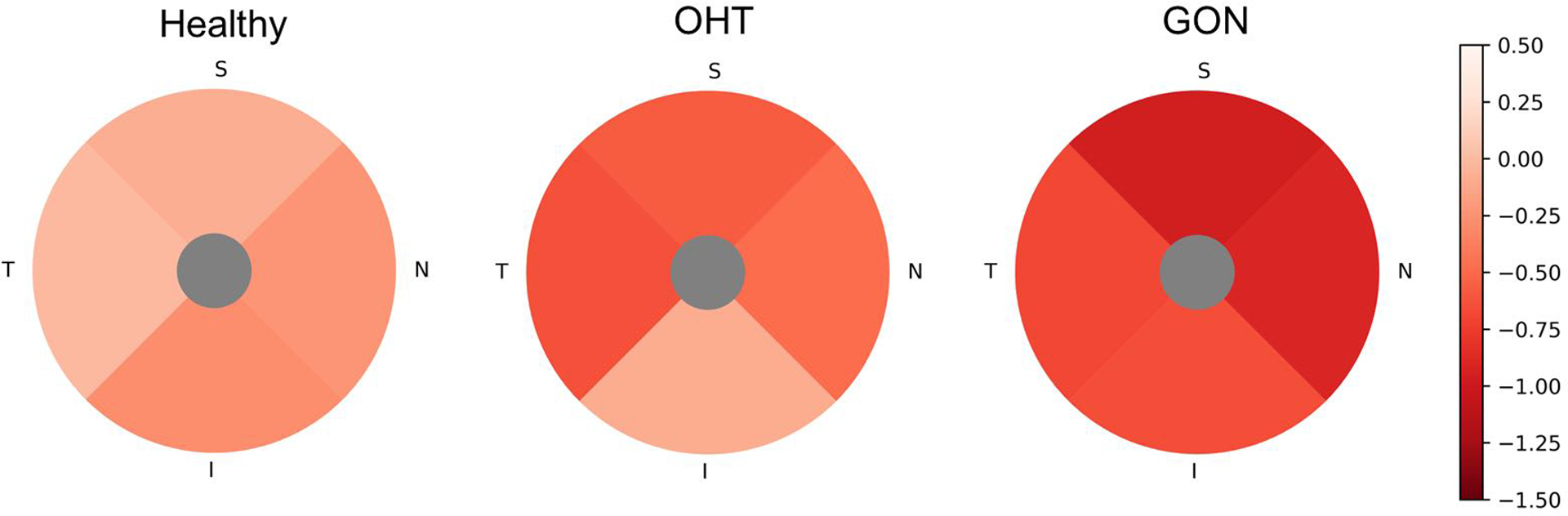
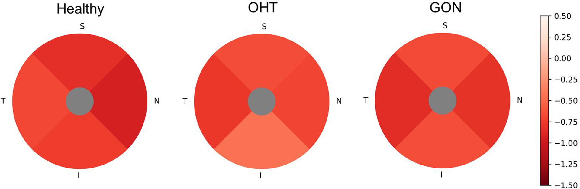
Results: Significant mean rates of both GCC thinning and vessel density loss were detectable in OHT and GON groups. Of the individual suspect eyes, 49.1% showed significant loss (P < 0.05) with either vessel density or GCC thickness. Of the GON eyes, 31.0% showed both significant GCC loss and vessel density loss, 51.7% showed only significant GCC loss, whereas 17.2% showed only significant vessel density loss. Vessel density loss was faster than GCC thinning in half of the suspect eyes based on percent loss analysis. The age and scan quality-adjusted GCC thinning rates of the OHT group (-0.59 μm/year; P = 0.025) and GON group (-0.79 μm/year; P = 0.058) were faster than those of the healthy group (-0.11 μm/year), whereas the rate of vessel density loss was not significantly different among the diagnostic groups (all P > 0.2). Higher mean intraocular pressure during follow-up was associated with faster GCC thinning in the OHT group (P = 0.065) and GON groups (P = 0.015), but was not associated with the rate of vessel density decrease.
Conclusions: Whereas the rate of GCC thinning was faster on average in suspect eyes than in healthy eyes, some suspect eyes showed significant loss of vessel density and faster vessel density loss than GCC thinning. OCT and OCTA are complementary and useful for evaluating eyes with OHT or GON.
Keywords: Glaucoma; Longitudinal; Macula; OCT; OCT angiography.
Copyright © 2021 American Academy of Ophthalmology. Published by Elsevier Inc. All rights reserved.
Figures