. 2021 Aug;10(8):474-487.
doi: 10.1302/2046-3758.108.BJR-2021-0086.

The role of TGF-β2 in cartilage development and diseases

Affiliations

The role of TGF-β2 in cartilage development and diseases

Mengmeng Duan et al. Bone Joint Res. 2021 Aug.

Abstract

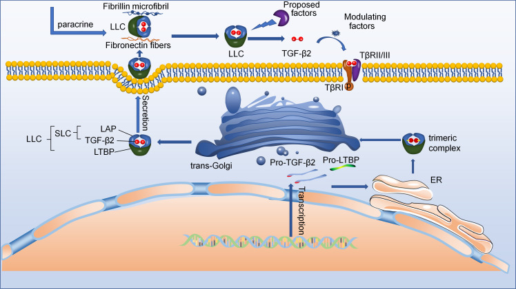
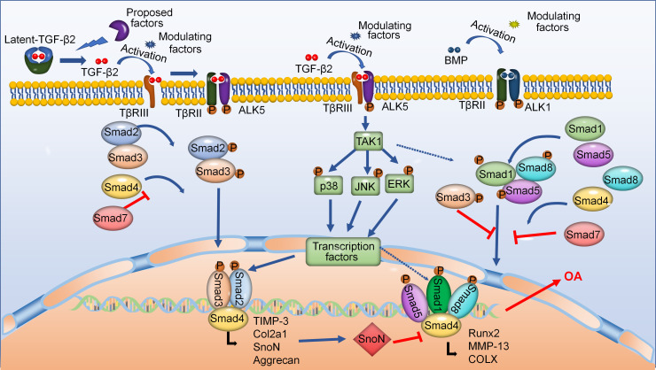
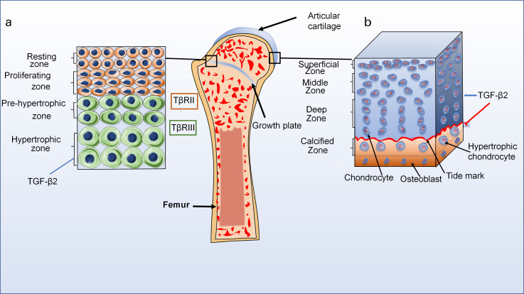
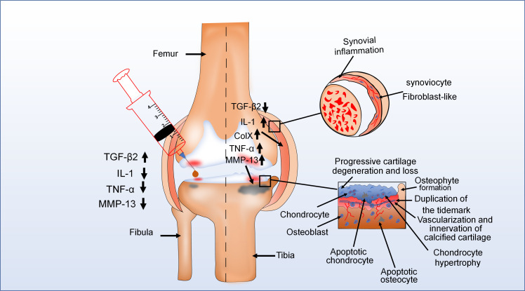
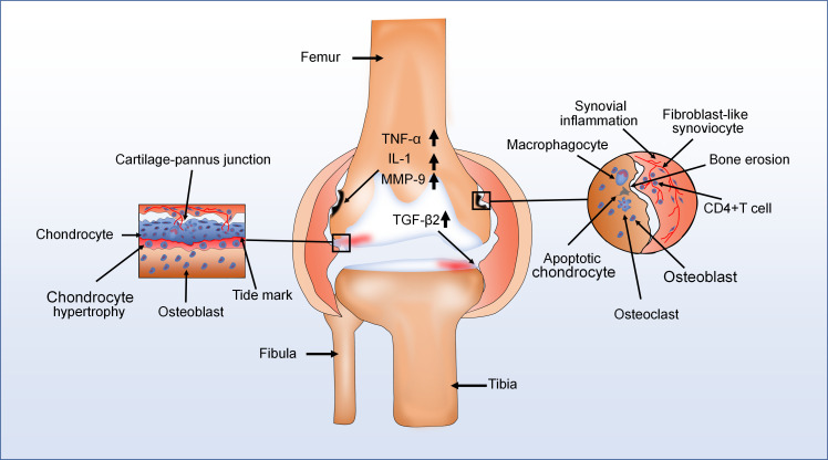
Transforming growth factor-beta2 (TGF-β2) is recognized as a versatile cytokine that plays a vital role in regulation of joint development, homeostasis, and diseases, but its role as a biological mechanism is understood far less than that of its counterpart, TGF-β1. Cartilage as a load-resisting structure in vertebrates however displays a fragile performance when any tissue disturbance occurs, due to its lack of blood vessels, nerves, and lymphatics. Recent reports have indicated that TGF-β2 is involved in the physiological processes of chondrocytes such as proliferation, differentiation, migration, and apoptosis, and the pathological progress of cartilage such as osteoarthritis (OA) and rheumatoid arthritis (RA). TGF-β2 also shows its potent capacity in the repair of cartilage defects by recruiting autologous mesenchymal stem cells and promoting secretion of other growth factor clusters. In addition, some pioneering studies have already considered it as a potential target in the treatment of OA and RA. This article aims to summarize the current progress of TGF-β2 in cartilage development and diseases, which might provide new cues for remodelling of cartilage defect and intervention of cartilage diseases.

Keywords: Cartilage development and diseases; Chondrocyte; TGF-β2; apoptosis; blood vessels; cartilage defects; cartilage diseases; cartilage tissue; chondrocytes; cytokines; growth factors; mesenchymal progenitor cells; osteoarthritis (OA).


Figures

Fig. 1
Fig. 1
The schematic diagram showing active transforming growth factor-beta2 (TGF-β2) secretion. Starting at the bottom right: New synthetic pro-TGF-β2 forms dimer/trimeric complexes in the endoplasmic reticulum (ER) with the help of latent TGF-β2-binding protein (LTBP). These dimer/trimeric complexes are then further processed in the trans-Golgi network to form large latent complexes (LLCs). After they are secreted, the LLC may bind to various fibres in the extracellular matrix (ECM) with the help of LTBP. Eventually, the LLC is activated by a number of ECM factors, resulting in the formation of active TGF-β2. LAP, latency-associated peptide.
Fig. 2
Fig. 2
Transforming growth factor-beta2 (TGF-β2) regulates cartilage homeostasis and disease progression via its canonical Smad-dependent and noncanonical mitogen-activated protein kinases (MAPK) signalling pathways. During the initiation of the canonical Smad-dependent signalling pathway, active TGF-β2 is binded to its receptors, then activates downstream Smad-dependent signalling pathway to regulate homeostasis of chondrocytes. Moreover, TGF-β2 signals can also transmit to noncanonical TGF-β-activated kinase1 (TAK1)-mediated pathways. Additionally, TGF-β2 signals are involved in bone morphogenetic protein (BMP) signalling pathways. ALK1, activin receptor-like kinase; Col2a1, collagen, type II, alpha 1; COLX, collagen type x; MMP-13, matrix metalloproteinase-13; OA, osteoarthritis; TβR, transforming growth beta receptor; TIMP-3, tissue inhibitor of metalloproteinase 3.
Fig. 3
Fig. 3
The distribution of transforming growth factor-beta2 (TGF-β2) in normal articular cartilage. a) Normal structure diagram of growth plate (left). The growth plate cartilage is divided into four zones: resting zone, proliferative zone, pre-hypertrophic zone, and hypertrophic zone. TGF-β2 can be expressed in all zones during cartilage development, but the highest levels are in hypertrophy zone. Interestingly, TGF-β2 has low affinity for transforming growth factor beta receptor (TβR)II but a strong affinity for TβRIII. b) Normal structure diagram of articular cartilage (right). The articular cartilage is also divided into four zones: superficial zone, middle zone, deep zone, and calcified zone. TGF-β2 signalling pathways can maintain chondrocyte phenotype, and inhibit pre-hypertrophic and hypertrophic differentiation in articular cartilage.
Fig. 4
Fig. 4
The pathological change of knee joint in the osteochondral unit during the evolution of osteoarthritis (OA). OA is typically characterized by cartilage damage, osteophyte formation, and thickening of the joint capsule. For example, in one diagram (upper right), the epithelial lining of the joint capsule is thickened because of synovial inflammation. In another (bottom right), during advanced stages of OA, there are pathological changes in different areas of articular cartilage. Moreover, in the left diagram, injection of transforming growth factor-beta 2 (TGF-β2) into the joint cavity of patients with OA reduced the expression of some proinflammatory factors and the clinical symptoms of OA. This means that TGF-β2 plays an important role in the development of OA. ColX, collagen type X; IL-1, interleukin 1; MMP, matrix metalloproteinase; TNF-α, tumour necrosis factor alpha.
Fig. 5
Fig. 5
The major pathological characteristics of rheumatoid arthritis (RA) are chronic synovitis with hyperplasia, pannus formation, and immune cell infiltration. For instance, in one diagram (left), various cell types in the cartilage-pannus of RA secrete lots of proinflammatory factors to induce cartilage damage and degradation of collagen. In another (right), there are also numerous immune cells infiltration and development of new blood vessels in the synovium of RA with hyperplasia, and the expression of transforming growth factor-beta2 (TGF-β2) also increases. Moreover, bone erosion appeared in the vicinity of the thickened synovium. CD4, cluster of differentiation 4; IL-1, interleukin 1; MMP, matrix metalloproteinase; TNF-α, tumour necrosis factor alpha.

References

    1. Watabe T, Miyazono K. Roles of TGF-β family signaling in stem cell renewal and differentiation. Cell Res. 2009;19(1):103–115. - PubMed
    1. Wojciech S, Ahmad R, Belaid-Choucair Z, et al. The orphan GPR50 receptor promotes constitutive TGFβ receptor signaling and protects against cancer development. Nat Commun. 2018;9(1):1216. - PMC - PubMed
    1. Seoane J, Gomis RR. TGF-β Family Signaling in Tumor Suppression and Cancer Progression. Cold Spring Harb Perspect Biol. 2017;9(12):a022277. - PMC - PubMed
    1. Manokawinchoke J, Pavasant P, Sawangmake C, et al. Intermittent compressive force promotes osteogenic differentiation in human periodontal ligament cells by regulating the transforming growth factor-β pathway. Cell Death Dis. 2019;10:761. - PMC - PubMed
    1. Su Y, Feng W, Shi J, Chen L, Huang J, Lin T. circRIP2 accelerates bladder cancer progression via miR-1305/Tgf-β2/smad3 pathway. Mol Cancer. 2020;19(1):23. - PMC - PubMed