. 2021 Aug 31;12(4):e0159021.
doi: 10.1128/mBio.01590-21. Epub 2021 Aug 3.

Dynamics of SARS-CoV-2 Spike Proteins in Cell Entry: Control Elements in the Amino-Terminal Domains

Affiliations

Dynamics of SARS-CoV-2 Spike Proteins in Cell Entry: Control Elements in the Amino-Terminal Domains

Enya Qing et al. mBio. .

Abstract

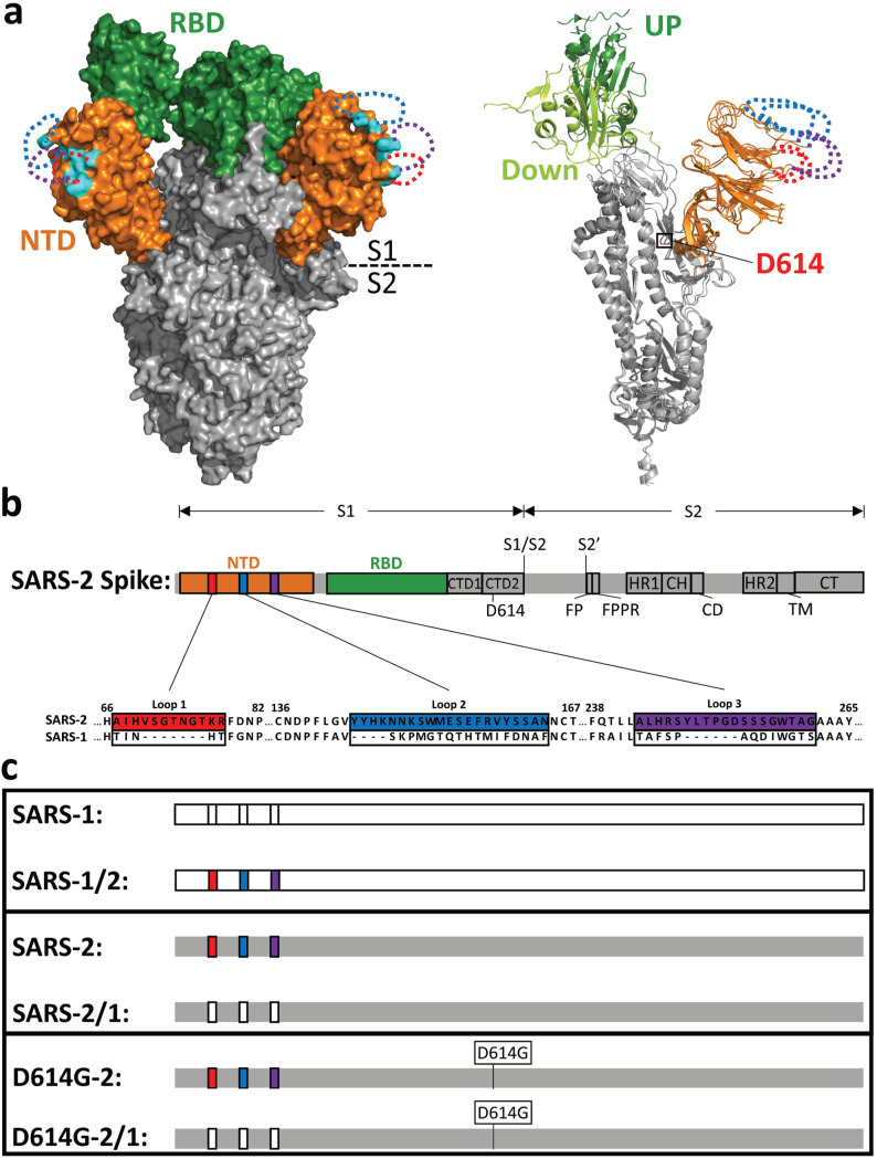
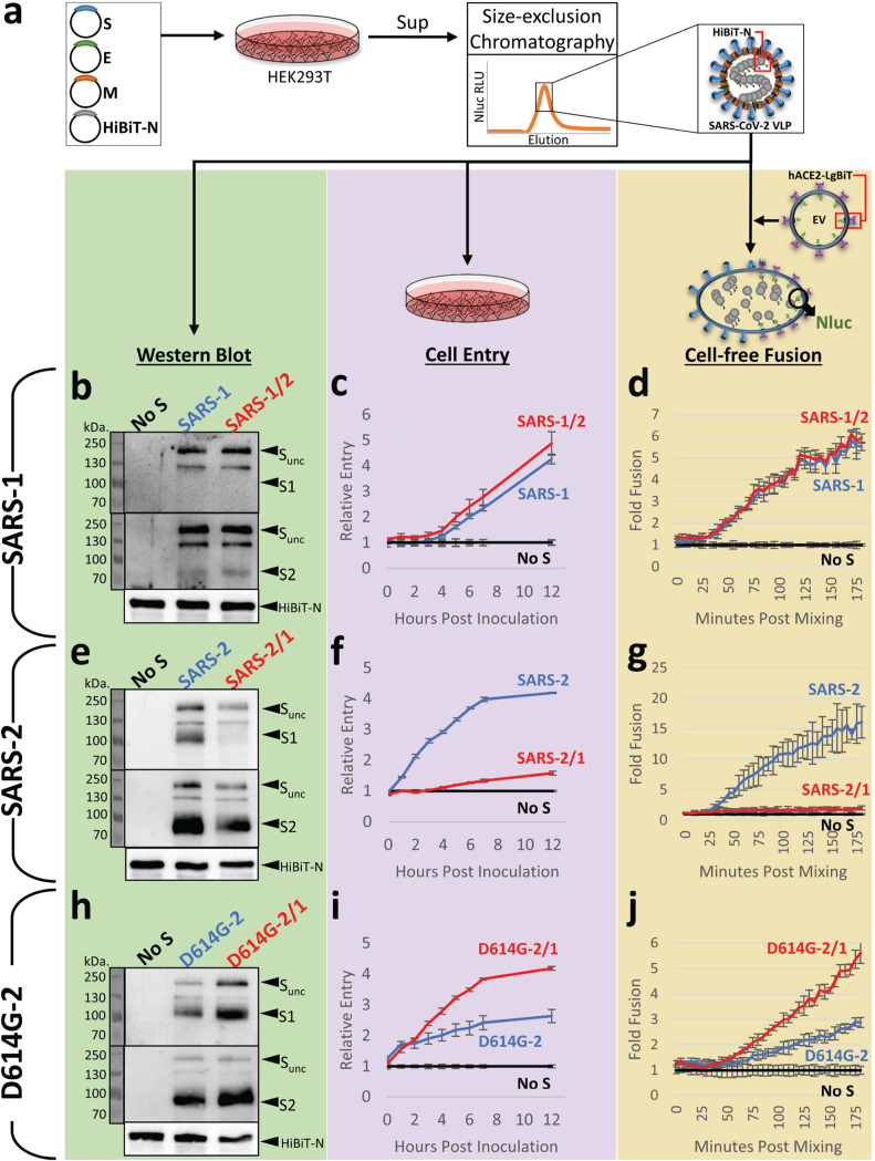
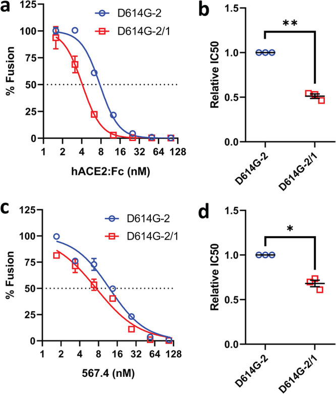
Selective pressures drive adaptive changes in the coronavirus spike proteins directing virus-cell entry. These changes are concentrated in the amino-terminal domains (NTDs) and the receptor-binding domains (RBDs) of complex modular spike protein trimers. The impact of this hypervariability on virus entry is often unclear, particularly with respect to sarbecovirus NTD variations. Therefore, we constructed indels and substitutions within hypervariable NTD regions and used severe acute respiratory syndrome coronavirus 2 (SARS-CoV-2) virus-like particles and quantitative virus-cell entry assays to elucidate spike structures controlling this initial infection stage. We identified NTD variations that increased SARS-CoV-2 spike protein-mediated membrane fusion and cell entry. Increased cell entry correlated with greater presentation of RBDs to ACE2 receptors. This revealed a significant allosteric effect, in that changes within the NTDs can orient RBDs for effective virus-cell binding. Yet, those NTD changes elevating receptor binding and membrane fusion also reduced interdomain associations, leaving spikes on virus-like particles susceptible to irreversible inactivation. These findings parallel those obtained decades ago, in which comparisons of murine coronavirus spike protein variants established inverse relationships between membrane fusion potential and virus stability. Considerable hypervariability in the SARS-CoV-2 spike protein NTDs also appear to be driven by counterbalancing pressures for effective virus-cell entry and durable extracellular virus infectivity. These forces may selectively amplify SARS-CoV-2 variants of concern. IMPORTANCE Adaptive changes that increase SARS-CoV-2 transmissibility may expand and prolong the coronavirus disease 2019 (COVID-19) pandemic. Transmission requires metastable and dynamic spike proteins that bind viruses to cells and catalyze virus-cell membrane fusion. Using newly developed assays reflecting these two essential steps in virus-cell entry, we focused on adaptive changes in SARS-CoV-2 spike proteins and found that deletions in amino-terminal domains reset spike protein metastability, rendering viruses less stable yet more poised to respond to cellular factors that prompt entry and subsequent infection. The results identify adjustable control features that balance extracellular virus stability with facile virus dynamics during cell entry. These equilibrating elements warrant attention when monitoring the evolution of pandemic coronaviruses.

Keywords: SARS-CoV-2; coronavirus; coronavirus spike protein; membrane fusion; virus entry; virus receptors.


Figures

FIG 1
FIG 1
NTD loops on the SARS-CoV-2 spike protein. (a, left) SARS-CoV-2 S cryo-EM structure (PDB accession no. 6VSB) in surface representation. The N-terminal domain (NTD; orange) and receptor-binding domain (RBD; green) are depicted in the context of the trimer ectodomain (gray). Unresolved NTD loops are depicted by dotted lines (red, blue, and purple). Resolved residues nearest to the NTD loops are in cyan. (Right) A single S monomer is depicted in a ribbon diagram with RBD-up (dark green) superimposed on RBD-down (light green). D614 is labeled in red. (b) Linear depiction of the complete SARS-CoV-2 S protein. The NTD (orange), the RBD (green), C-terminal domain 1 (CTD1), C-terminal domain 2 (CTD2), D614, S1/S2 priming, the S2′-activating cleavage sites, the fusion peptide (FP), the fusion peptide-proximal region (FPPR), heptad repeat 1 (HR1), the central helix (CH), the connector domain (CD), heptad repeat 2 (HR2), the transmembrane span (TM), and the cytoplasmic tail (CT) are depicted. Three NTD loops are highlighted in colored boxes (red, blue, and purple) and enlarged to reveal the amino acid sequences of SARS-CoV-2-S (GenBank accession no. NC_045512.2) and corresponding SARS-CoV-S (GenBank accession no. AY278741.1), which were exchanged. (c) Recombinant spike protein pairs. (Top) SARS-1 in comparison with SARS1/2 (SARS-2 NTD loops in color); (middle) SARS-2 in comparison with SARS-2/1 (SARS-1 NTD loops not in color); (bottom) SARS-2 and SARS-2/1 in the D614G background.
FIG 2
FIG 2
Hypervariable NTD loops control S protein stability and membrane fusion potential. (a) Plasmids encoding SARS-CoV-2 spike (S), envelope (E), membrane (M), and amino-terminal HiBiT-tagged nucleoprotein (HiBiT-N) were expressed in HEK293T cells, supernatants (sup) were harvested after 2 days, and HiBiT-VLPs were purified by size exclusion chromatography. VLPs were compared as isogenic pairs: SARS-1 versus SARS-1/2 (panels b to d), SARS-2 versus SARS-2/1 (panels e to g), and D614G-2 versus D614G-2/1 (panels h to j). Each pair was evaluated by Western blot (left), cell entry (middle), and cell-free fusion (right) assays. Western blot assays detected uncleaved S (S-unc), S1, S2, and HiBiT-N. Cell entry assays detected HiBiT-VLP entry into ACE2-LgBiT/hTMPRSS2 target cells. The cell entry data are presented relative to the cell entry of control inoculations of spikeless (No S) VLPs. Cell-free fusion data are presented as HiBiT-VLP:ACE2-LgBiT EV fusion levels relative to data under control conditions with spikeless VLPs. For the cell entry and cell-free fusion data, the error bars present standard deviations (SD) from three technical replicates (n = 3), with data being representative of three biological repeats.
FIG 3
FIG 3
Hypervariable NTD loops control RBD exposure. (a and c) Serial dilutions of hACE2-Fc (a) or RBD MAb 567.4 (c) were incubated with D614G-2 or D614G-2/1 VLPs for 30 min at 37°C, and cell-free VLP-EV fusions were then measured. Data trendlines were normalized to vehicle fusion levels. Error bars present SD. (b and d) Experiments were repeated three times, and IC50 values were calculated from the three fitted normalized response trendlines. Relative IC50 data are presented for hACE2-Fc (c) and RBD MAb 567.4 (d). Statistical analyses were assessed by an unpaired Student t test (*, P < 0.05; **, P < 0.01).
FIG 4
FIG 4
NTD loops control protease-triggered membrane fusion. (a) D614G-2 VLPs were incubated with hACE2-negative or hACE2-positive EVs for 30 min at 37°C in the presence of the indicated trypsin concentrations, and trypsin cleavage products were identified by Western blotting. Uncleaved S (S-unc), S2, and S2′ cleavage products are indicated. (b) The indicated SARS-CoV-2 VLPs were evaluated in cell-free VLP-EV fusion assays at the indicated trypsin concentrations. Fusion readouts were taken after 3 h at 37°C, and plotted data trendlines were normalized to the highest measured fusion levels. Error bars present standard errors (SE) of the means. (c) Cell-free VLP-EV fusion assays were repeated three times and trypsin EC50 values calculated from the fitted normalized response trendlines. Statistical analyses were assessed by an unpaired Student t test (***, P < 0.001; ns, not significant).
FIG 5
FIG 5
NTD loops control SARS-2 spike stability. (a) VLPs were pelleted through 20% sucrose, resuspended, and evaluated by Western blotting. Western blot assays detected uncleaved S (S-unc), S1, S2, and HiBiT-N. (b) The same resuspended VLPs were evaluated in cell entry assays. Time course cell entry data are presented relative to those of control inoculations of spikeless (No S) VLPs. (c) The same resuspended VLPs were evaluated in cell-free VLP-EV fusion assays. Time course fusion data are presented as fold changes from the no-S condition containing “spikeless” VLPs. (b and c) Error bars present standard deviations (SD) from three technical replicates (n = 3). Data shown are representative of three biological repeats.
FIG 6
FIG 6
NTDs require loop structures to interfere with S-directed transduction and cell-cell fusion. (a) Western blot analysis of purified NTD, RBD, and CEACAM:Fc proteins (20 μl/lane at 40 nM). (b) VSV-Fluc PPs bearing SARS-CoV-2 S were inoculated onto Calu-3 cells together with the vehicle or the indicated Fc constructs (2 μM). After 18 h, Fluc levels were measured to reflect PP cell entry and are presented as percent entry relative to that during vehicle control conditions. Each data point represents averages (n = 4 replicates) from independent experiments (n = 4 experiments). Error bars present standard errors (SE) of the means. Statistically significant deviations from vehicle control values were assessed by an unpaired Student t test (***, P < 0.001). (c and d) Cell-cell fusion assays were established with HeLa-hACE2 (c) or HeLa (d) target cells in the presence of the indicated Fc constructs (10 μM). Rluc levels were measured to reflect cell-cell fusion and are presented as percent cell fusion relative to that under vehicle control conditions. Each data point represents averages (n = 4 replicates) of Rluc levels measured at 4, 12, and 22 h after cocultivation. Error bars present standard errors (SE) of the means. Statistically significant deviations from vehicle control data were assessed by an unpaired Student t test (*, P < 0.05; **, P < 0.01). Data are representative of two biological repeats.

References

    1. Zhou P, Yang XL, Wang XG, Hu B, Zhang L, Zhang W, Si HR, Zhu Y, Li B, Huang CL, Chen HD, Chen J, Luo Y, Guo H, Jiang RD, Liu MQ, Chen Y, Shen XR, Wang X, Zheng XS, Zhao K, Chen QJ, Deng F, Liu LL, Yan B, Zhan FX, Wang YY, Xiao GF, Shi ZL. 2020. A pneumonia outbreak associated with a new coronavirus of probable bat origin. Nature 579:270–273. doi:10.1038/s41586-020-2012-7. - DOI - PMC - PubMed
    1. Andersen KG, Rambaut A, Lipkin WI, Holmes EC, Garry RF. 2020. The proximal origin of SARS-CoV-2. Nat Med 26:450–452. doi:10.1038/s41591-020-0820-9. - DOI - PMC - PubMed
    1. Korber B, Fischer WM, Gnanakaran S, Yoon H, Theiler J, Abfalterer W, Hengartner N, Giorgi EE, Bhattacharya T, Foley B, Hastie KM, Parker MD, Partridge DG, Evans CM, Freeman TM, de Silva TI, Sheffield C-GG, McDanal C, Perez LG, Tang H, Moon-Walker A, Whelan SP, LaBranche CC, Saphire EO, Montefiori DC, Sheffield COVID-19 Genomics Group . 2020. Tracking changes in SARS-CoV-2 spike: evidence that D614G increases infectivity of the COVID-19 virus. Cell 182:812–827.e19. doi:10.1016/j.cell.2020.06.043. - DOI - PMC - PubMed
    1. Zhang L, Jackson CB, Mou H, Ojha A, Peng H, Quinlan BD, Rangarajan ES, Pan A, Vanderheiden A, Suthar MS, Li W, Izard T, Rader C, Farzan M, Choe H. 2020. SARS-CoV-2 spike-protein D614G mutation increases virion spike density and infectivity. Nat Commun 11:6013. doi:10.1038/s41467-020-19808-4. - DOI - PMC - PubMed
    1. Plante JA, Mitchell BM, Plante KS, Debbink K, Weaver SC, Menachery VD. 2021. The variant gambit: COVID-19's next move. Cell Host Microbe 29:508–515. doi:10.1016/j.chom.2021.02.020. - DOI - PMC - PubMed

Publication types

Substances