. 2021 Aug 31;12(4):e0139221.
doi: 10.1128/mBio.01392-21. Epub 2021 Aug 3.

The Mammalian Membrane Microenvironment Regulates the Sequential Attachment of Bacteria to Host Cells

Affiliations

The Mammalian Membrane Microenvironment Regulates the Sequential Attachment of Bacteria to Host Cells

Xavier Pierrat et al. mBio. .

Abstract

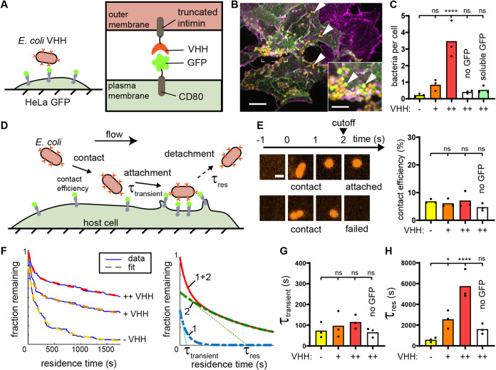
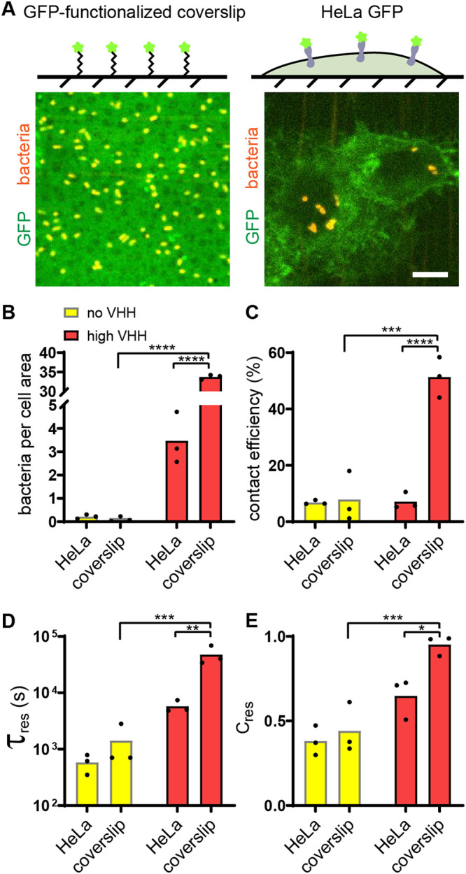
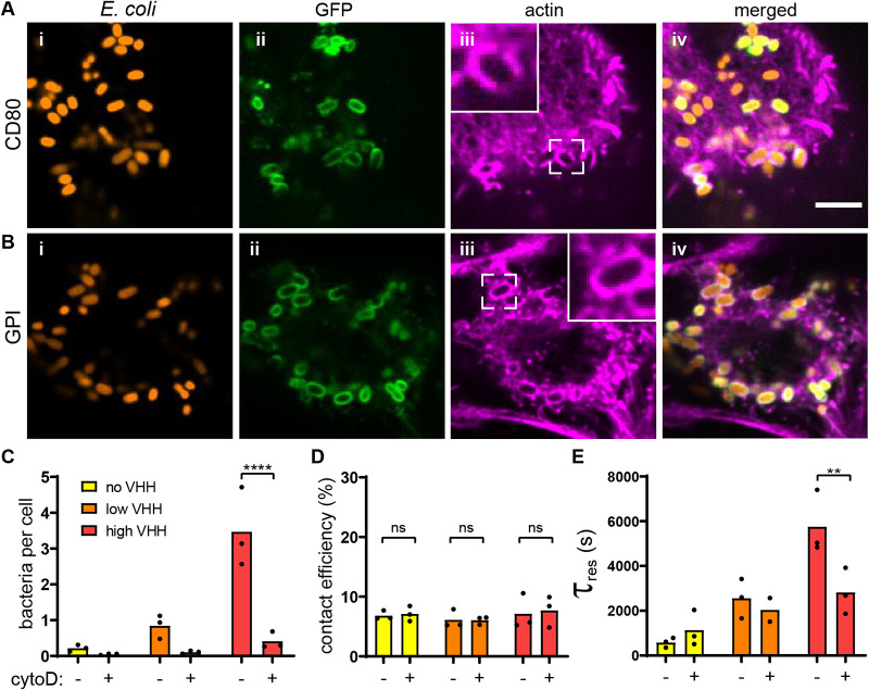
Pathogen attachment to host tissue is critical in the progress of many infections. Bacteria use adhesion in vivo to stabilize colonization and subsequently regulate the deployment of contact-dependent virulence traits. To specifically target host cells, they decorate themselves with adhesins, proteins that bind to mammalian cell surface receptors. One common assumption is that adhesin-receptor interactions entirely govern bacterial attachment. However, how adhesins engage with their receptors in an in vivo-like context remains unclear, in particular under the influence of a heterogeneous mechanical microenvironment. We here investigate the biophysical processes governing bacterial adhesion to host cells using a tunable adhesin-receptor system. By dynamically visualizing attachment, we found that bacterial adhesion to host cell surface, unlike adhesion to inert surfaces, involves two consecutive steps. Bacteria initially attach to their host without engaging adhesins. This step lasts about 1 min, during which bacteria can easily detach. We found that at this stage, the glycocalyx, a layer of glycosylated proteins and lipids, shields the host cell by keeping adhesins away from their receptor ligand. In a second step, adhesins engage with their target receptors to strengthen attachment for minutes to hours. The active properties of the membrane, endowed by the actin cytoskeleton, strengthen specific adhesion. Altogether, our results demonstrate that adhesin-ligand binding is not the sole regulator of bacterial adhesion. In fact, the host cell's surface mechanical microenvironment mediates the physical interactions between host and bacteria, thereby playing an essential role in the onset of infection. IMPORTANCE Microbial adhesion to host cells is the initial step toward many infections. Despite playing a pivotal role in the onset of disease, we still know little about how bacteria attach in an in vivo-like context. By employing a biophysical approach where we investigated host-microbe physical interactions at the single-cell level, we unexpectedly discovered that bacteria attach to mammalian cell membranes in two successive steps. We found that mechanical factors of the cell microenvironment regulate each of these steps, and even dominate biochemical factors, thereby challenging preconceptions on how pathogens interact with their hosts.

Keywords: adhesins; autotransporter proteins; cell adhesion; cell membranes; cytoskeleton; glycocalyx; membrane biophysics; microfluidics.


Figures

FIG 1
FIG 1
A synthetic adhesin-receptor system reveals a two-step mechanism of bacterial attachment to host cells. (A) Schematic of the synthetic adhesin-receptor system. E. coli cells display nanobody targeting GFP (VHH) fused to a truncated intimin autotransporter scaffold. HeLa cells display GFP receptors by fusion with the membrane-anchored CD80 scaffold (HeLa GFP). (B) In a mixed population of GFP+ (green) and GFP (purple) HeLa cells, E. coli (orange, indicated with white arrowheads) specifically binds to GFP+ cells. Actin stained with phalloidin (purple). Bars, 10 μm (main) and 5 μm (inset). (C) Bacterial count per HeLa cell increases with E. coli nanobody density. E. coli expressing VHH at low density or expressing VHH at high density but preincubated with soluble GFP only rarely binds to HeLa cells displaying GFP (“−,” “+,” and “++” correspond to no, low, and high VHH induction, respectively). (D) Dynamic visualizations of bacterial adhesion to HeLa cells under flow allow us to simultaneously monitor attachment and detachment events at multiple timescales. (E) Bacterial contact efficiency is independent of VHH density and GFP display. High-speed confocal imaging at 1 frame per second highlights bacterial populations that detach rapidly after contact. We considered bacteria attached if they stayed on the HeLa cell surface for more than 2 s. Bar, 2 μm. (F) We constructed residence time distributions using long-timescale tracking of attached bacteria (1 h). Bacteria adhering during the first 30 min were followed for 30 supplementary min in order to avoid artificial cropping of the data (see Materials and Methods). Bare E. coli and E. coli displaying low and high VHH levels have largely different residence time distributions. We fit these distributions using the sum of two exponentials to highlight two characteristic timescales, τtransient and τres (right illustrative graph). The single exponentials are shown in dashed green and blue, and their sum is the continuous red line. (G) The model parameter τtransient is independent of the adhesin displayed. (H) In contrast, the characteristic residence time τres increases with nanobody density. Statistical tests: one-way analysis of variance (ANOVA) followed by Dunnett’s post hoc test (****, P < 10−4; *, P < 0.05; ns, not significant).
FIG 2
FIG 2
Attachment of bacteria to abiotic surface is a single-step process. (A) (Top) Controlled GFP-functionalized coverslips permit visualization of specific adhesion to hard, abiotic surface and quantitative comparison with adhesion to mammalian cells. (Bottom) Representative confocal microscopy images of bacterial binding to GFP-coated coverslips (left) and HeLa-GFP (right). Bar, 10 μm. (B) Final bacterial count per cell area is about 10-fold larger on GFP-coated coverslips than on HeLa cells in the presence of VHH. (C) Bacterial contact efficiency is higher on GFP-coated coverslips than on HeLa cells in the presence of VHH. (D) The characteristic residence time τres shows the VHH-dependent binding to coverslips is stronger than that to HeLa cells. (E) Relative contribution of short- and long-timescale exponential fits shows that 95% of E. coli VHH bacteria strongly bind to GFP-coated coverslips. Statistical tests: two-way ANOVA and Sidak post hoc test (****, P < 10−4; ***, P < 0.001; **, P < 0.01; *, P < 0.05).
FIG 3
FIG 3
Regulation of bacterial adhesion by host cytoskeleton. (A) Actin rearranges around attached bacteria. After static incubation with E. coli VHH (orange), HeLa cells displaying GFP with a CD80 anchor (green) were stained for actin (purple). Bar, 5 μm. (B) Bacteria promote actin embeddings in the absence of any cytosolic component in the mammalian cell. After static coculture with E. coli VHH (red), HeLa cells displaying GFP with a glycosylphosphatidylinositol (GPI), which does not harbor any cytosolic signaling domain, also show strong actin remodeling around attached bacteria. (C) HeLa cell treatment with the actin polymerization inhibitor cytochalasin D (cytoD) reduces the bacterial count per HeLa cell. (D) Bacterial contact efficiency is independent of actin polymerization. (E) The characteristic residence time τres decreases in the presence of cytochalasin D at high VHH density. Statistical tests: two-way ANOVA and Sidak post hoc test (****, P < 10−4; **, P < 0.01; ns, not significant).
FIG 4
FIG 4
The membrane glycocalyx inhibits bacterial attachment. (A) Enzymatic deglycosylation of HeLa cell surface proteins increases bacterial binding. The right image shows two deglycosylated HeLa cells covered by E. coli VHH while the negative control under otherwise identical conditions has a low bacterial count. Bar, 10 μm. (B to D) Comparison of bacterial adhesion dynamics between untreated cells (native) and deglycosylated cells (deglyco). (B) Final E. coli VHH count per HeLa cell is higher in deglycosylated cells. (C) Glycocalyx removal increases the contact efficiency of E. coli VHH. (D) Comparison of the characteristic residence time τres with or without deglycosylation mix. Statistical tests: two-way ANOVA and Sidak post hoc test (**, P < 0.01).
FIG 5
FIG 5
Flagella and flow attenuate the glycocalyx shield. (A) Schematic of the experimental setup. Flagellated E. coli VHH (blue) was compared to nonflagellated E. coli VHH (“+” and “−,” respectively). E. coli VHH contact efficiency is increased in the presence of flagellum in flow. (B) The presence of flagella decreases the characteristic residence time in flow. (C) Comparison of the preexponential factor of the characteristic transient binding time τtransient in the presence or absence of flagella shows that the proportion of bacteria strongly binding to HeLa GFP is lower with flagella. (D) Schematic of the experimental setup. We measured the attachment dynamics of E. coli VHH in increasing shear stresses. Bacterial contact efficiency increases with flow intensity. (E) The characteristic residence time τres increases with flow intensity. (F) Strong flows decrease the contact frequency despite a higher number of bacteria crossing the channel. Statistical tests for panels A to C: two-tailed unpaired t test (**, P < 0.01; *, P < 0.05). Statistical tests for panels D to F: one-way ANOVA and Tukey post hoc test (*, P < 0.05; ****, P < 10−4).
FIG 6
FIG 6
A model for mechanically-regulated, two-step bacterial attachment to host cells. Upon contact of a bacterium with a host cell (1), the glycocalyx blocks attachment by sterically shielding the membrane. This short-timescale interaction does not involve short-range adhesins or mammalian membrane receptors. Strong shear forces and bacterial flagellum can increase the transient binding efficiency, in part by attenuating the glycocalyx shield. The bacterium subsequently binds adhesins onto host receptors to promote specific adhesion (2). This increased adhesin density, affinity to the receptor ligand, flow, and actin polymerization promote the specific adhesion step, while the flagella and soluble antigen repress it, promoting bacterial detachment.

Similar articles

Cited by

References

    1. Nadell CD, Drescher K, Foster KR. 2016. Spatial structure, cooperation and competition in biofilms. Nat Rev Microbiol 14:589–600. doi:10.1038/nrmicro.2016.84. - DOI - PubMed
    1. Pizarro-Cerdá J, Cossart P. 2006. Bacterial adhesion and entry into host cells. Cell 124:715–727. doi:10.1016/j.cell.2006.02.012. - DOI - PubMed
    1. Viela F, Mathelié-Guinlet M, Viljoen A, Dufrêne YF. 2020. What makes bacterial pathogens so sticky? Mol Microbiol 113:683–690. doi:10.1111/mmi.14448. - DOI - PubMed
    1. Petrova OE, Sauer K. 2012. Sticky situations: key components that control bacterial surface attachment. J Bacteriol 194:2413–2425. doi:10.1128/JB.00003-12. - DOI - PMC - PubMed
    1. Berne C, Ellison CK, Ducret A, Brun YV. 2018. Bacterial adhesion at the single-cell level. Nat Rev Microbiol 16:616–627. doi:10.1038/s41579-018-0057-5. - DOI - PubMed

Publication types

LinkOut - more resources