. 2021 Oct;32(10):2579-2594.
doi: 10.1681/ASN.2020030263. Epub 2021 Aug 2.

Cilastatin Ameliorates Rhabdomyolysis-induced AKI in Mice

Affiliations

Cilastatin Ameliorates Rhabdomyolysis-induced AKI in Mice

Katsuyuki Matsushita et al. J Am Soc Nephrol. 2021 Oct.

Abstract

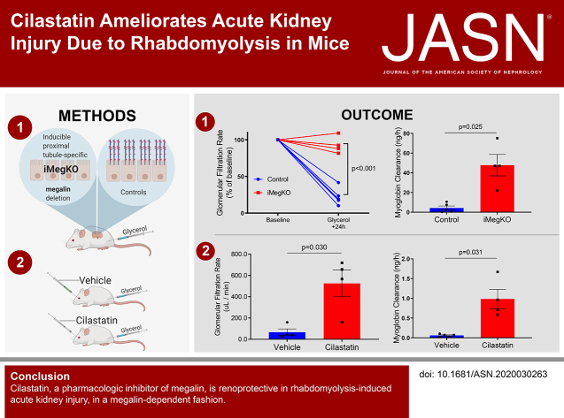
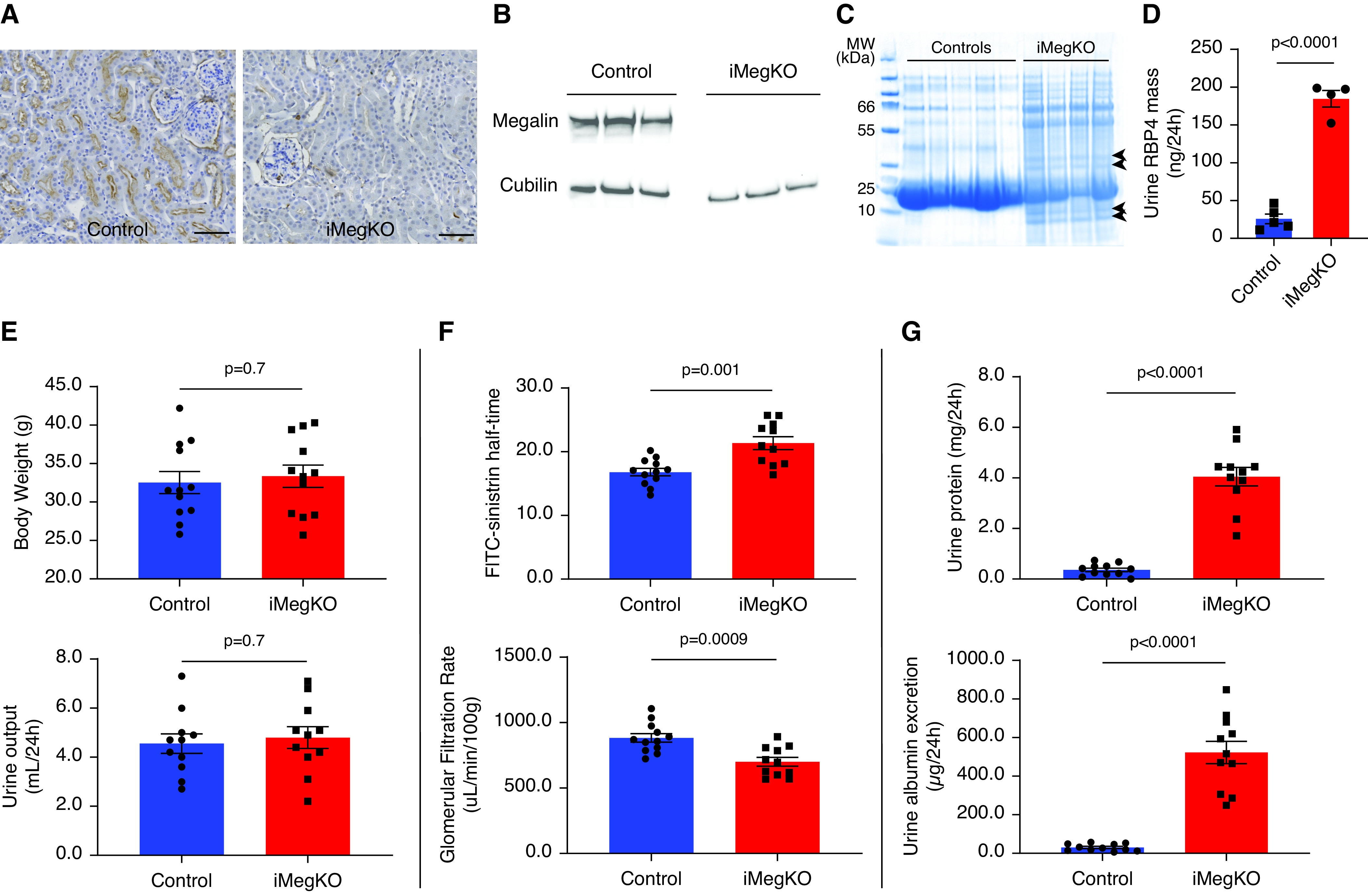
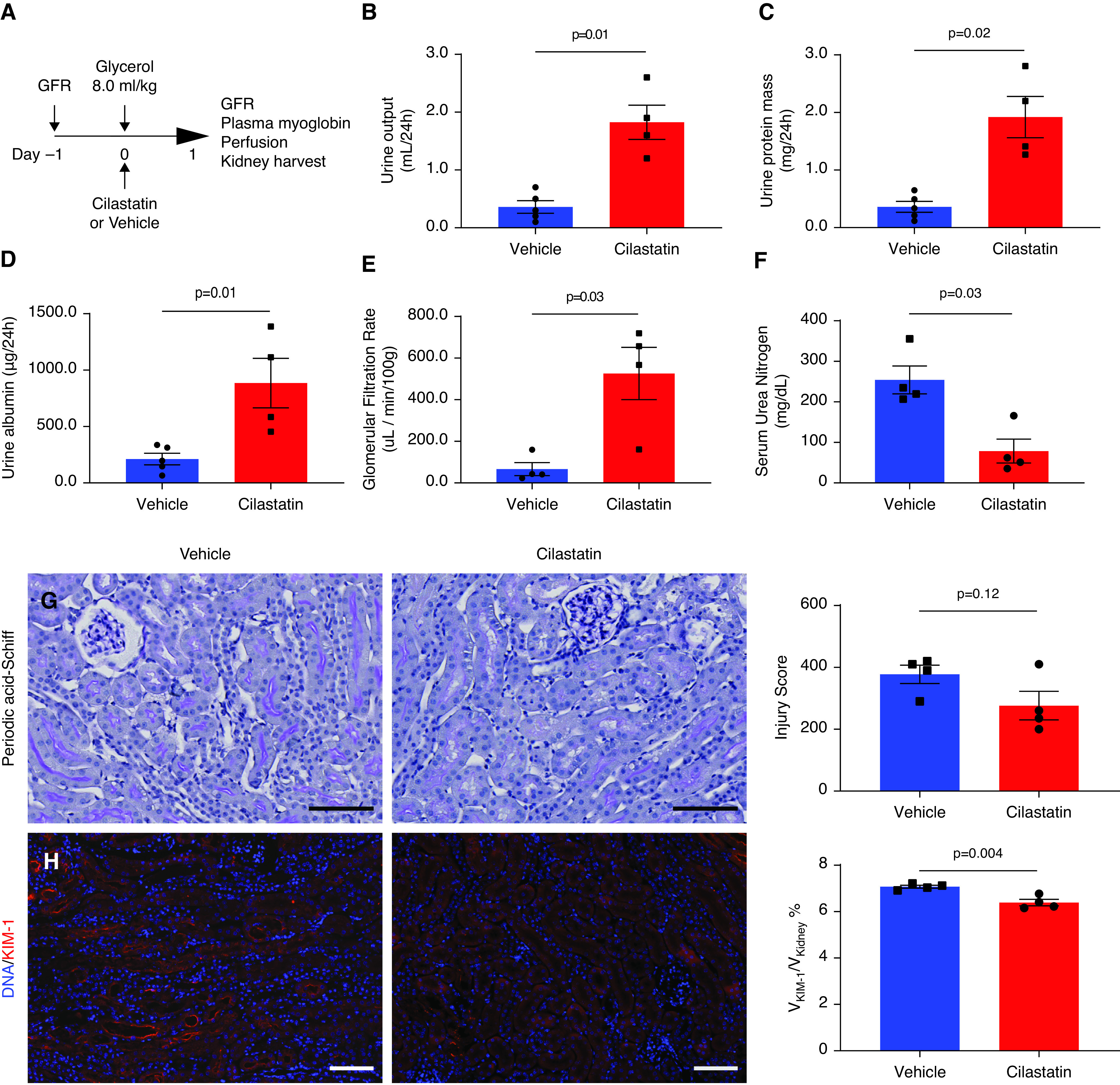
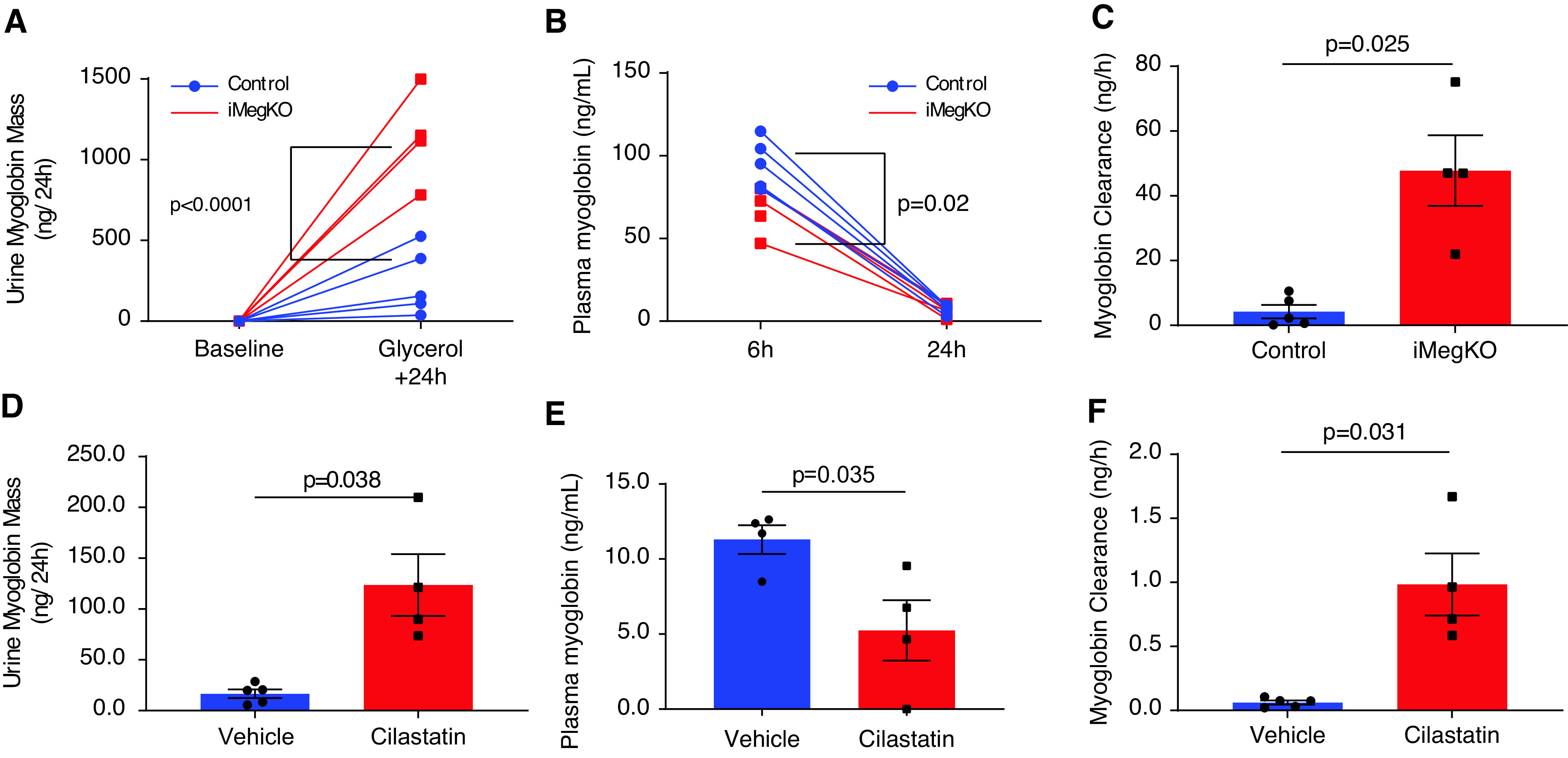
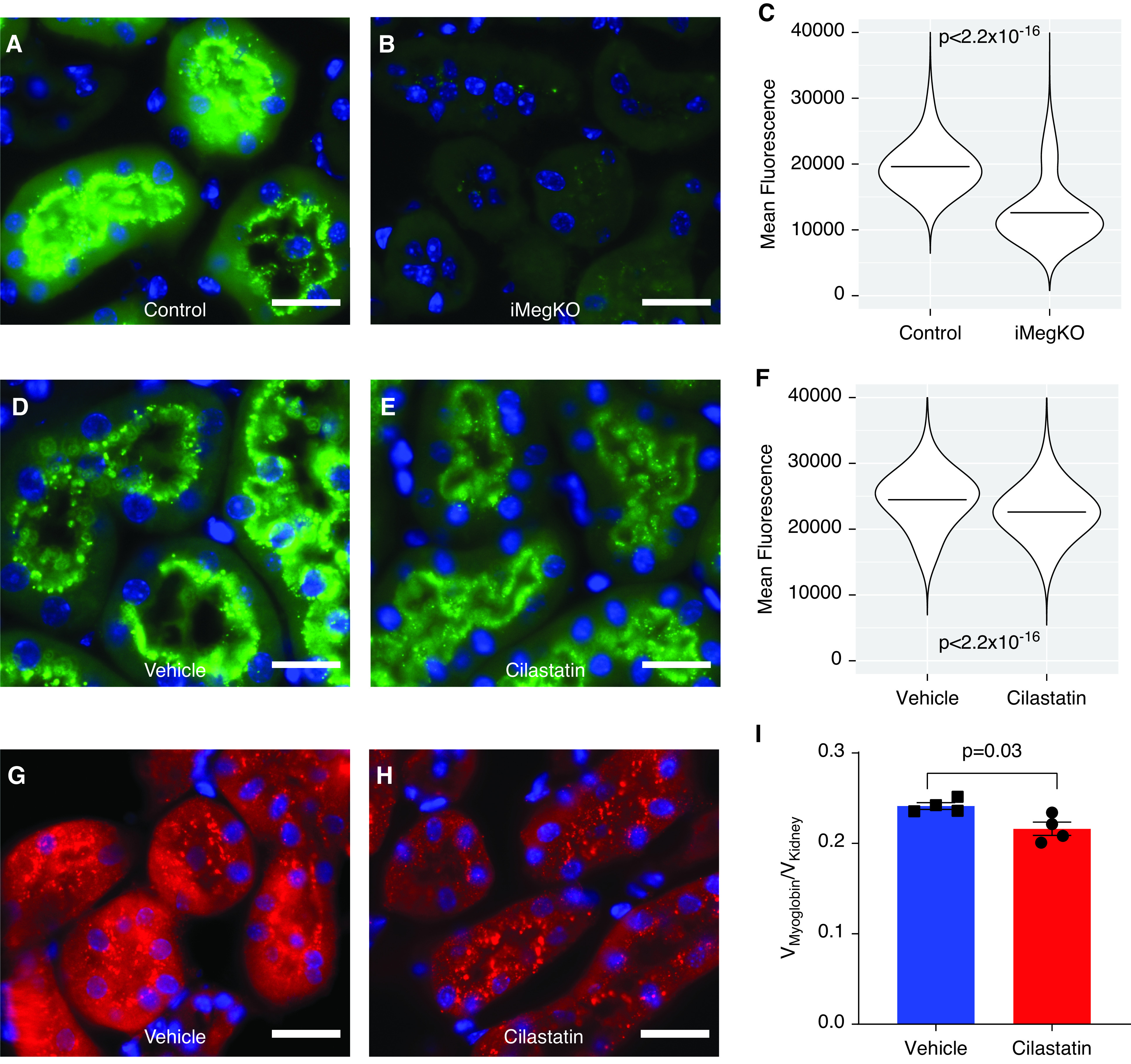
Background: Rhabdomyolysis, the destruction of skeletal muscle, is a significant cause of AKI and death in the context of natural disaster and armed conflict. Rhabdomyolysis may also initiate CKD. Development of specific pharmacologic therapy is desirable because supportive care is nearly impossible in austere environments. Myoglobin, the principal cause of rhabdomyolysis-related AKI, undergoes megalin-mediated endocytosis in proximal tubule cells, a process that specifically injures these cells.

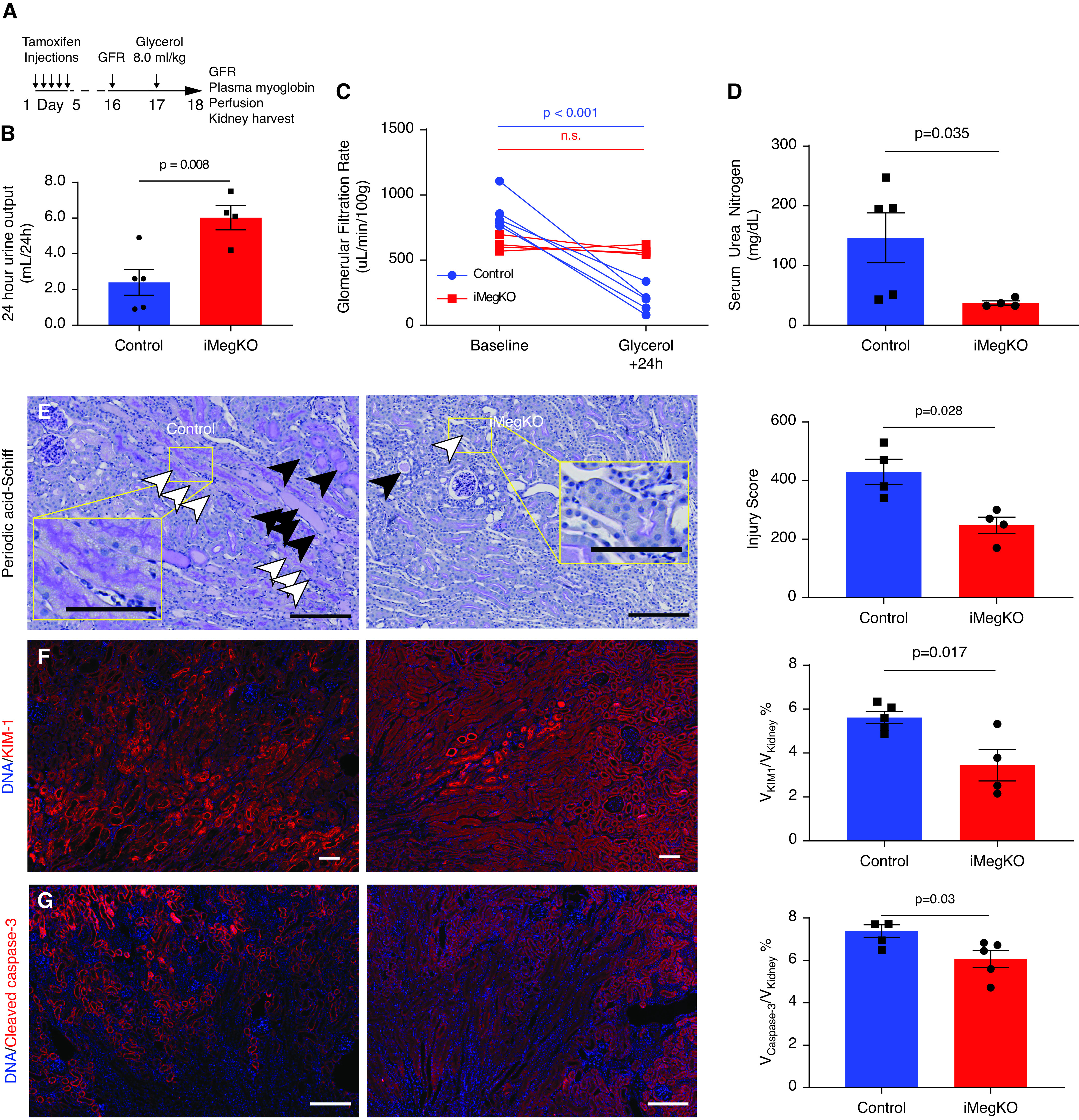
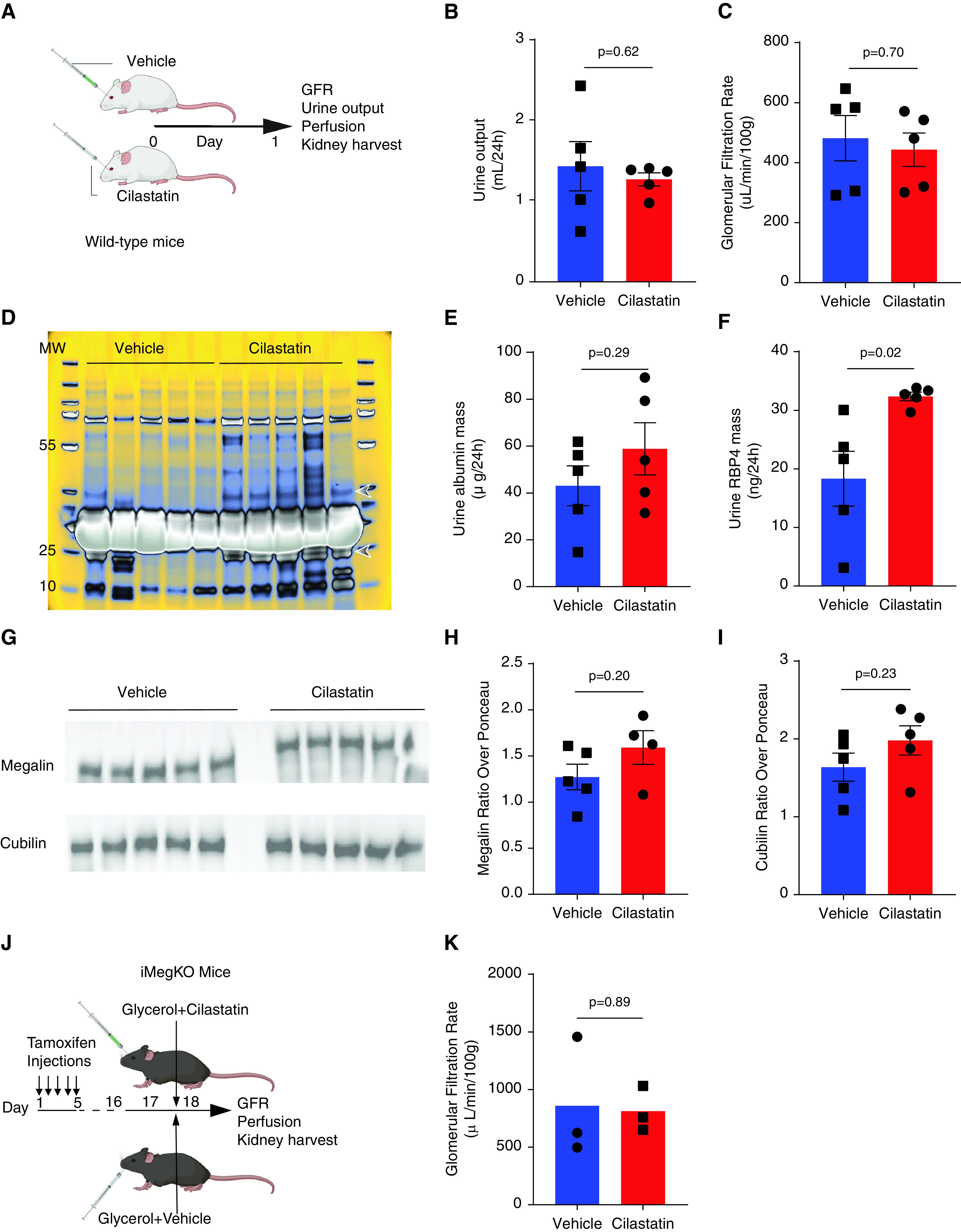
Methods: To investigate whether megalin is protective in a mouse model of rhabdomyolysis-induced AKI, we used male C57BL/6 mice and mice (14-32 weeks old) with proximal tubule-specific deletion of megalin. We used a well-characterized rhabdomyolysis model, injection of 50% glycerol in normal saline preceded by water deprivation.

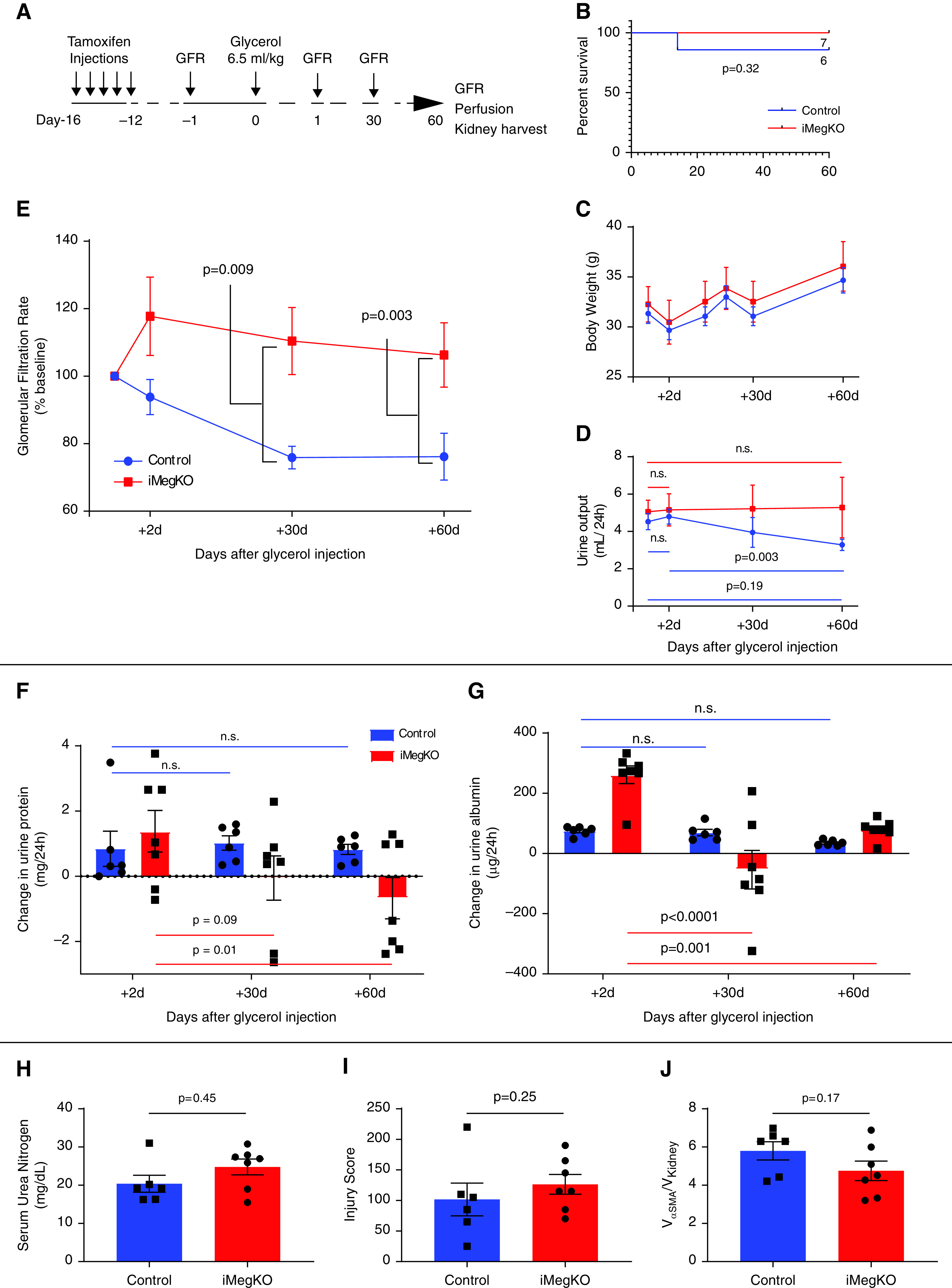
Results: Inducible proximal tubule-specific deletion of megalin was highly protective in this mouse model of rhabdomyolysis-induced AKI. The megalin knockout mice demonstrated preserved GFR, reduced proximal tubule injury (as indicated by kidney injury molecule-1), and reduced renal apoptosis 24 hours after injury. These effects were accompanied by increased urinary myoglobin clearance. Unlike littermate controls, the megalin-deficient mice also did not develop progressive GFR decline and persistent new proteinuria. Administration of the pharmacologic megalin inhibitor cilastatin to wild-type mice recapitulated the renoprotective effects of megalin deletion. This cilastatin-mediated renoprotective effect was dependent on megalin. Cilastatin administration caused selective proteinuria and inhibition of tubular myoglobin uptake similar to that caused by megalin deletion.

Conclusions: We conclude that megalin plays a critical role in rhabdomyolysis-induced AKI, and megalin interference and inhibition ameliorate rhabdomyolysis-induced AKI. Further investigation of megalin inhibition may inform translational investigation of a novel potential therapy.

Keywords: acute renal failure; chronic kidney disease; endocytosis; renal protection; rhabdomyolysis.


Figures

None
Graphical abstract
Figure 1.
Figure 1.
Animal and experimental models. (A–G) Results of tamoxifen treatment in Lrp2fl/fl; Ndrg1CreERT2+ (iMegKO) and cre- littermates (control). (A) High-power micrographs obtained 14 days after the first of five daily tamoxifen injections demonstrate that control mice exhibit robust immunostaining for megalin at proximal tubule brush borders, whereas iMegKO mice demonstrate near-complete absence of megalin. (B) Megalin is absent in immunoblots performed on renal homogenate of iMegKO mice, whereas cubilin is not affected by iMegKO status. (C) The urine of iMegKO mice contains low molecular-weight proteins (arrowheads), which are not present in the urine of controls as demonstrated by Coomassie-stained electrophoresis of equal volumes of urine obtained from 24 hours collection. (D) Urine RBP4, a megalin ligand, is greatly upregulated by iMegKO. (E) Body weight and 24 hours urine output are not altered by megalin deletion. (F) Clearance of FITC-conjugated sinistrin (FITC-sinistrin) is reduced by megalin deletion, indicating a reduction in GFR. (G) Urine protein and urine albumin excretion are greatly increased by megalin deletion. Scale bars: 100 µm. Statistical test used for all comparisons: t test.
Figure 2.
Figure 2.
Deletion of proximal tubular megalin mediates rhabdomyolysis-induced AKI 24 hours after glycerol injection. (A) Experimental design. Tamoxifen induction started 15 days before glycerol injection; mice with inducible proximal tubule–specific megalin deletion (iMegKO) and littermate control mice received identical tamoxifen regimens. (B–G) At 24 hours after glycerol injection, control mice demonstrated AKI, which was attenuated in iMegKO mice. (B) Control mice were oliguric, whereas iMegKO mice demonstrated urine output that was greater than baseline. (C) Control mice demonstrate severe loss of GFR, whereas in iMegKO, GFR is not different from baseline value. (D) Serum urea nitrogen is much greater in control mice than iMegKO mice. (E) Photomicrographs of periodic acid–Schiff stained sections in control and iMegKO mice are distinguished by extensive proteinaceous material in distal tubule and collecting ducts (black arrowheads) and cell swelling and luminal effacement (white arrowheads) in control, with more normal architecture in iMegKO. Composite injury score, right, is greater in control mice. (F) KIM-1 stain is greatly attenuated in iMegKO mice, and accordingly, apoptosis, indicated by cleaved caspase-3 staining (G), is also reduced by megalin interference. Scale bars are 100 µM. Statistical test used for all comparisons: t test.
Figure 3.
Figure 3.
Megalin mediates progressive kidney disease due to rhabdomyolysis. (A) Experimental design. (B) Survival plot. One control mouse died during the 60-day experiment. (C) Body weight after glycerol injection was not mediated by induced proximal tubule-specific megalin deletion (iMegKO). (D) Control mice developed relative oliguria at 60 days when compared with 2 days after glycerol injection, whereas iMegKO mice demonstrated unchanged urine output throughout the experiment. (E) GFR progressively declined in control mice, whereas in iMegKO mice, GFR did not change from baseline. (F and G) Because megalin interference causes proteinuria (see Figure 1), urine protein (F) and urine albumin (G) are displayed as change from baseline. Control mice demonstrated increased proteinuria and albuminuria, compared with baseline, which persisted through the full 60-day experimental course, whereas in iMegKO mice, both proteinuria and albuminuria were reduced at 60 days compared with 2 days after glycerol injection. iMegKO mice did not have significantly increased proteinuria at day 60, whereas controls did. (H) Serum urea nitrogen, pathologic injury score (I), and α smooth muscle actin deposition (αSMA, an indicator of renal fibrosis) were not mediated by proximal tubule megalin status 60 days after glycerol injection. Statistical analysis presented is Mantell-Cox logrank test (B), repeated measures ANOVA (C–G), and t test (H–J).
Figure 4.
Figure 4.
Pharmacologic treatment with megalin inhibitor cilastatin recapitulates AKI amelioration by proximal tubule-specific megalin deletion. (A) Experimental design. Cilastatin was administered immediately after glycerol. (B–D) In accordance with findings in iMegKO mice, cilastatin administration at the time of rhabdomyolysis induction resulted in increased urine output (B), increased acute proteinuria (C), and increased urine albumin (D), compared with vehicle. Cilastatin administration also prevented severe loss of GFR observed in vehicle-treated mice (E), and prevented the highly elevated serum urea nitrogen seen in vehicle-treated mice (F). Periodic acid–Schiff stained sections were also generally in accordance with findings in control and iMegKO mice (G), although histopathologic injury scoring was not different between cilastatin- and vehicle-treated mice. KIM-1 was elevated in both groups (H), but significantly reduced in cilastatin-treated mice compared with vehicle-treated mice. Scale bars are 100 µm. Statistical analysis presented is derived from the t test.
Figure 5.
Figure 5.
Myoglobin clearance is similarly altered by proximal tubule megalin interference and cilastatin administration. (A) Induced proximal tubule–specific megalin deletion (iMegKO) status conferred much greater excretion of myoglobin in the urine. (B) Plasma myoglobin concentration was elevated in controls relative to iMegKO mice with 6 hours, but not 24 hours, after glycerol injection, with rapid decline in plasma myoglobin occurring in both groups. (C) Myoglobin clearance was significantly greater in iMegKO mice than controls. (D–F) Treatment with the pharmacologic inhibitor of renal megalin, cilastatin, had similar actions to proximal tubule–specific deletion of megalin. (D) Urine myoglobin was increased by cilastatin treatment. (E) After 24 hours plasma myoglobin was reduced by cilastatin treatment. (F) Myoglobin clearance was increased approximately 16× by cilastatin treatment. Statistical analysis presented is derived from (A, B) repeated measures ANOVA (C–F) and t test.
Figure 6.
Figure 6.
Cilastatin and megalin deletion have similar effects on renal function. (A) Experimental design for results (B–I). Wild-type mice received cilastatin (200 mg/kg) or vehicle injection as in prior experiments, without injection of glycerol. The 24 hours later, outcomes were assessed. (B and C) Urine output and GFR were not altered by cilastatin administration. (D) Coomassie-stained urine electrophoresis (loaded with identical volumes of urine from each animal). Bands at approximately 23 kD and approximately 40 kD marked by arrowheads appear differentially expressed in cilastatin versus vehicle samples. The same bands may be seen in the urine of iMegKO mice in Figure 1. Unaltered gel image shown in pseudo-color to better visualize peak protein density. (E) Urine albumin was not significantly increased. (F) RBP4, a plasma protein that is a known megalin ligand, and is greatly increased in the urine of megalin-deleted mice, was nearly doubled by cilastatin administration. (G–I) Immunoblots performed on kidney lysate 24 hour after vehicle or cilastatin injection. Megalin expression was reduced after cilastatin administration, whereas cubilin was not significantly altered. (J) Experimental design. iMegKO mice received cilastatin or vehicle with injection of glycerol. Then 24 hours later, mean GFR was identical between groups (K), indicating that cilastatin-dependent protection from rhabdomyolysis-induced AKI is absent in mice without proximal tubule megalin. Statistical analysis presented is derived from the t test.
Figure 7.
Figure 7.
Similar interference in myoglobin uptake in iMegKO and cilastatin-treated mice. To characterize the role of megalin in myoglobin endocytosis, iMegKO mice and controls were injected with FITC-myoglobin (0.5 mg). Then 15 minutes later, mice were killed and kidneys prepared for histologic examination. (A) Controls demonstrated abundant tubular epithelial FITC signal, primarily organized in punctae within the apical brush border and the adjacent cytoplasm, whereas (B), iMegKO mice demonstrated attenuated FITC-myoglobin signal, and far fewer punctae. (C) Mean fluorescence of all puncta (n=371,156) quantified by strain demonstrates that puncta from iMegKO mice exhibit reduced fluorescence compared with those from controls. Horizontal lines depict the mean. (D–F) To characterize the effect of cilastatin on myoglobin endocytosis, wild-type mice were injected with FITC-myoglobin (0.5 mg). Then 15 minutes later, mice were killed and kidneys prepared for histologic examination. Vehicle-treated mice exhibited abundant FITC-myoglobin punctae, whereas (D) cilastatin-treated mice exhibited fewer punctae and overall reduced FITC-fluorescence. (H) Mean fluorescence of all puncta (n=2,298,499) compared by drug treatment demonstrates that puncta from cilastatin-treated mice exhibit reduced fluorescence compared with those from vehicle-treated mice. Horizontal lines depict the mean. (G–I) To determine whether cilastatin interfered with overall renal myoglobin uptake, wild-type mice received vehicle or cilastatin injection 1 hour before glycerol intramuscular injection. (G, H) Then 2 hours after glycerol injection, myoglobin-directed immunofluorescence demonstrates abundant myoglobin-positive punctae at the brush border and within the cytoplasm of proximal tubules of vehicle injected mice, this signal appeared attenuated in tubules from cilastatin-injected mice. (I) Unbiased stereology demonstrates reduced total intrarenal myoglobin content in cilastatin-treated mice. Scale bars are 20 µm. Statistical analysis presented is derived from the t test.

References

    1. Stewart IJ, Snow BD, Clemens MS, Sosnov JA, Ross JD, Howard JT, et al. : Hyperkalemia in combat casualties: Implications for delayed evacuation. Mil Med 182: e2046–e2051, 2017 - PubMed
    1. Stewart IJ, Faulk TI, Sosnov JA, Clemens MS, Elterman J, Ross JD, et al. : Rhabdomyolysis among critically ill combat casualties: Associations with acute kidney injury and mortality. J Trauma Acute Care Surg 80: 492–498, 2016 - PubMed
    1. Stewart IJ, Sosnov JA, Howard JT, Chung KK: Acute kidney injury in critically injured combat veterans: A retrospective cohort study. Am J Kidney Dis 68: 564–570, 2016 - PubMed
    1. Belliere J, Casemayou A, Ducasse L, Zakaroff-Girard A, Martins F, Iacovoni JS, et al. : Specific macrophage subtypes influence the progression of rhabdomyolysis-induced kidney injury. J Am Soc Nephrol 26: 1363–1377, 2015 - PMC - PubMed
    1. Stewart IJ, Sosnov JA, Howard JT, Orman JA, Fang R, Morrow BD, et al. : Retrospective analysis of long-rerm outcomes after combat injury: A hidden cost of war. Circulation 132: 2126–2133, 2015 - PubMed

Publication types

MeSH terms

LinkOut - more resources