. 2022 Feb;16(2):346-357.
doi: 10.1038/s41396-021-01073-y. Epub 2021 Aug 2.

A general approach to explore prokaryotic protein glycosylation reveals the unique surface layer modulation of an anammox bacterium

Affiliations

A general approach to explore prokaryotic protein glycosylation reveals the unique surface layer modulation of an anammox bacterium

Martin Pabst et al. ISME J. 2022 Feb.

Abstract

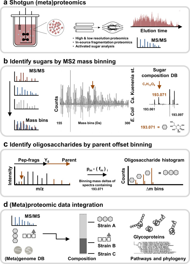
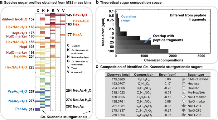
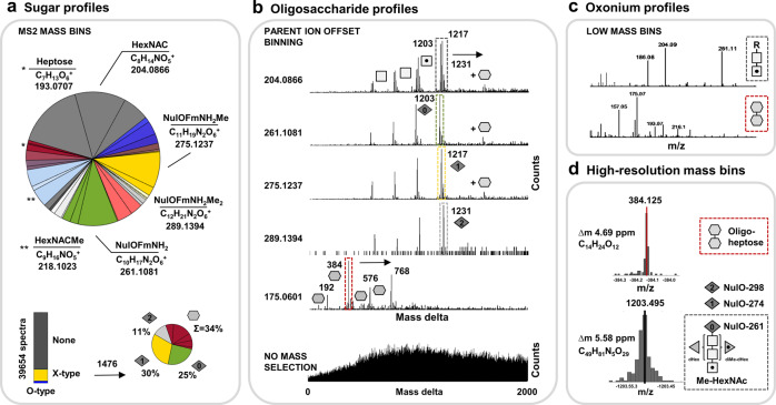
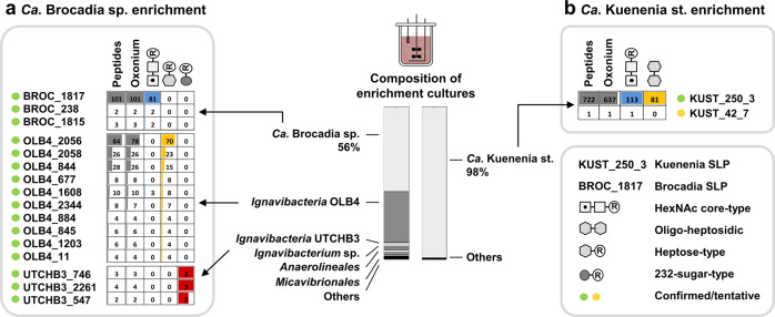
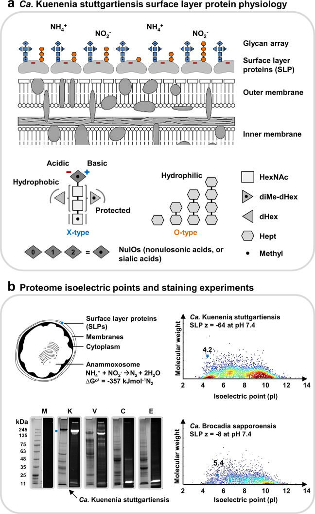
The enormous chemical diversity and strain variability of prokaryotic protein glycosylation makes their large-scale exploration exceptionally challenging. Therefore, despite the universal relevance of protein glycosylation across all domains of life, the understanding of their biological significance and the evolutionary forces shaping oligosaccharide structures remains highly limited. Here, we report on a newly established mass binning glycoproteomics approach that establishes the chemical identity of the carbohydrate components and performs untargeted exploration of prokaryotic oligosaccharides from large-scale proteomics data directly. We demonstrate our approach by exploring an enrichment culture of the globally relevant anaerobic ammonium-oxidizing bacterium Ca. Kuenenia stuttgartiensis. By doing so we resolve a remarkable array of oligosaccharides, which are produced by two seemingly unrelated biosynthetic routes, and which modify the same surface-layer protein simultaneously. More intriguingly, the investigated strain also accomplished modulation of highly specialized sugars, supposedly in response to its energy metabolism-the anaerobic oxidation of ammonium-which depends on the acquisition of substrates of opposite charges. Ultimately, we provide a systematic approach for the compositional exploration of prokaryotic protein glycosylation, and reveal a remarkable example for the evolution of complex oligosaccharides in bacteria.


Conflict of interest statement

The authors declare no competing interests.

Figures

Fig. 1
Fig. 1. The mass binning glycoproteomics approach to explore prokaryotic protein glycosylation.
a The approach identifies the chemical composition of the proteome specific carbohydrate components and establishes the related protein linked oligosaccharide chains from large-scale (meta)proteomics data. Optional in-source fragmentation and metabolomics experiments provide additional chemical information to guide the exploration of biosynthetic routes or phylogenetic relations. b The first step—establishing the proteome-specific sugar components—performs mass binning of the complete set of fragmentation spectra acquired at very high mass resolution. Binned spectra thereby show highly comparable pattern across proteomes/species because those are the products of the universal set of amino acids (mirror bar chart). Carbohydrate fragments, however, have a characteristic chemical composition and, therefore, (usually) lay outside the amino acid composition space. Signals are automatically matched against a constructed database, consisting of over 3300 theoretical carbohydrate compositions. The procedure is exemplified by the heptose fragment (193.071 m/z), observed in the Ca. Kuenenia stuttgartiensis enrichment (meta)proteome. The heptose signal (193.071 m/z) is unique to Kuenenia, but the neighboring mass peaks (−0.01 m/z and +0.026 m/z) are also observed in the non-glycosylated comparator proteome (small mirror bar chart) and are, therefore, amino acid related. c The second step—termed “parent ion offset binning”—establishes the actual oligosaccharide chains, which are linked to the proteins of individual strains. This is achieved by binning the mass deltas obtained after subtracting the fragment masses (fm) from their parent (ion) peptide mass (pm). Spectra containing the same carbohydrate fragments will, thereby, show repeatedly mass deltas consistent with the mass of the oligosaccharide modification. d The established oligosaccharide chains are then integrated into the metaproteomic database search (using e.g., a metagenomics constructed databases), to identify the target proteins and related strains.
Fig. 2
Fig. 2. Carbohydrate profiles obtained from the anammox enrichment cultures (and reference samples) using the MS2 mass binning approach.
a The graph (from left to right) shows the carbohydrate profiles (m/z values are distributed along the y-axis) from the C. jejuni sample (C, control), Ca. Kuenenia stuttgartiensis enrichment culture (K), HEK protein sample (H, control), Ca. Brocadia sapporoensis enrichment culture (b), S. cerevisiae sample (Y, control) and H. volcanii sample (V, control). The carbohydrate profiles of the control samples established by the MS2 mass binning approach reflected the known oligosaccharide compositions of the individual species. Only sugar components, which commonly provide only very low abundant or no carbohydrate related fragments (oxonium ions), such as deoxy hexoses, were not observed. NulO and HexNAc fragments are prominent in the C. jejuni proteomics sample, NeuAc/HexNAc and Hex related signals in the HEK derived sample, HexNAC- and hexose-related signals in the S. cerevisiae (yeast) proteomics sample, and hexuronic acid (HexA)- and hexose (Hex)-related signals in the H. volcanii sample. The latter was cultured at high salinity; therefore, the proteins appeared modified by only a single type of glycan structure. Fragments with the same color indicate water loss clusters (-H2O), which are a characteristic consequence of the oligosaccharide chain fragmentation process, and therefore indicators for the discovery process. The mass compositions and sugar type annotations for the Ca. Kuenenia stuttgartiensis enrichment proteome are detailed in the table on the right (c). The abbreviations “NulO” stand for nonulosonic acid, “NeuAc” for N-Acetyl neuraminic acid; “Pse” for pseudaminic acid; “HexNAc” for N-Acetyl-hexose amine; “Hept” for heptose; “Hex” for hexose; “HexA” for hexuronic acid; “dHex” for deoxyhexose and “Me” for methyl, respectively. The sugar symbols are depicted in generic white and gray shades because a further classification into specific types of monosaccharides, beyond sum formulae, chain length and modifications, cannot be obtained from accurate mass experiments. The different gray scales were simply chosen to make the individual sugar symbols more distinguishable within graphs and depicted oligosaccharide structures (b) The constructed carbohydrate chemical composition space. A large chemical composition space was constructed (>3300 theoretical compositions) used to assign chemical compositions to non-peptide related features. At high mass resolution (>100 K) and accuracy (<1 ppm), the mass overlap with amino acid-related fragments is considerably low. Mass recalibration using frequently observed amino acid fragments enables to operate at very high mass accuracy (blue arrow) “(color figure online)”.
Fig. 3
Fig. 3. Outline of identified sugar components and observed oligosaccharide profiles for the Ca. Kuenenia stuttgartiensis enrichment.
a The large pie chart outlines the proportions of the carbohydrate fragments identified in the Ca. Kuenenia stuttgartiensis enrichment proteome using the MS2 mass binning approach. The lower charts depict the proportions of spectra containing carbohydrate related signals (oxonium ions) in the sequencing spectra, and the frequency of individual glycoforms across all spectra. b The graphs outline the furthermore established oligosaccharide chains using parent ion offset binning of fragmentation spectra containing the identified carbohydrate fragments (graphs labeled with 204, 275, 289, and 175 m/z). Thereby, oligosaccharide chains appear in histograms as repeatedly occurring mass deltas. The same parent ion offset approach applied to the complete, non-carbohydrate-filtered dataset, does not reveal any identifiable systematically reoccurring mass deltas (bottom graph). This revealed two completely unrelated oligosaccharide chains. The fragments 204, 261, 275, and 289 m/z belong to variations of a complex type oligosaccharide with a HexNAc core structure (X-type). The fragment 175 (and 193 m/z) retrieved a second, fully unrelated heptose type oligosaccharide chain (O-type). Squares represent HexNAcs (methylations are depicted by a dot), diamonds represent NulO variants, hexagons represent heptoses, triangles are deoxyhexoses, and doted triangles are dimethyl-deoxyhexoses. Moreover, due to the predominant fragmentation of the oligosaccharide chains, nearly the complete sequence of the oligosaccharide can be derived. c The histograms outline the (intensity normalized) low mass bins of fragmentation spectra where the complex type oligosaccharide (upper graph) or the oligo-heptosidic chains (O-type, lower graph) were identified. The thereby-observed sugar fragments correlate with the proposed composition of the individual carbohydrate chains (e.g., 204/261 m/z for complex, or 175/193 m/z for oligo-heptosidic). d The histogram shows binning of mass deltas into very small bin sizes to establish the oligosaccharide compositions at very high mass accuracy (<7.5 ppm).
Fig. 4
Fig. 4. Glycosylated proteins and strains present in the explored anammox enrichment cultures.
The metaproteomic analysis of the Ca. Kuenenia stuttgartiensis enrichment showed that ~98% of identified peptide sequences derived from Ca. Kuenenia stuttgartiensis (right bar, SI-EXCEL-T7 and T9). The vast majority of glycosylated peptides moreover could be assigned to the surface layer protein (KUST_250_3) of Ca. Kuenenia stuttgartiensis (b, right table, SI-EXCEL-T8 and T10, SI-DOC-Table-S8). This confirmed that both types of oligosaccharides (complex-type and oligo-heptosidic) target the same surface layer protein simultaneously. Furthermore, the metaproteomic analysis of the Brocadia enrichment culture showed that ~56% of the peptide sequences derived from Ca. Brocadia sapporoensis, and significant other proportions to at least three different Ignavibacteria strains (left bar, SI-EXCEL-T12 and T14). A modification search, including the identified oligosaccharide chains, confirmed that the putative surface layer protein of Ca. Brocadia sapporoensis is modified by a HexNAc core type oligosaccharide (204, squares). A second type of oligosaccharide chain (161/193 = hexagons) was assigned to multiple proteins from Ignavibacteria bacterium OLB4, and a third type of oligosaccharide (232, dark gray circles) to several proteins from Ignavibacteria bacterium UTCHB3 (a, left table, SI-EXCEL-T13, SI-DOC-Table-S9). Only the top matches for every database search are shown in the graph. Proteins with peptide matches at all three levels (i–iii) were considered as confirmed (green circle; i=database search, ii=oxonium ions and iii=oligosaccharide mass deltas). Peptides indicates the number of variable modification search matches (VM); oxonium indicates the number of VM matches with additional oxonium ion identifications; squares show the number of HexNAc core type oligosaccharide matches assigned to the same VM matches; gray hexagon counts the number of heptose-type oligosaccharide matches that were also assigned to VM matches; dark gray circle counts the number of 232 sugar-type oligosaccharide matches that were also assigned to the same VM matches.
Fig. 5
Fig. 5. Physiology of the Ca. Kuenenia stuttgartiensis surface layer protein (SLP) and oligosaccharides.
a The surface layer protein (SLP) of Ca. Kuenenia stuttgartiensis is densely covered by two entirely different types of oligosaccharides (“X-type” and “O-type”, SI-DOC-Fig-S16). The dense layer supposedly provides shielding of the very acidic SLP. Interestingly, the investigated Ca. Kuenenia stuttgartiensis strain produces nonulosonic acids (NulOs), which possess an unmasked amine. Those have the potential to counterbalance the carboxylic acid groups. The sugar symbols are depicted in generic white and gray shades because a further classification into specific types of monosaccharides, beyond sum formulae, chain length and modifications, cannot be obtained from accurate mass experiments. The different shades of gray were simply chosen to make the individual sugars more distinguishable within the oligosaccharide structures. The oligosaccharide structures depicted on the cell surface layer (top graph) are colored in blue if those structures represent a X-type (complex type) structure, or in orange if they represent an O-type (oligo-heptosidic) oligosaccharide. The colors do not provide any further indications on the types of monosaccharides. b The Ca. Kuenenia stuttgartiensis surface layer protein shows a predicted pI (isoelectric point) of ~4.25 and a net charge of ~−60 at physiological pH. In fact, the surface layer protein is one of the most acidic proteins of the complete Ca. Kuenenia stuttgartiensis proteome. On the other hand, the putative surface layer protein of Ca. Brocadia sapporoensis has a predicted pI of only 5.4 and a substantially lower net charge of ~−8 at physiological pH. Moreover, Ca. Brocadia, uses also only a related form of the complex-type oligosaccharide to cover its much less acidic surface layer protein. The SDS-PAGE analyzes show protein and sugar staining for the protein extracts from Ca. Kuenenia stuttgartiensis and the additional control strains H. volcanii (glycan-positive control), C. jejuni (glycan-positive control) and E. coli K12 (glycan-negative control). The left lanes each show the total protein staining (P; Brilliant Blue G staining solution), whereas the right lanes each show the carbohydrate staining (C; Pro-Q 488 Emerald staining kit) “(color figure online)”.

Similar articles

Cited by

References

    1. Prabakaran S, Lippens G, Steen H, Gunawardena J. Post‐translational modification: nature’s escape from genetic imprisonment and the basis for dynamic information encoding. Wiley Interdiscip Rev Syst Biol Med. 2012;4:565–83. - PMC - PubMed
    1. Khoury GA, Baliban RC, Floudas CA. Proteome-wide post-translational modification statistics: frequency analysis and curation of the swiss-prot database. Sci Rep. 2011;1:90. - PMC - PubMed
    1. den Ridder M, Daran-Lapujade P, Pabst M. Shot-gun proteomics: why thousands of unidentified signals matter. FEMS Yeast Res. 2020;20:foz088. - PubMed
    1. Spoel SH. Orchestrating the proteome with post-translational modifications. Oxford University Press UK. 2018;19:4499–4503. - PMC - PubMed
    1. Varki A, Cummings RD, Esko JD, Freeze HH, Stanley P, Bertozzi CR, et al. Essentials of glycobiology. 3rd edition. (Cold Spring Harbor Laboratory Press, New York, 2015–2017). - PubMed

Publication types

Substances