Review
. 2021 Oct;22(10):653-670.
doi: 10.1038/s41580-021-00387-1. Epub 2021 Aug 2.

Nuclear compartmentalization as a mechanism of quantitative control of gene expression

Affiliations
Review

Nuclear compartmentalization as a mechanism of quantitative control of gene expression

Prashant Bhat et al. Nat Rev Mol Cell Biol. 2021 Oct.

Abstract

Gene regulation requires the dynamic coordination of hundreds of regulatory factors at precise genomic and RNA targets. Although many regulatory factors have specific affinity for their nucleic acid targets, molecular diffusion and affinity models alone cannot explain many of the quantitative features of gene regulation in the nucleus. One emerging explanation for these quantitative properties is that DNA, RNA and proteins organize within precise, 3D compartments in the nucleus to concentrate groups of functionally related molecules. Recently, nucleic acids and proteins involved in many important nuclear processes have been shown to engage in cooperative interactions, which lead to the formation of condensates that partition the nucleus. In this Review, we discuss an emerging perspective of gene regulation, which moves away from classic models of stoichiometric interactions towards an understanding of how spatial compartmentalization can lead to non-stoichiometric molecular interactions and non-linear regulatory behaviours. We describe key mechanisms of nuclear compartment formation, including emerging roles for non-coding RNAs in facilitating their formation, and discuss the functional role of nuclear compartments in transcription regulation, co-transcriptional and post-transcriptional RNA processing, and higher-order chromatin regulation. More generally, we discuss how compartmentalization may explain important quantitative aspects of gene regulation.


Conflict of interest statement

Competing interests

The authors declare no competing interests.

Figures

Fig. 1 |
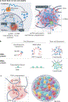
Fig. 1 |. Mechanisms of nuclear compartments formation.
a | Nuclear compartments contain high local concentration of specific molecules in 3D space. Upward arrows represent higher concentration and downward arrows represent lower concentration. b | Stoichiometric interactions between two molecules, M1 (an RNA) and M2 (a protein), show a linear increase in the number of M2 molecules bound per unit increase in M1. By contrast, non-stoichiometric interactions lead to an exponential increase in binding of the target molecule (M2) as M1 increases. c | Stoichiometric interactions between molecules and cooperative interactions between multivalent intrinsically disordered regions (IDRs) of proteins can be found within a sample of complexes. The low-affinity interactions between IDRs are often mediated by stretches of charged and polar amino acids (aa). d | Image of polypyrimidine tract-binding protein 1 (PTBP1) molecules undergoing concentration-dependent condensate formation with RNA in vitro (top). When protein concentration is high, multivalent interactions can promote ‘de-mixing’ (bottom). e | Phase diagram describing whether molecules will be present in a diffusive state or a compartmentalized state. Formation of these structures is controlled by local concentration leading to sharp transition behaviours (blue line). f | Condensates can exhibit properties of liquids, including the ability to split (fission), merge (fusion) and undergo rapid internal rearrangement (diffusion). g | Cartesian plane highlighting the relationship between affinity, avidity and physical manifestations of different types of molecular associations. Well-mixed solution of two soluble proteins with little to no affinity for one another, such as GAPDH and enolase (bottom left). U1 small nuclear ribonucleoprotein (U1 snRNP) is an example of a macromolecular complex composed of high-affinity interactions between a fixed stoichiometric ratio of interacting component molecules (bottom right). Nuclear bodies are composed of self-interacting proteins, for example, SC35 (also known as SRSF2) proteins in nuclear speckles (top left). Solid aggregates of tau are observed in neurodegenerative diseases (top right). Image courtesy of Inna-Marie Strazhnik/Caltech. Part d adapted from REF., Springer Nature Limited.
Fig. 2 |
Fig. 2 |. Spatially constrained non-coding RNAs can drive compartmentalization in the nucleus.
a | Model of how non-coding RNAs (ncRNAs) can drive compartmentalization. Sites of ncRNA transcription can support high ncRNA concentrations and, thus, seed the formation of high-concentration territories (seed); ncRNAs can bind to diffusible proteins or ncRNAs through stoichiometric interactions (bind) and, in this manner, lead to enrichment of diffusible factors within a spatially defined region (recruit). In some cases, the recruited proteins can recruit other proteins and/or form condensates through homotypic and heterotypic interactions (not shown). b | Stoichiometric RNA–protein or DNA–protein interactions can involve single or multiple binding events, but the number of proteins recruited is limited to the number of available sites on the nucleic acid. By contrast, non-stoichiometric interactions enable binding of more proteins than available binding sites. c | Space filling model of localization of hundreds of ncRNAs (coloured areas) across the nucleus in mouse embryonic stem cells. Pol II, polymerase II; XIST, X-inactive specific transcript. Image courtesy of Inna-Marie Strazhnik/Caltech.
Fig. 3 |
Fig. 3 |. Enhancers, promoters and transcription factors can form condensates that may facilitate rapid target search in the genome.
a | Example of multiple enhancers (super-enhancer) confined to a topologically associating domain (TAD; dotted red line) that contains the promoter they regulate. Comparison of a simple chromatin looping model of enhancers and promoters (left) with a condensate model (right) shows how multiple enhancers can occupy the same territory by forming cooperative interactions. b | Two distinct transcription factors (TF1 and TF2) can have the same affinity for the same DNA sequences, but occupy different genomic locations. Transcription factors lacking intrinsically disordered regions (IDRs) cannot distinguish between these high-affinity sites (‘non-specific’), but transcription factors containing IDRs are targeted to specific genomic regions (‘specific’). c | Comparison of diffusion–affinity models that involve stochastic movement of proteins to target sites (left) with the facilitated diffusion model, which involves a combination of 3D movement with sliding (right). Pol II, polymerase II. Image courtesy of Inna-Marie Strazhnik/Caltech. Part b adapted with permission from REF., AAAS.
Fig. 4 |
Fig. 4 |. compartmentalization and chromatin regulation.
a | Stoichiometric interactions occur at defined ratios of components, whereas non-stoichiometric interactions occur without fixed ratios and can exceed the binding capacity of any individual molecule. Increasing the concentration of the long non-coding RNA (ncRNA) X-inactive specific transcript (XIST) along the X chromosome increases in the concentration of the XIST-binding protein SHARP, which eventually results is silencing of most genes on the chromosome. Spatial amplification of SHARP (which exceeds XIST concentration) requires its intrinsically disordered regions (IDRs) and may be achieved through concentration-dependent condensate formation. b | Concentration thresholds may explain the ‘switch-like’ control of gene repression within and outside ncRNA compartments. ncRNAs can achieve their highest concentration at the centre of a spatial territory. If the ncRNA concentration at the central area reaches a critical threshold, enough proteins to mediate compartment formation and to achieve gene repression can be recruited. In this model, ncRNA molecules can still diffuse outside the silencing compartment, but not at levels high enough to recruit sufficient regulatory molecules to change gene expression. As a result, the observed effect on gene expression is binary: ‘on’ within the compartment and ‘off’ outside it. c | Schematic of the Kcnq1ot1 compartment, which also recruits SHARP to silence imprinted genes on the paternal allele. Dashed arrow represents a cohesin complex promoting the formation of the Kcnq1ot1 compartment. d | Schematic showing heterochromatin protein 1 (HP1)-mediated compartmentalization of centromeric heterochromatin, which is marked by trimethylated histone H3 Lys9 (H3K9me3). Pol II, polymerase II. Image courtesy of Inna-Marie Strazhnik/Caltech.
Fig. 5 |
Fig. 5 |. Spatial and kinetic coupling of RNA polymerase II transcription and mRNA splicing.
a | Different models of co-transcriptional splicing. Random diffusion of splicing factors (for example, SR proteins) that have high affinity for target pre-mRNAs (left). In this model, splicing levels are independent of transcription and constant (part b). Stoichiometric model of co-transcriptional splicing, showing molecular interactions between the phosphorylated carboxy-terminal domain (CTD-P) ‘tail’ of RNA polymerase II (Pol II) and the spliceosome, which facilitates splicing as nascent pre-mRNAs are extruded from the polymerase (middle). In this model, splicing increases linearly with transcription rate (part b). Condensate model of co-transcriptional splicing, showing cooperative interactions between the CTD of Pol II, phosphorylated (P) splicing factors and a reservoir of unphosphorylated splicing factors in close proximity to nascent pre-mRNAs (right). b | A condensate model of co-transcriptional splicing would lead to an ‘economy of scale’ dynamic, in which splicing increases non-linearly with increased transcription. c | Pol II CTD phosphorylation mediates a switch from transcriptional condensates of the Mediator complex during transcription initiation, to condensates of splicing factors during transcription elongation. SRSF1, serine/arginine-rich splicing factor 1. Image courtesy of Inna-Marie Strazhnik/Caltech.
Fig. 6 |
Fig. 6 |. Involvement of nuclear bodies in RNA processing.
a | Schematic of a cell nucleus highlighting many RNA processing hubs, including nuclear speckles (compartments of mRNA splicing factors), nucleoli (ribosome biogenesis), histone locus bodies (processing histone pre-mRNAs) and Cajal bodies (biogenesis of small nuclear RNAs (snRNAs)). b | Close-up schematics of different RNA processing hubs. A splicing hub is generally composed of two layers: an inner layer of splicing factors and an outer layer of genomic regions with active RNA polymerase II (Pol II), phosphorylated (P) splicing factors and nascent pre-mRNAs. The nucleolus is composed of three layers: inner fibrillar centre (FC), which is the site of Pol I-mediated pre-rRNA transcription; middle dense fibrillar component (DFC), where rRNA processing occurs by small nucleolar RNAs (snoRNAs) and RNase MRP (scissors); and outer granular component (GC), which is the site of ribosome assembly. Histone locus bodies are sites of histone pre-mRNA maturation, including by the U7 snRNA (U7). Cajal bodies are sites of snRNA maturation, including by small Cajal body RNAs (scaRNAs). Ψ, pseudouridylation. Image courtesy of Inna-Marie Strazhnik/Caltech.

References

    1. Mirny L et al. How a protein searches for its site on DNA: the mechanism of facilitated diffusion. J. Phys. A Math. Theor. 42, 434013 (2009).
    1. Jana T, Brodsky S & Barkai N Speed–specificity trade-offs in the transcription factors search for their genomic binding sites. Trends Genet. 37, 421–432 (2021). - PubMed
    1. Strom AR & Brangwynne CP The liquid nucleome—phase transitions in the nucleus at a glance. J. Cell Sci. 132, jcs235093 (2019). - PMC - PubMed
    1. Pombo A & Dillon N Three-dimensional genome architecture: players and mechanisms. Nat. Rev. Mol. Cell Biol. 16, 245–257 (2015). - PubMed
    1. Bonev B & Cavalli G Organization and function of the 3D genome. Nat. Rev. Genet. 17, 772–772 (2016). - PubMed

Publication types

LinkOut - more resources