. 2021 Oct;48(10):6226-6236.
doi: 10.1002/mp.15147. Epub 2021 Aug 18.

A novel use of biomechanical model-based deformable image registration (DIR) for assessing colorectal liver metastases ablation outcomes

Affiliations

A novel use of biomechanical model-based deformable image registration (DIR) for assessing colorectal liver metastases ablation outcomes

Brian M Anderson et al. Med Phys. 2021 Oct.

Abstract

Purpose: Colorectal cancer is the third most common form of cancer in the United States, and up to 60% of these patients develop liver metastasis. While hepatic resection is the curative treatment of choice, only 20% of patients are candidates at the time of diagnosis. While percutaneous thermal ablation (PTA) has demonstrated 24%-51% overall 5-year survival rates, assurance of sufficient ablation margin delivery (5 mm) can be challenging, with current methods of 2D distance measurement not ensuring 3D minimum margin. We hypothesized that biomechanical model-based deformable image registration (DIR) can reduce spatial uncertainties and differentiate local tumor progression (LTP) patients from LTP-free patients.

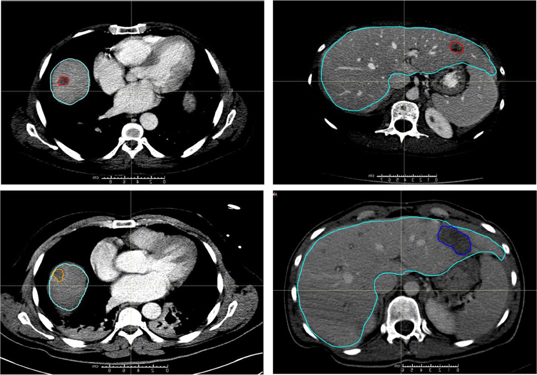
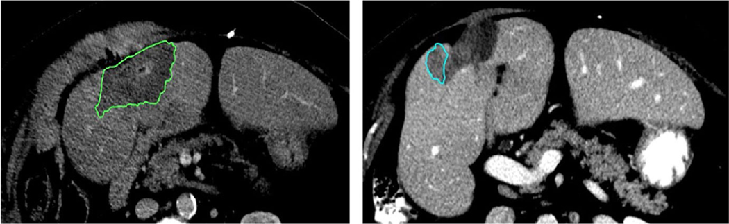
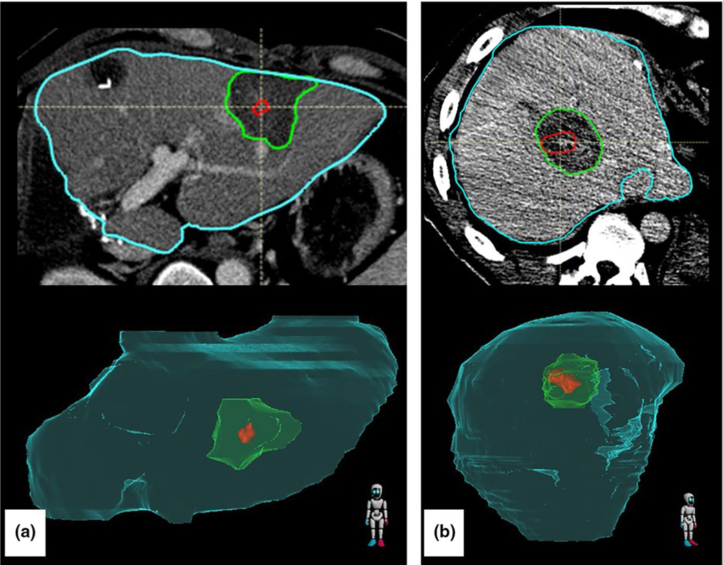
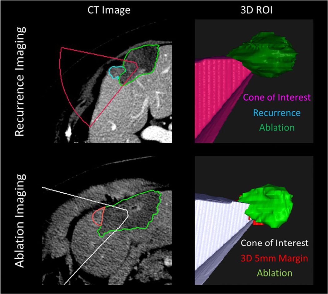
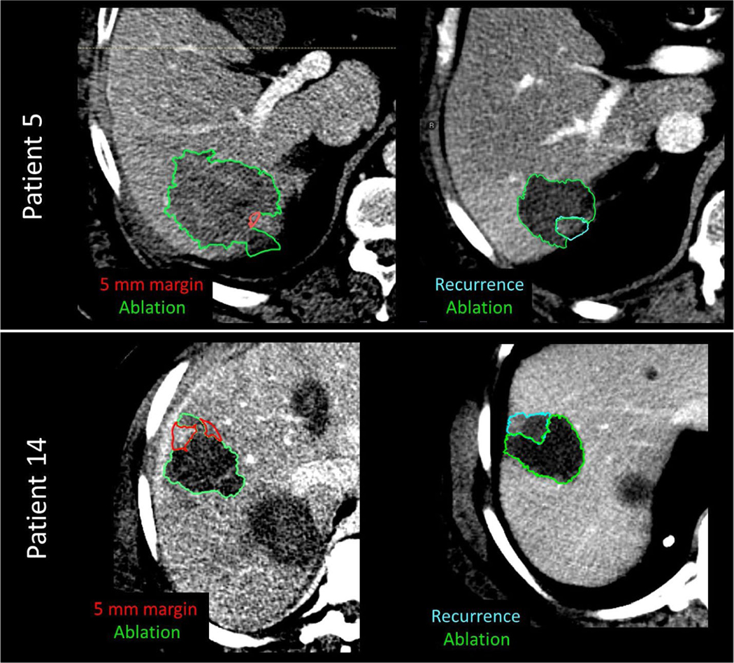
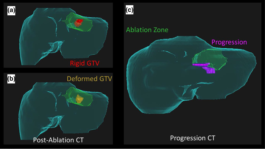
Methods: We retrospectively acquired 30 patients (16 LTP and 14 LTP-free) at our institution who had undergone PTA and had a contrast-enhanced pre-treatment and post-ablation CT scan. Liver, disease, and ablation zone were manually segmented. Biomechanical model-based DIR between the pre-treatment and post-ablation CT mapped the gross tumor volume onto the ablation zone and measured 3D minimum delivered margin (MDM). An in-house cone-tracing algorithm determined if progression qualitatively collocated with insufficient 5 mm margin achieved.

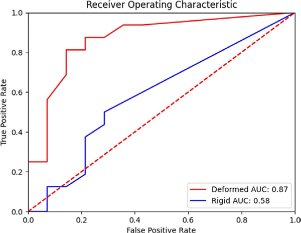
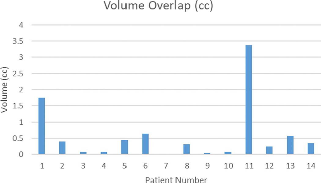
Results: Mann-Whitney U test showed a significant difference (p < 0.01) in MDM from the LTP and LTP-free groups. A total of 93% (13/14) of patients with LTP had a correlation between progression and missing 5 mm of margin volume.

Conclusions: Biomechanical DIR is able to reduce spatial uncertainty and allow measurement of delivered 3D MDM. This minimum margin can help ensure sufficient ablation delivery, and our workflow can provide valuable information in a clinically useful timeframe.

Keywords: biomechanical model-based deformable image registration; colorectal cancer; colorectal liver metastases; liver ablation therapy.


Conflict of interest statement

CONFLICT OF INTEREST

Dr. Brock reports grants from National Institutes of Health R01CA235564, R01CA221971, during the conduct of the study; grants from RaySearch Laboratories, outside the submitted work. Dr. Cazoulat reports grants from NIH, during the conduct of the study; grants from RaySearch Laboratories, outside the submitted work. Dr. Odisio reports grants from NIH, grants from Society of Interventional Radiology, during the conduct of the study. Dr. Bruno Odisio received grants from Siemens Healthineers and other incentives from Koo Foundation outside of the submitted work.

Figures

FIGURE 1
FIGURE 1
Computed tomography (CT) scans from two patients are shown, one in the left column and one in the right. (Top Row) Contouring of colorectal liver metastasis from diagnostic contrast-enhanced CT (red) and the liver contour (teal). (Bottom Row) Contouring of the ablation region (orange on the left and dark blue on the right) on the CT scan obtained immediately following the ablation
FIGURE 2
FIGURE 2
Left: Post-treatment ablation image with the ablation zone contoured in green. Right: Image from the same patient with recurrence contoured in blue. The difference in ablation zone volume from post-treatment to recurrence is 50%
FIGURE 3
FIGURE 3
Two patients demonstrating the gross tumor volume (red) deformedly propagated onto the post-treatment scan with ablation zone (green) with the liver (teal). (a) Image shows a uniform surrounding of the disease by the ablation zone. (b) Image shows potentially insufficient ablation zone in the lateral aspect of the disease
FIGURE 4
FIGURE 4
Top: Image of recurrence. Spherical coordinates of phi and theta intersecting the centroid of the recovered ablation zone (green) to each voxel of the recurrence (teal) are used to create a radiating cone (pink). Bottom: The recurrence phi and theta values are used to create another cone of interest (white) from the centroid of the post-treatment ablation zone (green) where the minimum margin has been identified (red). Because the cone intersects with the red 5-mm minimum margin ROI, we would claim that the minimum margin is in the same region that later turns into further progression
FIGURE 5
FIGURE 5
Receiver operator characteristic for identifying the development of local progression. The area under the curve (AUC) for the deformedly registered patients is 0.87, while for the rigidly registered patients is 0.58
FIGURE 6
FIGURE 6
Amount of volume overlap (cc) between projected recurrence cone and 5 mm margin outside of ablation
FIGURE 7
FIGURE 7
Example of 5-mm expansion on post-treatment CT images (left column) vs recurrence images (right column) for patient 5 and patient 14
FIGURE 8
FIGURE 8
Patient 7, where no overlap between 5mm margin and actual progression was present. The poor contrast scan makes it difficult to identify where exactly the boundary of sufficient ablation is located
FIGURE 9
FIGURE 9
Failings of rigid registration. (a) Rigid registration of the gross tumor volume (GTV, red) onto the post-ablation CT maps part of the tumor outside of the ablation zone (green), and gives a false sense of confidence in the delivered margin caudally. (b) Deformable registration of the GTV (gold) onto the post-ablation CT indicates potentially insufficient ablation margin in caudal aspect. (c) Development of disease progression (pink) in the caudal aspect of the recovered ablation zone (green) corresponds with insufficient margin in (b)

Similar articles

Cited by

References

    1. WHO Biennial Report 2012–2013. Accessed October 27, 2020. https://governance.iarc.fr/SC/SC50/BiennialReport2012-2013.pdf
    1. Siegel RL, Miller KD, Jemal A. Cancer statistics, 2019. CA Cancer J Clin. 2019;69(1):7–34. 10.3322/caac.21551 - DOI - PubMed
    1. Alper Sag A, Selcukbiricik F, Mandel NM. Evidence-based medical oncology and interventional radiology paradigms for liver-dominant colorectal cancer metastases. World J Gastroenterol. 2016;22(11):3127–3149. 10.3748/wjg.v22.i11.3127 - DOI - PMC - PubMed
    1. Tomlinson JS, Jarnagin WR, DeMatteo RP, et al. Actual 10-year survival after resection of colorectal liver metastases defines cure. J Clin Oncol. 2007;25(29):4575–4580. 10.1200/JCO.2007.11.0833 - DOI - PubMed
    1. Abdalla EK, Vauthey J-N, Ellis LM, et al. Recurrence and outcomes following hepatic resection, radiofrequency ablation, and combined resection/ablation for colorectal liver metastases. Ann Surg. 2004;239(6):818–827; discussion 825–7. 10.1097/01.SLA.0000128305.90650.71 - DOI - PMC - PubMed