. 2021 Dec;239(6):1336-1369.
doi: 10.1111/joa.13519. Epub 2021 Aug 3.

Roars, groans and moans: Anatomical correlates of vocal diversity in polygynous deer

Affiliations

Roars, groans and moans: Anatomical correlates of vocal diversity in polygynous deer

Roland Frey et al. J Anat. 2021 Dec.

Abstract

Eurasian deer are characterized by the extraordinary diversity of their vocal repertoires. Male sexual calls range from roars with relatively low fundamental frequency (hereafter fo ) in red deer Cervus elaphus, to moans with extremely high fo in sika deer Cervus nippon, and almost infrasonic groans with exceptionally low fo in fallow deer Dama dama. Moreover, while both red and fallow males are capable of lowering their formant frequencies during their calls, sika males appear to lack this ability. Female contact calls are also characterized by relatively less pronounced, yet strong interspecific differences. The aim of this study is to examine the anatomical bases of these inter-specific and inter-sexual differences by identifying if the acoustic variation is reflected in corresponding anatomical variation. To do this, we investigated the vocal anatomy of male and female specimens of each of these three species. Across species and sexes, we find that the observed acoustic variability is indeed related to expected corresponding anatomical differences, based on the source-filter theory of vocal production. At the source level, low fo is associated with larger vocal folds, whereas high fo is associated with smaller vocal folds: sika deer have the smallest vocal folds and male fallow deer the largest. Red and sika deer vocal folds do not appear to be sexually dimorphic, while fallow deer exhibit strong sexual dimorphism (after correcting for body size differences). At the filter level, the variability in formants is related to the configuration of the vocal tract: in fallow and red deer, both sexes have evolved a permanently descended larynx (with a resting position of the larynx much lower in males than in females). Both sexes also have the potential for momentary, call-synchronous vocal tract elongation, again more pronounced in males than in females. In contrast, the resting position of the larynx is high in both sexes of sika deer and the potential for further active vocal tract elongation is virtually absent in both sexes. Anatomical evidence suggests an evolutionary reversal in larynx position within sika deer, that is, a secondary larynx ascent. Together, our observations confirm that the observed diversity of vocal behaviour in polygynous deer is supported by strong anatomical differences, highlighting the importance of anatomical specializations in shaping mammalian vocal repertoires. Sexual selection is discussed as a potential evolutionary driver of the observed vocal diversity and sexual dimorphisms.

Keywords: acoustic variation; descended larynx; fallow deer; female contact calls; male sexual calls; polygynous deer; red deer; sexual dimorphism; sexual selection; sika deer; source-filter theory; vocal anatomy; vocal production; vocal repertoire.


Figures

FIGURE 1
FIGURE 1
Spectrograms of male mating calls and female contact calls. Calls include examples of the following: male red deer roar (a), male fallow deer groan (b), male sika moan (c), female red deer (d), female fallow deer (e) and female sika deer (f). Female fallow deer call was provided by A. McElligott, all other calls were provided by authors (see Audio S1–S6). Spectrograms were produced in Praat v. 6.0.24 (Boersma & Weenink, 2021), with 0.05 s window length, 16‐bit amplitude resolution and 44.1 kHz frequency sampling rate
FIGURE 2
FIGURE 2
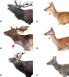
Images of Western European red deer (Cervus e. elaphus), male roaring (a), female (b); European fallow deer (Dama dama), male groaning (c), female (d); Japanese sika deer (Cervus n. nippon), male moaning (e), female (f). Not to scale, all individuals were brought to the same size approximately. In male and female fallow deer and in female red deer the larynx position is visible externally (red arrowheads). Sources: (a, b) © Megan Wyman; (c) © David Reby; (d) © Richard Steel 2011; (e) © Julian Dowse 2007 under a creative commons license at www.geograph.org.uk; (f) © Scandinavisk Dyrepark (Frank Vigh Larsen, director) 2014
FIGURE 3
FIGURE 3
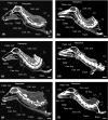
CT images of a male (a) and female (b) red deer, a male (c) and female (d) fallow deer, and a male (e) and female (f) sika deer. Not to scale, all specimens brought to the same size approximately. Note the pronouncedly low larynx resting position in male red and fallow deer, associated with an elongated pharynx and soft palate and a caudally shifted intrapharyngeal ostium. Scale bars 50 mm
FIGURE 4
FIGURE 4
MRI scans of a male (a) and female (b) red deer, a male (c) and female (d) fallow deer, and a male (e) and female (f) sika deer. Not to scale, all specimens were brought to the same size, approximately. Note the pronouncedly low larynx resting position in male red and fallow deer, less pronouncedly in female red and fallow deer. Larynx position is high and close to the root of the tongue in male and female sika deer. Red arrowheads point to the larynx, orange asterisks mark the soft palate. Green scale bars 100 mm
FIGURE 5
FIGURE 5
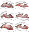
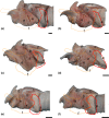
Dissection of male (a) and female (b) red deer, male (c) and female (d) fallow deer, and male (e) and female (f) sika deer, left lateral view. Not to scale, all specimens were brought to the same size, approximately. Scale bars 30 mm, respectively
FIGURE 6
FIGURE 6
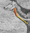
Estimation of permanent laryngeal descent as the ratio of the distance from the angle of the mandibula to the laryngeal prominence (red line) over the distance from the angle of the mandibula to the sternal manubrium (yellow line) exemplified in a male Iberian red deer (C. elaphus hispanicus) lacking a neck mane (modified from Frey et al., 2012)
FIGURE 7
FIGURE 7
Hyoid apparatuses of male (a) and female (b) red deer, male (c) and female (d) fallow deer, and male (e) and female (f) sika deer, left lateral view. Not to scale, all specimens brought to the same size, approximately. Scale bars 10 mm, respectively
FIGURE 8
FIGURE 8
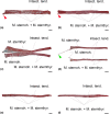
Left infrahyoid strap muscles (a, b, d, e, f) in left lateral view and left and right strap muscles (c) in ventral view of male (a) and female (b) red deer, male (c) and female (d) fallow deer, and male (e) and female (f) sika deer. In the male fallow deer the muscles of both sides were removed simultaneously and demonstrate the full set of the main retractors of the larynx. In all species and sexes the caudal portions of the sternohyoid and sternothyroid muscles are fused and separated from the rostral portions by a transverse tendinous intersection. The rostral portions of the sternohyoid and sternothyroid muscles are mostly separate in fallow deer but inseparably fused in red and sika deer. Remarkably, the left sternohyoid muscle of the female fallow deer retained its ancestral termination on the basihyoid (green arrowhead). In male and female red deer a slender ventral bundle possibly represents the termination of the sternohyoid muscle (red arrowheads). Scale bars 30 mm, respectively
FIGURE 9
FIGURE 9
Head dimensions and proportions. This figure illustrates the distances taken for the comparison between the sexes and the species (see Table 4). B–B: baseline; a: condylobasal length; b: osseous snout length; c: length of nasal soft tissue rostral to incisive bone; d: condylobasal length plus rostral soft tissue length; e: maximal height of the skull; background: CT scan of sika female W2. Scale bars 100 mm
FIGURE 10
FIGURE 10
Right halves of the excised larynges, medial view, of male (a) and female (b) red deer, male (c) and female (d) fallow deer, and male (e) and female (f) sika deer. Subfigures to scale, scale bar 10 mm. Slight sexual dimorphism of larynx size in red deer, no sexual dimorphism in sika deer and pronounced sexual dimorphism in fallow deer by evolutionary enlargement of the male larynx
FIGURE 11
FIGURE 11
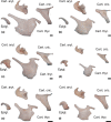
The laryngeal cartilages of male (a) and female (b) red deer, male (c) and female (d) fallow deer, and male (e) and female (f) sika deer. The cartilages are not to scale between species but they are to scale between the sexes allowing the depiction of sex‐related size differences. Sexual dimorphism increases from sika deer, via red deer to fallow deer. All scale bars 10 mm
FIGURE 12
FIGURE 12
The intrinsic laryngeal muscles of male (a) and female (b) red deer, male (c) and female (d) fallow deer, and male (e) and female (f) sika deer. The six sub‐illustrations are overlays reconstructed from several original dissection photos because it was not possible to show all intrinsic laryngeal muscles in one photo. The overlays are not to scale but presented at about the same size to facilitate inter‐specific comparison of the intrinsic laryngeal muscles. The contour of the removed thyroid cartilage is shown in light orange; the contour of the removed cricothyroid muscle is shown in bright red (portion lateral to the thyroid cartilage) and in light red (portion medial to the thyroid cartilage). The distance between the ventral edge of the thyroarytenoid muscle and the ventral contour of the thyroid cartilage is artificial and results from separating this muscle from the thyroid cartilage when removing the latter. a = lateral portion of cricothyroid muscle; a′ = medial portion of cricothyroid muscle; b = lateral cricoarytenoid muscle; c = dorsal cricoarytenoid muscle; d = transverse arytenoid muscle; e = thyroarytenoid muscle; f = contour of thyroid cartilage. All scale bars 10 mm
FIGURE 13
FIGURE 13
Transverse sections of the mid‐region of one vocal fold of an adult male red deer (a, d, g), fallow deer (b, e, h) and sika deer (c, f, i) stained with Hematoxylin/Eosin (a, b, c), Masson Trichrome (d, e, f), and Elastica/Van Gieson (g, h, i). Samples were sliced perpendicular to the dorsoventral length of the vocal fold. This overview reveals the similarity in basic structure of the male red and sika deer vocal fold and the distinct shape of the male fallow deer vocal fold. EP = Epithelium, FT = Fat, IS = Infraglottic space, MC = Mucosa, SG = Seromucous glands, TA = Thyroarytenoid muscle, and VL = Vocal ligament
FIGURE 14
FIGURE 14
Graphical reconstructions of relevant parts of the vocal organs in male (a) and female (b) red deer, male (c) and female (d) fallow deer, and male (e) and female (f) sika deer. Not to scale, all specimens have been brought to the same size approximately. The reconstructions comprise findings on vocal gesture in males, head, neck and rostral thoracic skeleton, hyoid apparatus, larynx position, oral cavity and tongue, pharynx and vocal tract length, hyoid and extrinsic laryngeal muscles, trachea and oesophagus. (a, c, e) In addition to filter‐relevant anatomical features, male sexual calls are affected by behavioural features (neck extension, head angle, lip and tongue configuration)
FIGURE 15
FIGURE 15
Average minimum fundamental frequency (minf o) against vocal fold size (mm) in red deer, fallow deer and sika deer. This log‐log graph depicts the measured log10 dorsoventral vocal fold length of each specimen (Table 5) versus the log10 minf o of vocalizations produced by males and females of each species based on published literature (Table 1)

Similar articles

References

    1. Apollonio, M. , Festa‐Bianchet, M. , Mari, F. , Mattioli, S. & Sarno, B. (1992) To lek or not to lek: mating strategies of male fallow deer. Behavioral Ecology, 3, 25–31.
    1. Azorit, C. , Analla, M. & Muñoz‐Cobo, J. (2003) Variation of mandible size in red deer Cervus elaphus hispanicus from southern Spain. Acta Theriologica, 48, 221–228.
    1. Berthommier, F. , Boë, L.‐J. , Meguerditchian, A. , Sawallis, T.R. & Captier, G. (2017). Comparative anatomy of the baboon and human vocal tracts: renewal of methods, data, and hypotheses. In: Boë, L.‐J. , Fagot, J. , Perrier, P. & Schwartz, J.‐L. (Eds.) Origins of human language: continuities and discontinuities with nonhuman primates, pp. 101–135. Speech production and perception, Berlin: Peter Lang GmbH, Internationaler Verlag der Wissenschaften. 78‐3‐631‐73726‐2. 10.3726/b12405. hal‐01649499v2. - DOI
    1. Blant, M. & Gaillard, J.M. (2004) Use of biometric body variables as indicators of roe deer (Capreolus capreolus) population density changes. Game and Wildlife Science, 21, 21–40.
    1. Boë, L.‐J. , Berthommier, F. , Legou, T. , Captier, G. , Kemp, C. , Sawallis, T.R. et al. (2017) Evidence of a vocalic proto‐system in the baboon (Papio papio) suggests pre‐hominin speech precursors. PLoS ONE, 12(1), e0169321. 10.1371/journal.pone.0169321 - DOI - PMC - PubMed

Publication types