. 2022 Jan;66(1):38-52.
doi: 10.1165/rcmb.2020-0408OC.

Endothelial-Specific Loss of Sphingosine-1-Phosphate Receptor 1 Increases Vascular Permeability and Exacerbates Bleomycin-induced Pulmonary Fibrosis

Affiliations

Endothelial-Specific Loss of Sphingosine-1-Phosphate Receptor 1 Increases Vascular Permeability and Exacerbates Bleomycin-induced Pulmonary Fibrosis

Rachel S Knipe et al. Am J Respir Cell Mol Biol. 2022 Jan.

Abstract

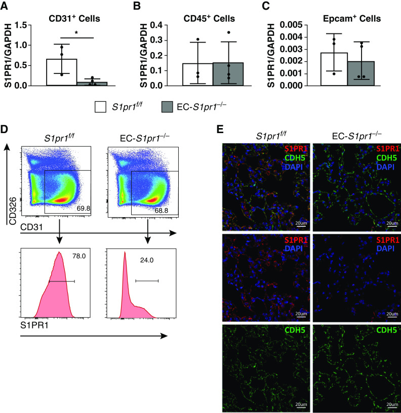
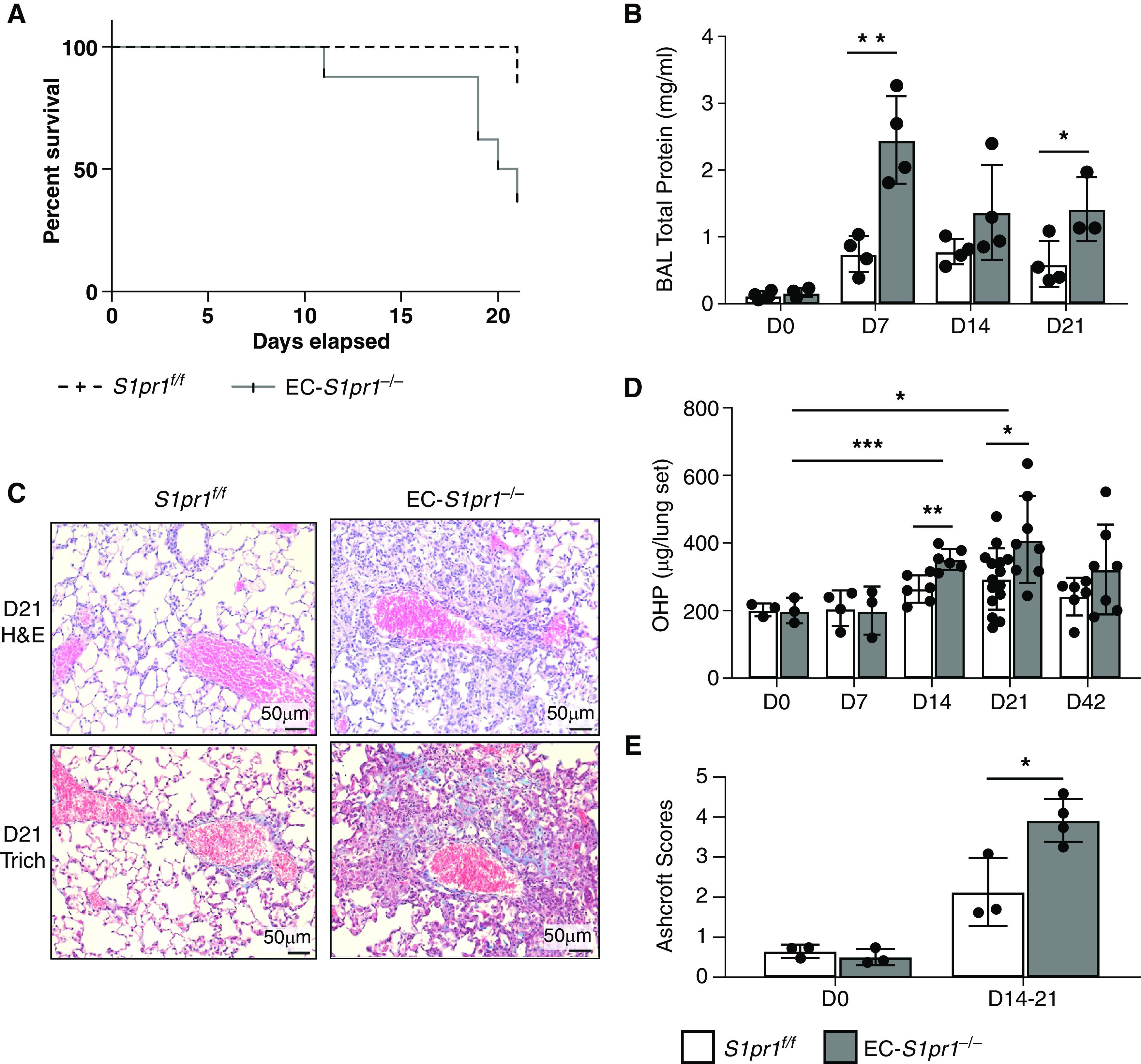
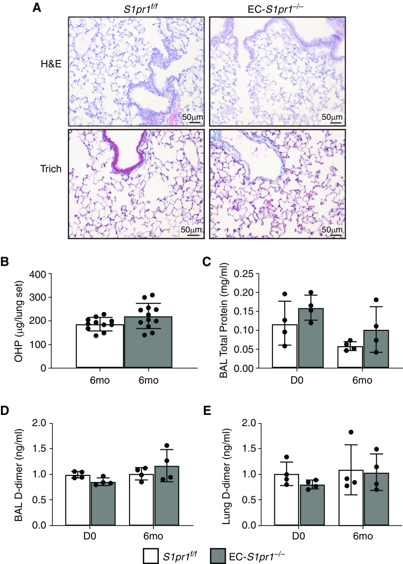
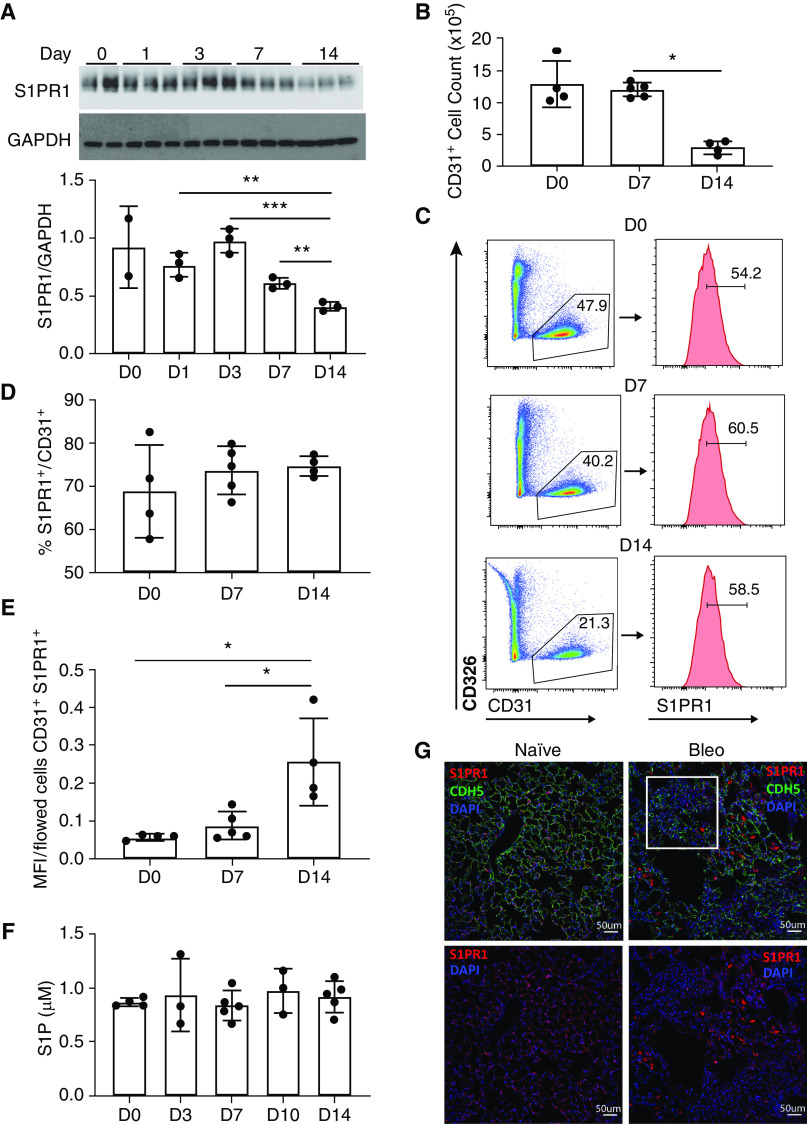
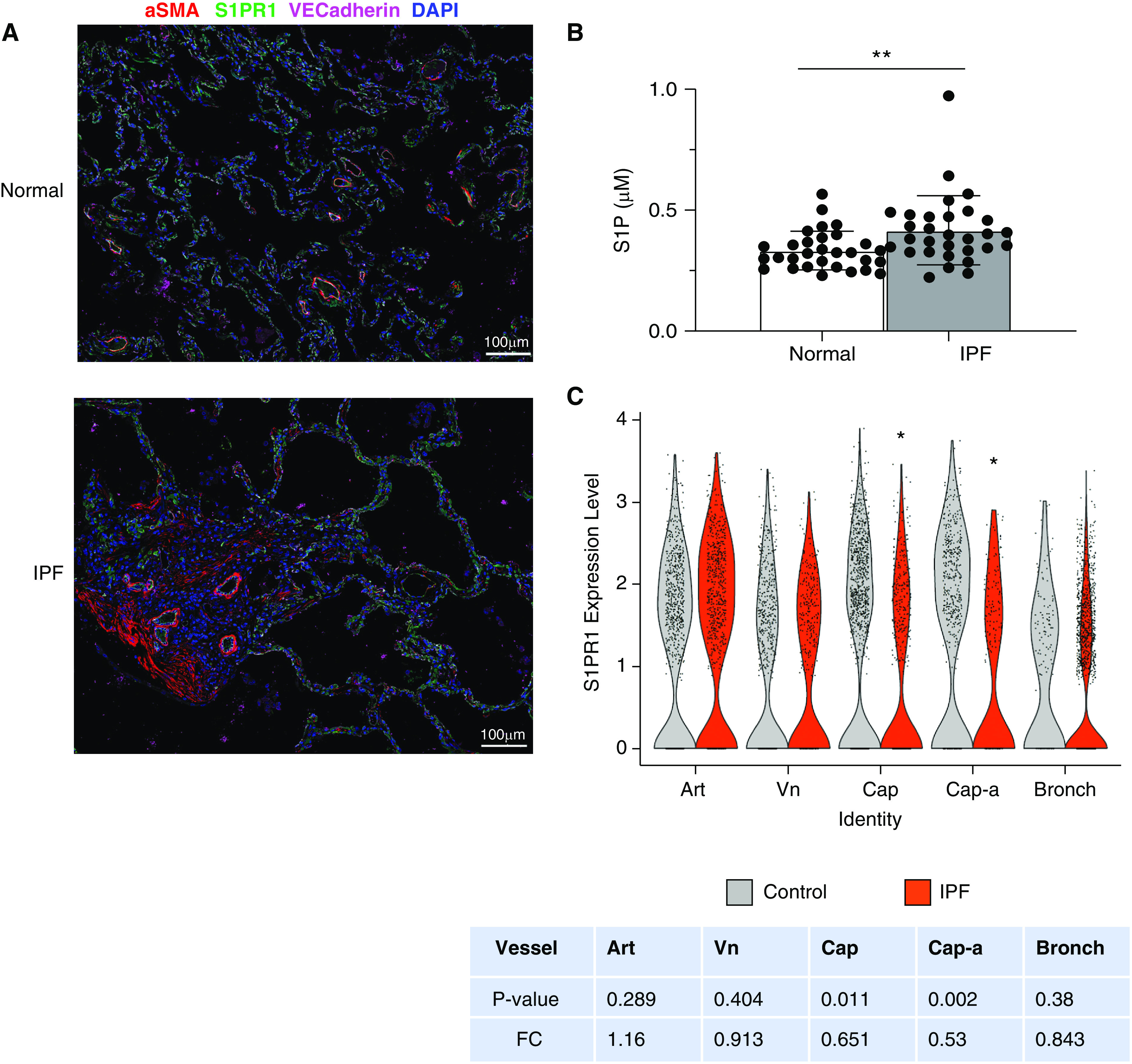
Idiopathic pulmonary fibrosis (IPF) is a chronic, progressive disease which leads to significant morbidity and mortality from respiratory failure. The two drugs currently approved for clinical use slow the rate of decline in lung function but have not been shown to halt disease progression or reverse established fibrosis. Thus, new therapeutic targets are needed. Endothelial injury and the resultant vascular permeability are critical components in the response to tissue injury and are present in patients with IPF. However, it remains unclear how vascular permeability affects lung repair and fibrosis following injury. Lipid mediators such as sphingosine-1-phosphate (S1P) are known to regulate multiple homeostatic processes in the lung including vascular permeability. We demonstrate that endothelial cell-(EC) specific deletion of the S1P receptor 1 (S1PR1) in mice (EC-S1pr1-/-) results in increased lung vascular permeability at baseline. Following a low-dose intratracheal bleomycin challenge, EC-S1pr1-/- mice had increased and persistent vascular permeability compared with wild-type mice, which was strongly correlated with the amount and localization of resulting pulmonary fibrosis. EC-S1pr1-/- mice also had increased immune cell infiltration and activation of the coagulation cascade within the lung. However, increased circulating S1P ligand in ApoM-overexpressing mice was insufficient to protect against bleomycin-induced pulmonary fibrosis. Overall, these data demonstrate that endothelial cell S1PR1 controls vascular permeability in the lung, is associated with changes in immune cell infiltration and extravascular coagulation, and modulates the fibrotic response to lung injury.

Keywords: lung fibrosis; sphingosine-1-phosphate; sphingosine-1-phosphate 1 receptor; vascular permeability.


Figures

Figure 1.
Figure 1.
Endothelial-specific S1pr1 expression is reduced in EC-S1pr1−/− mice. S1pr1 mRNA expression in isolated (A) endothelial cells (CD31+), (B) hematopoietic cells (CD45+), and (C) epithelial cells (Epcam+). n = 3 mice per group. (D) Flow cytometry of mouse lung endothelial cells in EC-S1pr1−/− mice (S1PR1f/f × VECad-CreERT2) compared with littermate controls (S1pr1f/f). (E) Immunofluorescence confirming deletion of EC S1PR1 after tamoxifen. Representative images shown; n = 3 mice per group. *P < 0.05 comparing expression of S1pr1 in EC-S1pr1−/− mice as compared with S1pr1f/f mice. Scale bars, 20 μm.
Figure 2.
Figure 2.
Standard-dose bleomycin increases pulmonary vascular permeability in EC-S1pr1−/− mice. (A) Evans blue dye assay was performed on Day 0 and Day 7 after standard-dose intratracheal (IT) bleomycin (1.0 U/kg). (B) Quantification of vascular permeability was calculated as an Evans blue (EB) index defined as the ratio of lung Evans blue to plasma Evans blue. n = 3–7 mice per group. (C) Hematoxylin and eosin and Masson’s trichrome staining of lungs was performed on Day 7 after standard dose bleomycin. Representative images are shown from n = 3 mice per group. Scale bars, 50 μm. (D) Survival of mice receiving standard-dose (1.0 U/kg) bleomycin. n = 5 mice per group. *P < 0.05 comparing survival of EC-S1pr1−/− mice to littermate controls S1pr1f/f. ***P < 0.001 comparing EB index in EC-S1pr1−/− mice to littermate controls S1pr1f/f. H&E = hematoxin and eosin; Trich = trichrome.
Figure 3.
Figure 3.
Low-dose bleomycin induces fibrosis in EC-S1pr1−/− mice. (A) Survival of mice receiving low-dose (0.5 U/kg) bleomycin. n = 5 mice per group. (B) Total protein content in the BAL was quantified at Days 0, 7, 14, and 21 after low-dose bleomycin. BAL from EC-S1pr1−/− mice was compared with intact littermate controls S1pr1f/f. n = 3–5 mice per group. (C) Hematoxylin and eosin and Masson’s trichrome staining of lungs were performed on Day 21 after low-dose bleomycin. Representative images are shown from n = 3 mice per group. Scale bars, 50 μm. (D) Fibrosis was quantified at Days 0, 7, 14, 21, and 42 after low-dose IT bleomycin by measuring hydroxyproline levels in the mouse lungs. n = 3–15 mice per group. (E) Ashcroft scoring performed on trichrome stained lung sections from EC-S1pr1−/− mice at Days 0 and 14 or 21 was compared with intact littermate controls S1pr1f/f. n = 3–4 mice per group. *P < 0.05, **P < 0.01, and ***P < 0.001 for comparisons of total protein or hydroxyproline in EC-S1pr1−/− mice compared with intact littermate control mice S1pr1f/f. OHP = hydroxyproline.
Figure 4.
Figure 4.
Endothelial-specific S1pr1 deletion leads to reduced intercellular junction proteins and increased extravascular coagulation. (A) Mice were injected with FITC-labeled albumin to identify albumin extravasated into the lung tissue after bleomycin. Sequential sectioning and staining of the lung tissue were performed with Masson’s trichrome stain. Representative images are shown from n = 3 mice per group. Scale bars, 100 μm. (B) D-dimer ELISA was performed on BAL fluid collected from mice on days 0, 7, 14, and 21 after low-dose IT bleomycin (0.5 U/kg). n = 4 mice per group. (C) D-dimer ELISA was performed on homogenized lung tissue collected from mice on days 0, 7, 14, and 21 after low-dose IT bleomycin (0.5 U/kg). n = 4 mice per group. (D) Junctional proteins were assessed through immunoblotting for VE-Cadherin in lung tissue homogenates harvested at days 0 and 14 after low-dose bleomycin challenge. Densitometry was performed to quantify differences in protein expression at each time point. n = 2–3 mice per group. *P < 0.05 and ***P < 0.001 for comparisons of D-dimer in the BAL and lung tissue of EC-S1pr1−/− mice as compared with intact littermate controls S1pr1f/f.
Figure 5.
Figure 5.
Immune cell populations in the BAL of mice lacking endothelial S1pr1 after bleomycin. (A) Total cell counts were quantified from BAL obtained on Day 0 and Day 7 after low-dose IT bleomycin. n = 3–4 mice per group. Flow cytometry was performed on BAL collected 7 days after bleomycin challenge in EC-S1pr1−/− and control mice to assess the number of (B) alveolar macrophages, (C) dendritic cells, (D) NK cells, (E) monocyte-derived dendritic cells (DCs), (F) inflammatory monocytes, (G) CD4+ T cells, (H) CD8+ T cells, and (I) T regulatory cells. In contrast, no differences were detected in the quantities of BAL (J) B cells, (K) eosinophils, (L) γδ T cells, or (M) neutrophils. n = 3–4 mice per group. *P < 0.05, **P < 0.01, and ***P < 0.001 for comparisons of BAL cells in EC-S1pr1−/− mice as compared with intact control mice S1pr1f/f. γδ T cells = GD T cells.
Figure 6.
Figure 6.
Endothelial S1pr1 deletion is insufficient to induce fibrosis in the absence of lung injury. (A) For longitudinal studies, mice were treated with tamoxifen to induce EC-S1pr1 deletion and then maintained in the mouse facility for 6 months. After 6 months, hematoxylin and eosin and Masson’s trichrome staining were performed on lung histology from EC-S1pr1−/− mice and littermate controls S1pr1f/f. Representative images are shown from n = 3 mice per group. Scale bars, 50 μm. (B) OHP assay was performed on lung tissue from mice after 6 months. n = 11–12 mice per group. (C) Total protein in the BAL fluid of mice after 6 months was compared. n = 4 mice per group. (D) BAL D-dimer levels were measured by ELISA. n = 4 mice per group. (E) Lung D-dimer levels were measured by ELISA. n = 4 mice per group.
Figure 7.
Figure 7.
Bleomycin-induced lung injury reduces endothelial cell populations in the lung. (A) Immunoblotting was performed using protein from lung homogenates of C57BL/6 wild-type mice (WT) at Days 0, 1, 3, 7, and 14 after IT bleomycin (0.5 U/kg). Blots were probed for S1PR1, and protein expression levels were compared using GAPDH as a loading control. n = 2–3 mice per group. Densitometry was performed using Image J software (NIH). (B) Flow cytometry to identify CD31+ cells was performed on lung tissue from WT mice on Days 0, 7, and 14 after standard-dose IT bleomycin. n = 4 mice per group. (C) Flow cytometry of CD31+ cells expressing S1PR1. n = 4 mice per group. (D) The percentage of endothelial cells expressing S1PR1 at each time point after bleomycin was quantified. (E) Mean fluorescence intensity of S1PR1 in CD31+ cells after bleomycin was quantified. (F) Plasma S1P levels in WT mice were quantified at Days 0, 3, 7, 10, and 14 after IT bleomycin. n = 3–4 mice per group. (G) Immunoflourescence co-staining on lungs from naive and bleomycin treated mice at Day 14 was performed for S1PR1 (red), endothelial CDH5/VE-cadherin (green), and nuclear DAPI (blue). White box indicates a fibrotic region. Scale bars, 50 μm. *P < 0.05, **P < 0.01, and ***P < 0.001 for comparisons of cells in WT mice at various time points after bleomycin injury.
Figure 8.
Figure 8.
Increased circulating S1P is insufficient to protect mice from bleomycin-induced pulmonary fibrosis. (A) Transgenic mice overexpressing human apolipoprotein M (ApoM Tg+) were bred, and Evans blue dye assay was performed at Day 7 after standard-dose IT bleomycin. (B) Evans blue indices were calculated using Evans blue dye indices in the lungs and plasma of ApoM Tg+ mice and littermate controls (WT). n = 4 mice per group. (C) BAL total protein was quantified at Day 14 after standard dose bleomycin. (D) Histology at Days 0 and 14 after standard-dose IT bleomycin were stained with hematoxylin and eosin and Masson’s trichrome staining. Representative images are shown from n = 3 mice per group. Scale bars, 100 μm. (E) Quantification of fibrosis was performed with hydroxyproline assay. (F) S1P levels were quantified by mass spectrometry from circulating plasma at baseline (Day 0), Day 7, and Day 14 after standard-dose bleomycin. n = 3–6 mice per group. n = 3–8 mice per group. ***P < 0.001, and ****P < 0.0001 for comparisons of S1P between ApoM-Tg+ and control mice.
Figure 9.
Figure 9.
Clinical relevance of S1pr1 modulation in pulmonary fibrosis. (A) Lung tissue from patients with idiopathic pulmonary fibrosis (IPF) and healthy age-matched controls were stained for endothelial marker VE-Cadherin and S1PR1. White box indicates an area of fibrosis. Scale bars, 100 μm. (B) Plasma from patients with IPF and sex- and age-matched healthy controls were analyzed by mass spectrometry to quantify S1P levels. n = 30 patients per group. (C) Violin plot of S1PR1 expression in subtypes of lung vascular endothelial cells from 38 control donors (3,458 total cells; 2,458 cells from Kropski paper, 1,000 cells from Kaminski paper) and 44 IPF patients (3,663 total cells; 1,720 cells from Kropski paper, 1,943 cells from Kaminski paper). Cells from scRNA-seq accessions GSE135893 and GSE136831 were merged and re-clustered (see supplemental Figure E6). Fold-change values indicate the expression ratio of [IPF/Control]. See Figure E6. *P > 0.05 and **P > 0.01. Art = artery; Bronch = bronchial; Cap = capillary; Cap-a = capillary A; FC = fold change; Vn = vein.

References

    1. Raghu G, Weycker D, Edelsberg J, Bradford WZ, Oster G. Incidence and prevalence of idiopathic pulmonary fibrosis. Am J Respir Crit Care Med . 2006;174:810–816. - PubMed
    1. King TE, Jr, Bradford WZ, Castro-Bernardini S, Fagan EA, Glaspole I, Glassberg MK, et al. ASCEND Study Group A phase 3 trial of pirfenidone in patients with idiopathic pulmonary fibrosis. N Engl J Med . 2014;370:2083–2092. - PubMed
    1. Richeldi L, du Bois RM, Raghu G, Azuma A, Brown KK, Costabel U, et al. INPULSIS Trial Investigators Efficacy and safety of nintedanib in idiopathic pulmonary fibrosis. N Engl J Med . 2014;370:2071–2082. - PubMed
    1. Selman M, King TE, Pardo A, American Thoracic Society; European Respiratory Society; American College of Chest Physicians Idiopathic pulmonary fibrosis: prevailing and evolving hypotheses about its pathogenesis and implications for therapy. Ann Intern Med . 2001;134:136–151. - PubMed
    1. Brown LF, Dvorak AM, Dvorak HF. Leaky vessels, fibrin deposition, and fibrosis: a sequence of events common to solid tumors and to many other types of disease. Am Rev Respir Dis . 1989;140:1104–1107. - PubMed

Publication types

MeSH terms