Review
. 2021 Nov 22;117(13):2624-2638.
doi: 10.1093/cvr/cvab259.

Cardiac allograft vasculopathy: current review and future research directions

Affiliations
Review

Cardiac allograft vasculopathy: current review and future research directions

Jordan S Pober et al. Cardiovasc Res. .

Abstract

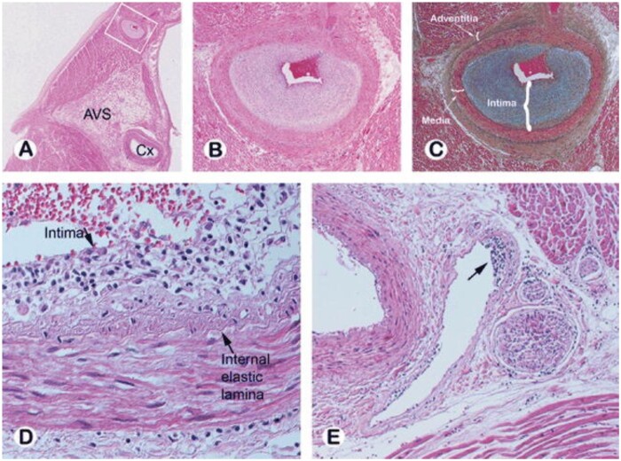
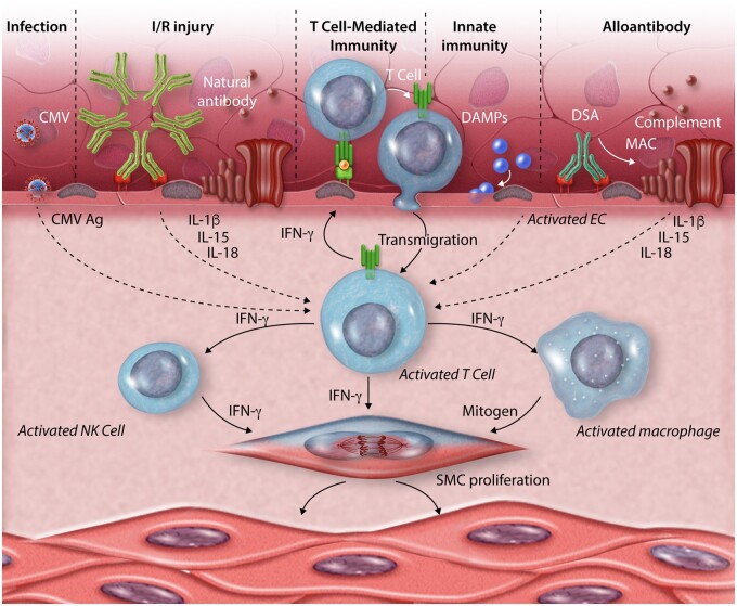
Cardiac allograft vasculopathy (CAV) is a pathologic immune-mediated remodelling of the vasculature in transplanted hearts and, by impairing perfusion, is the major cause of late graft loss. Although best understood following cardiac transplantation, similar forms of allograft vasculopathy occur in other vascularized organ grafts and some features of CAV may be shared with other immune-mediated vasculopathies. Here, we describe the incidence and diagnosis, the nature of the vascular remodelling, immune and non-immune contributions to pathogenesis, current therapies, and future areas of research in CAV.

Keywords: chronic rejection; endothelial cells; heart transplantation; innate and adaptive immunity; vascular smooth muscle cells.


Figures

Figure 1
Figure 1
Histopathology of cardiac allograft vasculopathy. (A) Photomicrograph of the left atrium and ventricle with the left circumflex (Cx) artery noted at the atrioventricular sulcus (AVS) from a patient who died of CAV 3 years post-transplant. (B, C) Intramyocardial branch of circumflex artery (from inset A) supplying the atrium shows marked intimal proliferation with preservation of the elastic lamina and normal medial layer [haematoxylin-eosin (B) and Movat pentachrome (C)]. (D, E) Subendothelial lymphocytic infiltration is seen in a small epicardial artery (D) and in an intramyocardial vein (arrow E). Reprinted with permission from Ref.
Figure 2
Figure 2
Invasive coronary imaging for CAV assessment: coronary angiography is the current gold standard for identifying epicardial disease and grading severity (top left); coronary physiologic measurements including coronary flow reserve, index of microcirculatory resistance and fractional flow reserve (top right) provide functional information on epicardial and microvascular disease; intracoronary imaging using intravascular ultrasound (IVUS, bottom left) or optical coherence tomography (OCT, bottom right) enables detailed imaging of the coronary intima to detect early intimal hyperplasia as well as plaque characterization.
Figure 3
Figure 3
Schematic representation of pathogenic mechanisms that lead to CAV. T cells that recognize graft antigens expressed on the endothelium are central to the process. These cells may undergo transmigration into the graft in response to antigen recognition or to chemokines made by and expressed on the endothelial cell surface in response to activating signals such as damage-associated molecular patterns (DAMPs) as part of an innate immune response. Endothelium may also be activated by IgG donor specific alloantibodies or by natural antibodies, the latter targeting antigens exposed following ischaemia/reperfusion (I/R) injury. These antibody responses are mediated by proteins of the complement system that form membrane attack complexes (MACs). Other sources of antigens (Ag) that stimulate T cells can come from infections such as cytomegalovirus (CMV). The T-cell response is augmented by cytokines produced by the activate endothelial cells such as interleukins (IL)-1β, IL-15 or IL-18. Cytokines produced by the activated T cells within the vessel wall, notably IFN-γ but likely others as well, induce smooth muscle cell (SMC) proliferation and also activate infiltrating natural killer (NK) cells and macrophages to make additional mitogens that also drive intimal smooth muscle cell proliferation and matrix deposition. The net effect is the expand the vessel intima and comproimise the lumen.

Similar articles

Cited by

References

    1. Bruno S, Remuzzi G, Ruggenenti P. Transplant renal artery stenosis. J Am Soc Nephrol 2004;15:134–141. - PubMed
    1. Kollar B, Kamat P, Klein HJ, Waldner M, Schweizer R, Plock JA. The significance of vascular alterations in acute and chronic rejection for vascularized composite allotransplantation. J Vasc Res 2019;56:163–180. - PubMed
    1. Harifi G, Nour-Eldine W, Noureldine MHA, Berjaoui MB, Kallas R, Khoury R, Uthman I, Al-Saleh J, Khamashta MA. Arterial stenosis in antiphospholipid syndrome: update on the unrevealed mechanisms of an endothelial disease. Autoimmun Rev 2018;17:256–266. - PubMed
    1. Gori T. Coronary vasculitis. Biomedicines 2021;9: 622. - PMC - PubMed
    1. Khush KK, Cherikh WS, Chambers DC, Harhay MO, Hayes D Jr, Hsich E, Meiser B, Potena L, Robinson A, Rossano JW, Sadavarte A, Singh TP, Zuckermann A, Stehlik J; International Society for Heart and Lung Transplantation. The International Thoracic Organ Transplant Registry of the International Society for Heart and Lung Transplantation: thirty-sixth adult heart transplantation report - 2019; focus theme: donor and recipient size match. J Heart Lung Transplant 2019;38:1056–1066. - PMC - PubMed

Publication types

MeSH terms