Review
. 2021 Aug 4;15(1):388.
doi: 10.1186/s13256-021-02990-4.

Coincidental light chain induced proximal tubulopathy with lupus nephritis: a case report and review of the literature

Affiliations
Review

Coincidental light chain induced proximal tubulopathy with lupus nephritis: a case report and review of the literature

Wael Mostafa Hamza et al. J Med Case Rep. .

Abstract

Background: We report a case of light chain proximal tubulopathy associated with lupus nephritis in a patient known to have systemic lupus erythematosus. The kidney can be injured in several ways in any of these disorders. Light chain proximal tubulopathy is a rare form of renal tubular injury that may occur in and complicate plasma cell dyscrasia, characterized by cytoplasmic inclusions of the monoclonal light chain within proximal tubular cells. Lupus nephritis is a common form of renal injury as it occurs in about 25-50% of adult patients with systemic lupus erythematosus.

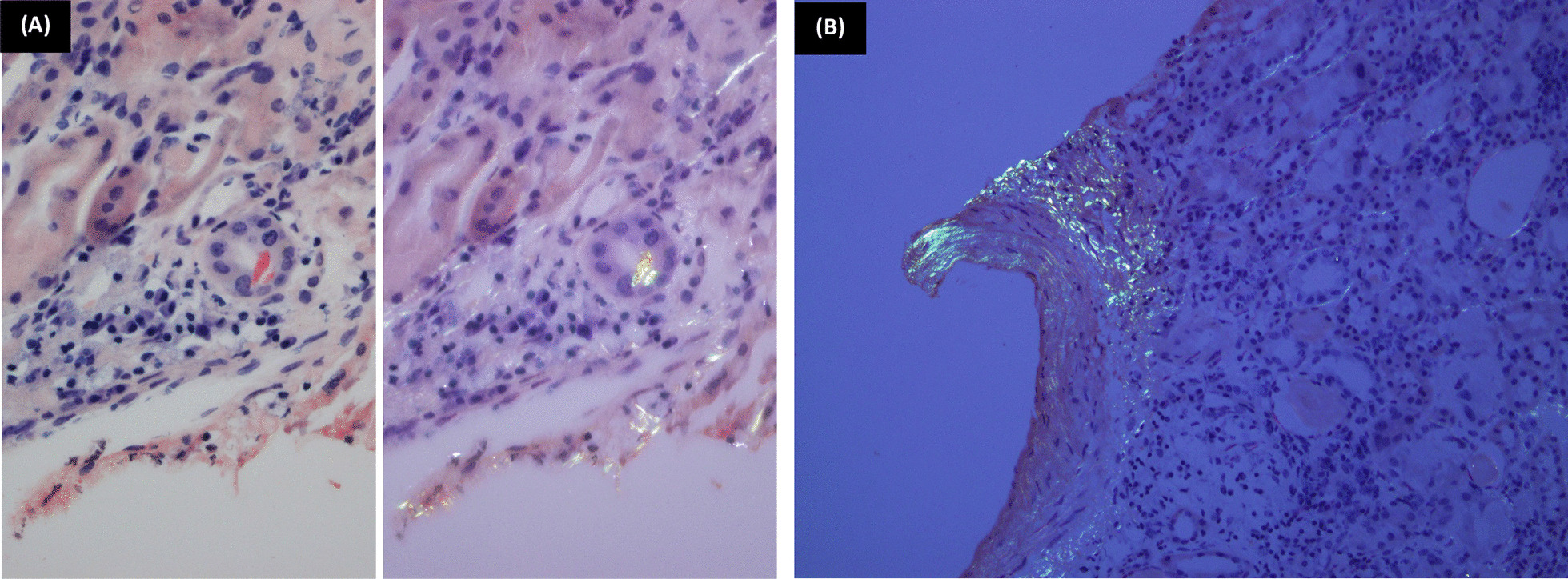
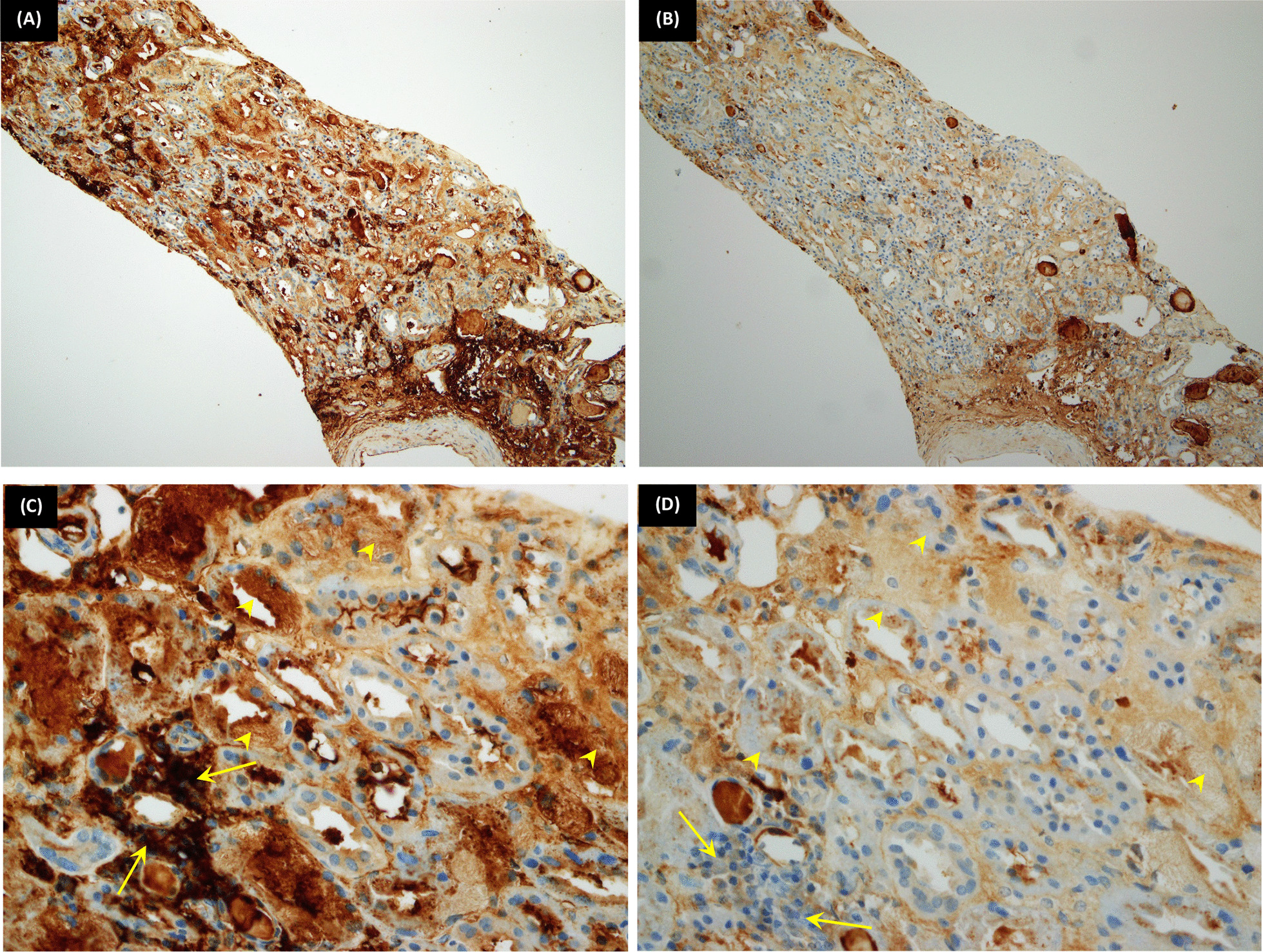
Case presentation: We present a 57-year-old African patient known to have systemic lupus erythematosus and hypertension presented with a new complaint of microscopic hematuria. A renal biopsy was performed and revealed lupus nephritis class II concurrently associated with light chain induced proximal tubulopathy. A subsequent bone marrow biopsy was performed, which revealed multiple myeloma.

Conclusions: We report a case of coincidental lupus nephritis and proximal tubulopathy featuring a combined constellation of rare histopathological features that might add to the relationship between systemic lupus and paraproteinemia.

Keywords: Free light chain; Lupus nephritis; Paraproteinemia; Plasma cell dyscrasia; Proximal tubulopathy.


Conflict of interest statement

The authors declare that they have no competing interests.

Figures

Fig. 1
Fig. 1
Glomerular findings (by LM and IF). A [Hematoxylin and eosin (H&E) ×400]: A glomerulus showing expanded mesangial matrix associated with mesangial hypercellularity. B [Immunofluorescence immunoglobulin G (IF-IgG) ×400]: Positive mesangial granular deposits; score 2. C (IF-IgA ×400): Positive mesangial granular deposits; score 1
Fig. 2.
Fig. 2.
Proximal tubular lesions. A (H&E ×100): Renal cortical tissue showing mononuclear (plasmacytic infiltrate) and apparently tubular cytoplasmic inclusions. B (H&E ×200): The tubular epithelium showing textured intracytoplasmic inclusions. C (H&E ×1000): Monoclonal plasma cells; a Mott cell is seen “arrow head.” D (H&E ×1000): The proximal tubular epithelium showing textured fibrilloid inclusions of monoclonal light chains. E (Trichrome ×400), F [Jones' Methenamine Silver (JMS) ×400]: The tubular inclusions are fuchsinophilic and silver negative
Fig. 3.
Fig. 3.
Amyloidal cast and deposits. A (Congo red ×200): Single orangeophilic cast seen giving apple-green birefringence under polarized light. B (Congo red ×200): An interlobular sized artery showing amyloidal deposits, as well as the adjacent interstitium
Fig. 4.
Fig. 4.
Immunoperoxidase stains (kappa and lambda); tubular findings. A, B (Immunoperoxidase technique-A: Kappa light chain, B: Lambda light chain ×100): The same core as in Fig. 2A; the plasmacytic infiltrate and inclusions showing positivity for kappa light chains rather than lambda. C, D (IP-C: Kappa light chain, D: Lambda light chain ×200): The arrowheads are pointing to intratubular cytoplasmic inclusions that are positive for Kappa (Fig. 2C) and negative for Lambda (Fig. 2D). The arrows are pointing to interstitial plasmacytic infiltrate that is also positive for Kappa (Fig. 2C) and negative for Lambda (Fig. 2D)

References

    1. Rajkumar SV, Dimopoulos MA, Palumbo A, Blade J, Merlini G, Mateos M-V, et al. International Myeloma Working Group updated criteria for the diagnosis of multiple myeloma. Lancet Oncol. 2014;15(12):e538–e548. doi: 10.1016/S1470-2045(14)70442-5. - DOI - PubMed
    1. Heher EC, Goes NB, Spitzer TR, Raje NS, Humphreys BD, Anderson KC, et al. Kidney disease associated with plasma cell dyscrasias. Blood. 2010;116(9):1397–1404. doi: 10.1182/blood-2010-03-258608. - DOI - PMC - PubMed
    1. Herrera GA, Joseph L, Gu X, Hough A, Barlogie B. Renal pathologic spectrum in an autopsy series of patients with plasma cell dyscrasia. Arch Pathol Lab Med. 2004;128(8):875–879. doi: 10.5858/2004-128-875-RPSIAA. - DOI - PubMed
    1. Herrera GA. Renal lesions associated with plasma cell dyscrasias: practical approach to diagnosis, new concepts, and challenges. Arch Pathol Lab Med. 2009;133(2):249–267. doi: 10.5858/133.2.249. - DOI - PubMed
    1. Stokes MB, Valeri AM, Herlitz L, Khan AM, Siegel DS, Markowitz GS, et al. Light chain proximal tubulopathy: clinical and pathologic characteristics in the modern treatment era. J Am Soc Nephrol. 2016;27(5):1555–1565. doi: 10.1681/ASN.2015020185. - DOI - PMC - PubMed