. 2021 Dec 1;11(12):3214-3229.
doi: 10.1158/2159-8290.CD-21-0441.

Rlf-Mycl Gene Fusion Drives Tumorigenesis and Metastasis in a Mouse Model of Small Cell Lung Cancer

Affiliations

Rlf-Mycl Gene Fusion Drives Tumorigenesis and Metastasis in a Mouse Model of Small Cell Lung Cancer

Metamia Ciampricotti et al. Cancer Discov. .

Abstract

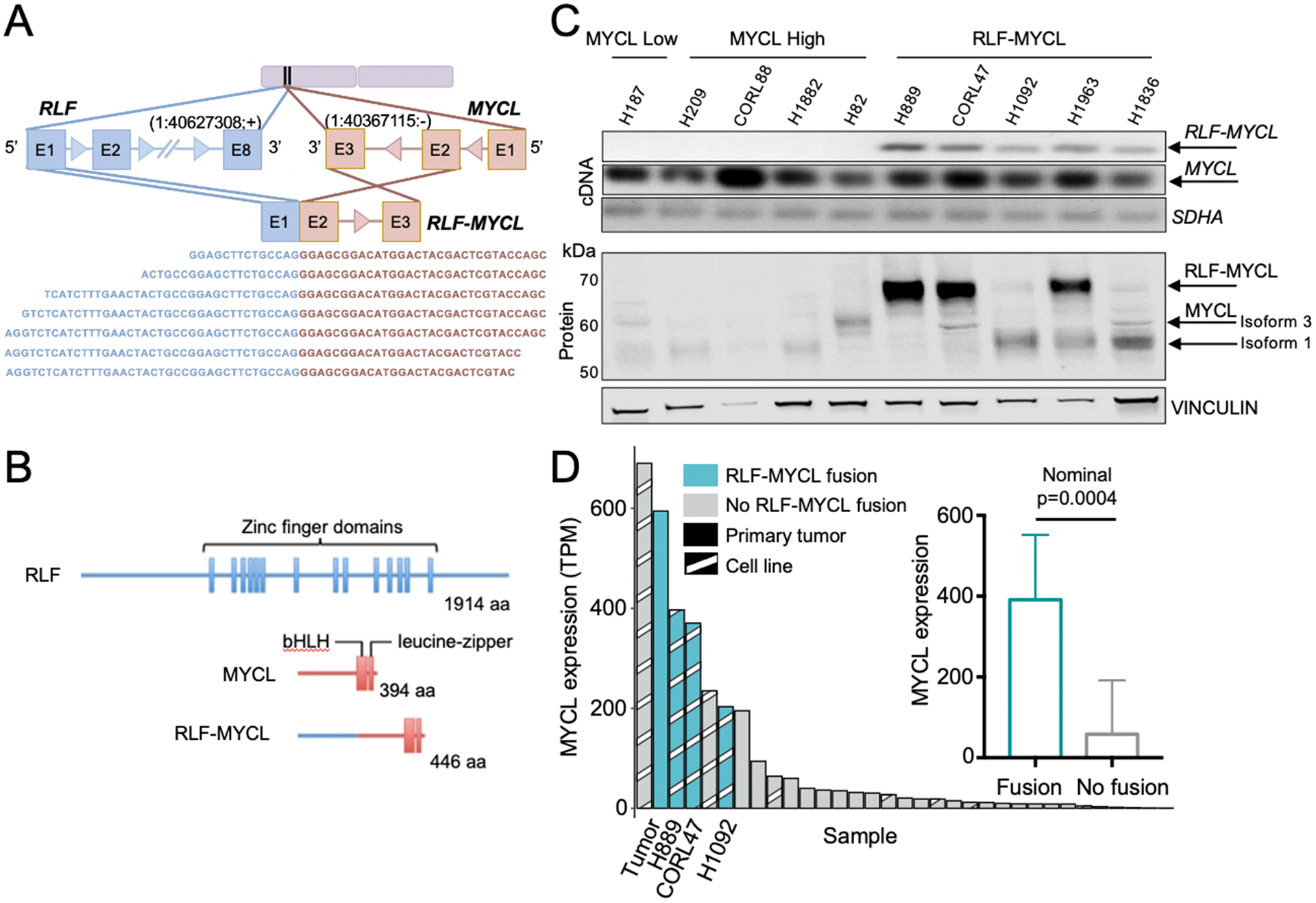
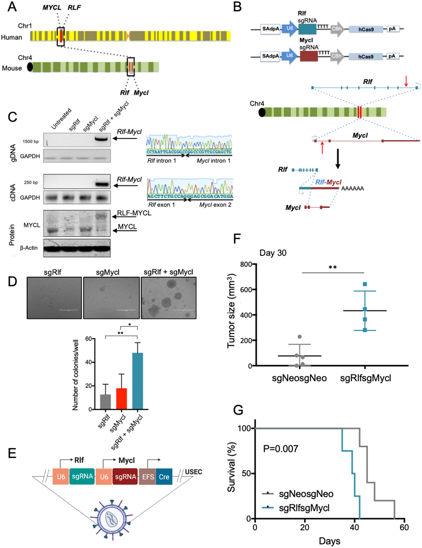
Small cell lung cancer (SCLC) has limited therapeutic options and an exceptionally poor prognosis. Understanding the oncogenic drivers of SCLC may help define novel therapeutic targets. Recurrent genomic rearrangements have been identified in SCLC, most notably an in-frame gene fusion between RLF and MYCL found in up to 7% of the predominant ASCL1-expressing subtype. To explore the role of this fusion in oncogenesis and tumor progression, we used CRISPR/Cas9 somatic editing to generate a Rlf-Mycl-driven mouse model of SCLC. RLF-MYCL fusion accelerated transformation and proliferation of murine SCLC and increased metastatic dissemination and the diversity of metastatic sites. Tumors from the RLF-MYCL genetically engineered mouse model displayed gene expression similarities with human RLF-MYCL SCLC. Together, our studies support RLF-MYCL as the first demonstrated fusion oncogenic driver in SCLC and provide a new preclinical mouse model for the study of this subtype of SCLC.

Significance: The biological and therapeutic implications of gene fusions in SCLC, an aggressive metastatic lung cancer, are unknown. Our study investigates the functional significance of the in-frame RLF-MYCL gene fusion by developing a Rlf-Mycl-driven genetically engineered mouse model and defining the impact on tumor growth and metastasis. This article is highlighted in the In This Issue feature, p. 2945.


Conflict of interest statement

Conflict of Interest

T.P. has received honoraria/consulting fees from Calithera Biosciences, Vividion Therapeutics and research support from Bristol Myers Squibb, Dracen Pharmaceutical and Agios Pharmaceuticals. C.M.R. has consulted regarding oncology drug development with Amgen, Astra Zeneca, Epizyme, Genentech/Roche, Ipsen, Jazz, Lilly, and Syros. CMR serves on the scientific advisory boards of Bridge Medicines, Earli, and Harpoon Therapeutics.

Figures

Figure 1.
Figure 1.. RLF-MYCL fusion gene samples in human SCLC are defined by high MYCL expression.
(A) Schematic of the in frame RLF-MYCL fusion identified using RNA-Seq. Multiple confirmatory junction reads from the cell line NCI-H889 are displayed. (B) Schematic representation of the human RLF-MYCL fusion protein. (C) RLF-MYCL gene and corresponding protein expression was assessed by cDNA PCR and Western blot of SCLC cell lines. (D) MYCL expression by transcripts per million (TPM) in RLF-MYCL fusion-positive primary tumors and cell lines (N=4) relative to all other SCLC-A subtype (N=29) SCLC samples from (8) (Wald test, nominal p=0.0004).
Figure 2.
Figure 2.. Induction of Rlf-Mycl fusion using CRISPR-Cas9 accelerates SCLC transformation.
(A) Schematic of the gene location of the MYCL and RLF loci on human chromosome 1 and of Mycl and Rlf on mouse chromosome 4. (B) Diagrams of pX330 expression vectors used and schematic for generation of the Rlf-Mycl fusion gene and transcript. Red arrows indicate the sites recognized by the sgRNAs. (C) PCR on genomic DNA and RNA, and Western blot on protein from NIH/3T3 cells transfected with the indicated pX330 constructs. Sanger sequence of the gDNA and cDNA PCR products confirming the expected Rlf-Mycl junction (right panels). (D) Representative images of targeted preSC cells with the indicated PX330 constructs in soft agar 5 weeks after seeding of 1×10⁵ cells/well (n=3). Scale bar = 2000 μm. Bottom: quantification of colonies > 0.1 mm in diameter. Unpaired Student’s t-test; *p=0.025, **p=0.008. (E) Schematic of the USEC lentivirus sgRlfsgMycl containing the sgRNAs to induce the Rlf-Mycl rearrangement. (F) Tumor sizes on day 30 in nude mice subcutaneous injected with LentiCas9-Blast + sgNeosgNeo-preSC cells (n=5) and with LentiCas9-Blast + sgRlfsgMycl-preSC cells (n=4) on day 30; 5×10⁵ cells per mouse. Unpaired Student’s t-test; **p= 0.004. (G) Kaplan-Meier survival curves of nude mice subcutaneously injected with LentiCas9-Blast + sgNeosgNeo-preSC cells (n=5) and LentiCas9-Blast + sgRlfsgMycl-preSC cells (n=4); 5×10⁵ cells per mouse. Mantel-Cox log-rank test; p=0.007.
Figure 3.
Figure 3.. Rlf-Mycl induction in Rb1/Trp53/Rbl2/Cas9 (RPR2C) mice accelerates primary SCLC tumor formation.
(A) Schematic of the experiment in chimeric RPR2C mice. Mice were euthanized 6 months after intratracheal administration of 0.5×10⁶ transduction units (TU) per mouse USEC lentivirus and analyzed by H&E staining microscopy; sgNeosgNeo (n=17), sgRlfsgMycl (n=20). (B) Tumor burden. Chi-square; *p=0.012. (C) Quantification of tumor area (mm2). Unpaired Student’s t-test; *p=0.041. (D) Representative H&E-stained lung section from a mouse of each group, scale bar = 100μm. (E) Schematic of the experiment in chimeric RPR2C mice; sgNeosgNeo (n=13) and sgRlfsgMycl (n=14) mice transduced with USEC lentiviruses (5×10⁶ TU per mouse). (F) Schematic of the experiment in fully transgenic RPR2C mice; sgNeosgNeo (n=19) and sgRlfsgMycl (n=13). (G) Representative MRI of lungs of chimeric mice at 6 months after intratracheal administration of USEC lentiviruses in each cohort. Lung tumors are indicated by arrows. (H) Quantification of MRI tumor volume (mm3) of chimeric RPR2C injected sgNeosgNeo (n=10) and sgRlfsgMycl (n=11) mice. Unpaired Student’s t-test; *p=0.047. (I) Representative MRI of GEMM mice at 6 months after intratracheal administration of USEC lentiviruses in each cohort. Lung tumors are indicated by arrows. (J) Quantitative tumor volume (mm3) determined by MRI of GEMM sgNeosgNeo (n=13) and sgRlfsgMycl (n=12) transduced mice. Unpaired Student’s t-test; *p=0.016.
Figure 4.
Figure 4.. Induction of Rlf-Mycl drives metastasis formation in chimeric and fully transgenic RPR2C mice.
(A) Chimeric and fully transgenic mice with overt metastasis at the survival endpoint. Chimeric sgNeosgNeo (n=13) and sgRlfsgMycl (n=14), chi-square; ****p<0.0001; transgenic sgNeosgNeo (n=18) and sgRlfsgMycl (n=12); chi-square; **p=0.004. (B) Mice with metastases in multiple organs at the survival end point. Chimeric chi-square; ***p=0.001; fully transgenic chi-square; **p=0.004. (C) Distribution of overt metastasis in sgRlfsgMycl mice in chimeric and fully transgenic cohorts. (D) Representative H&E-stained sections of end stage livers in each cohort of chimeric and fully transgenic mice. Scale bar = 1000μm. (E) Weight in grams (gm) of endpoint livers of fully transgenic sgNeosgNeo (n=18) and sgRlfsgMycl (n=12) mice. Unpaired Student’s t-test; *p=0.043.
Figure 5.
Figure 5.. Transcription factor expression in Rlf-Mycl primary tumors resembles SCLC-A.
Quantitative measurement of neuroendocrine transcription factors (A) ASCL1, (B) INSM1 and (C) NEUROD1 in RPR2C transgenic sgRlfsgMycl, sgNeosgNeo, RPR2 and RPM mice (5 mice/group) using H-Score method. Unpaired Student’s t-test; ****p<0.0001. Representative stained sections of end stage lung tumors. Scale bar = 100 μm.
Figure 6.
Figure 6.. Rlf-Mycl tumor gene expression pattern resembles that of human RLF-MYCL SCLC.
(A) Mycl gene expression in primary tumors of sgNeosgNeo mice (n=7) and of fusion-detected sgRlfsgMycl mice (n=4). (Wald test, nominal p<0.05). (B) Heat map of top differentially expressed genes between RLF-MYCL fusion-positive and fusion negative SCLC-A samples from (8). Z-score for expression of each gene was calculated and plotted. (C) Expression by transcripts per million (TPM) corrected for sample type of PPT1, PPIE and RLF genes in human RLF-MYCL fusion samples (N=4) vs. non-fusion SCLC-A samples (N=29) from (8)(**q<0.05). Expression by TPM of Ppt1, Ppie and Rlf genes in mouse sgRlfsgMycl (n=4) vs. sgNeosgNeo (n=7) primary lung samples. (Wald test, nominal p<0.05). (D) GSEA enrichment plot of KEGG pathways differentially enriched in RLF-MYCL fusion-positive (N=4) versus fusion-negative (N=29) human SCLC-A samples. (E) GSEA enrichment plot of KEGG pathways differentially enriched between sgRlfsgMycl (n=4) versus sgNeosgNeo (n=7) primary tumor samples in mouse. Shared KEGG pathways identified in both sgRlfsgMycl mice and human RLF-MYCL samples include negative enrichment for cell adhesion molecules, ECM-receptor interaction, cytokine-cytokine receptor interaction, allograft rejection, and complement and coagulation cascades. In both (D) and (E) the top portion plots the running enrichment scores for each pathway and bottom portion shows value of the ranking metric in the ordered dataset.

References

    1. Rudin CM, Brambilla E, Faivre-Finn C, Sage J. Small-cell lung cancer. Nat Rev Dis Primers 2021;7(1):3 doi 10.1038/s41572-020-00235-0. - DOI - PMC - PubMed
    1. Poirier JT, George J, Owonikoko TK, Berns A, Brambilla E, Byers LA, et al. New Approaches to SCLC Therapy: From the Laboratory to the Clinic. J Thorac Oncol 2020;15(4):520–40 doi 10.1016/j.jtho.2020.01.016. - DOI - PMC - PubMed
    1. Rudin CM, Awad MM, Navarro A, Gottfried M, Peters S, Csoszi T, et al. Pembrolizumab or Placebo Plus Etoposide and Platinum as First-Line Therapy for Extensive-Stage Small-Cell Lung Cancer: Randomized, Double-Blind, Phase III KEYNOTE-604 Study. J Clin Oncol 2020;38(21):2369–79 doi 10.1200/JCO.20.00793. - DOI - PMC - PubMed
    1. Gazdar AF, Bunn PA, Minna JD. Small-cell lung cancer: what we know, what we need to know and the path forward. Nat Rev Cancer 2017;17(12):725–37 doi 10.1038/nrc.2017.87. - DOI - PubMed
    1. Rudin CM, Poirier JT, Byers LA, Dive C, Dowlati A, George J, et al. Molecular subtypes of small cell lung cancer: a synthesis of human and mouse model data. Nat Rev Cancer 2019;19(5):289–97 doi 10.1038/s41568-019-0133-9. - DOI - PMC - PubMed

Publication types