. 2021 Sep 3;373(6559):1109-1116.
doi: 10.1126/science.abj3321. Epub 2021 Aug 6.

Broad betacoronavirus neutralization by a stem helix-specific human antibody

Affiliations

Broad betacoronavirus neutralization by a stem helix-specific human antibody

Dora Pinto et al. Science. .

Abstract

The spillovers of betacoronaviruses in humans and the emergence of severe acute respiratory syndrome coronavirus 2 (SARS-CoV-2) variants highlight the need for broad coronavirus countermeasures. We describe five monoclonal antibodies (mAbs) cross-reacting with the stem helix of multiple betacoronavirus spike glycoproteins isolated from COVID-19 convalescent individuals. Using structural and functional studies, we show that the mAb with the greatest breadth (S2P6) neutralizes pseudotyped viruses from three different subgenera through the inhibition of membrane fusion, and we delineate the molecular basis for its cross-reactivity. S2P6 reduces viral burden in hamsters challenged with SARS-CoV-2 through viral neutralization and Fc-mediated effector functions. Stem helix antibodies are rare, oftentimes of narrow specificity, and can acquire neutralization breadth through somatic mutations. These data provide a framework for structure-guided design of pan-betacoronavirus vaccines eliciting broad protection.


Figures

Fig. 1.
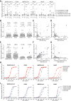
Fig. 1.. The S2P6 cross-reactive mAb broadly neutralizes betacoronaviruses from three subgenera.
(A) Binding avidity (EC50) of five mAbs to prefusion coronavirus S trimer ectodomains as determined by ELISA. (B) S2P6 ELISA binding curves showing the full titration. One representative experiment out of two is shown. OD, optical density. (C) Cladogram of representative alpha- (black) and betacoronavirus (red) S glycoprotein amino acid sequences inferred through maximum likelihood analysis. (D) Flow cytometry analysis of S2P6 binding (from 10 to 0.22 μg/ml) to a panel of 26 S glycoproteins representative of all sarbecovirus clades (left) and eight SARS-CoV-2 variants (right) displayed as EC50 values. (E) Neutralization of authentic SARS-CoV-2 by S2P6 determined using VeroE6-TMPRSS2 (left) or Vero-E6 (right) cells. The S309 mAb that binds RBD site IV (38) is included for comparison. The mean ± SD of triplicates from one representative experiment out of three is shown. (F) S2P6-mediated neutralization of SARS-CoV-2 B.1.1.7 (Alpha) S, B.1.351 (Beta) S, P.1 (Gamma) S, and B1.617.1 (Kappa) S VSV pseudotypes (mut) represented as IC50 fold change relative to wild-type (WT) (D614G) S VSV pseudotype. Individual values for each of the two replicates are shown as open circles, the mean as a colored bar, and the SD as error bars. (G) S2P6-mediated neutralization of VSV pseudotyped with various betacoronavirus S glycoproteins. Error bars indicate standard deviation of triplicates. IC50 values are 2.4 μg/ml, 1.4 μg/ml, 17.1 μg/ml, 1.3 μg/ml, and 0.02 μg/ml for SARS-CoV, SARS-CoV-2, MERS-CoV, OC43, and PANG/GD, respectively. pp, pseudoparticles. (H) Alignment of betacoronavirus stem helix amino acid sequences with the S2P6 epitope boxed. Residue numbering is shown according to SARS-CoV-2 S. The sequences of the peptides used in this study are shown in blue, and N-linked glycosylation sequons are highlighted in yellow. (I) Competition assay for S2P6 or B6 binding to the SARS-CoV-2 or SARS-CoV stem helix peptide (herein defined as SARS-CoV/-2). B6 binding in the presence of S2P6 (red line), B6 binding in the absence of S2P6 (black line), and the control lacking the SARS-CoV/-2 stem helix peptide (gray line) are shown. (J) Kinetics of S2P6 binding to a panel of biotinylated betacoronavirus stem helix peptides immobilized at the surface of biolayer interferometry biosensors.
Fig. 2.
Fig. 2.. Structural basis for the broad S2P6 cross-reactivity with the conserved coronavirus stem helix peptide.
(A) Composite model of the S2P6-bound SARS-CoV-2 S cryo-EM structure and of the S2P6-bound stem helix peptide crystal structure docked in the cryo-EM map (semitransparent gray surface). SARS-CoV-2 S protomers are colored pink, cyan, and gold; the S2P6 Fab heavy and light chains are colored purple and magenta; and the S2M11 Fab heavy and light chains are colored dark and light gray, respectively. (B) Crystal structure of the S2P6 Fab (surface rendering) in complex with the SARS-CoV-2 S stem helix peptide (yellow ribbon with side chains rendered as sticks). (C and D) Ribbon diagram in two orthogonal orientations of the S2P6 Fab bound to the SARS-CoV-2 S stem helix peptide showing the network of interactions. Only key interface residues and the S2P6 CDR loops are shown for clarity. Residues Q32 and H57, which are mutated during affinity maturation of the S2P6 heavy chain, are colored blue. Hydrogen bonds are indicated with dashed lines. Residues substituted in the escape mutants isolated are underlined. Single-letter abbreviations for the amino acid residues are as follows: A, Ala; C, Cys; D, Asp; E, Glu; F, Phe; G, Gly; H, His; I, Ile; K, Lys; L, Leu; M, Met; N, Asn; P, Pro; Q, Gln; R, Arg; S, Ser; T, Thr; V, Val; W, Trp; and Y, Tyr.
Fig. 3.
Fig. 3.. S2P6 binding disrupts the stem helix bundle and sterically inhibits membrane fusion.
(A) SARS-CoV-2 S binding to ACE2 in the presence of mAb S2P6 analyzed by ELISA. S2E12 was included as a positive control. (B) S2P6 inhibition of cell-cell fusion using VeroE6 cells transfected with SARS-CoV-2 S. S2M11 was included as a positive control. Inhibition of fusion values are normalized to the percentage of fusion without mAb (100%) and to that of fusion of nontransfected cells (0%). (C) Proposed mechanism of inhibition mediated by the S2P6 mAb. S2P6 binds to the hydrophobic core of the stem helix bundle and disrupts its quaternary structure. S2P6 binding likely prevents S2 subunit refolding from the pre- to the postfusion state and blocks viral entry.
Fig. 4.
Fig. 4.. S2P6 activates effector functions and reduces SARS-CoV-2 lung burden in Syrian hamsters.
(A) NFAT (nuclear factor of activated T cells)–driven luciferase signal induced in Jurkat cells stably expressing FcγRIIIa (V158, left) or FcγRIIa (V131, right) upon S2P6 binding to full-length wild-type SARS-CoV-2 S expressed at the surface of CHO target cells. S309 is included as positive control. RLU, relative luminescence unit. (B) mAb-mediated ADCC using SARS-CoV-2 CHO-K1 cells (genetically engineered to stably express a HaloTag-HiBit) as target cells and PBMC as effector cells. The magnitude of natural killer cell–mediated killing is expressed as a percentage of specific lysis. (C) mAb-mediated ADCP using cell trace violet–labeled PBMCs as a source of phagocytic cells (monocytes) and PKH67–fluorescently labeled S-expressing CHO cells as target cells. The y axis indicates the percentage of monocytes double-positive for anti-CD14 (monocyte) marker and PKH67. (D) Lysis of SARS-CoV-2 S stably transfected CHO cells by mAbs in the presence of complement. S309 was included as positive control; S309-GRLR with diminished FcγR binding capacity and an unrelated mAb (neg mAb) were used as negative controls. (E) Syrian hamsters were administered with the indicated amount of S2P6 mAb harboring either a hamster (Hm-S2P6) or a human (Hu-S2P6) constant region before intranasal challenge with prototypic SARS-CoV-2 (Wuhan-1 related). An irrelevant mAb (MGH2 against Plasmodium falciparum CSP) at 20 mg/kg was used as negative control (CTRL) (60). Viral RNA loads and replicating virus titers are shown on the left and right, respectively. TCID50, median tissue culture infectious dose. (F) Prophylactic administration of Hu-S2P6 at 20 mg/kg in hamsters challenged with SARS-CoV-2 B.1.351 (Beta) VOC. Viral RNA loads and replicating virus titers are shown on the left and right, respectively. *P < 0.05; **P < 0.01; Mann-Whitney test.
Fig. 5.
Fig. 5.. Stem helix–directed Abs are rare, of narrow specificities, and enhance their binding affinity and cross-reactivity through somatic mutations.
(A) Binding of prepandemic (P; n = 88), COVID-19 convalescent (C; n = 72), vaccinees immune (VI; n = 9), and vaccinees naïve (VN; n = 37) plasma Abs (diluted 1:10) to immobilized betacoronavirus stem helix peptides analyzed by ELISA. A cutoff of 0.7 was determined on the basis of the signal of prepandemic samples and binding to uncoated ELISA plates (horizontal dashed line). The fraction of samples for which binding above the cutoff was detected is indicated. (B to G) Analysis of memory B cell specificities for betacoronavirus stem helix peptides. Each dot represents a well containing oligoclonal B cell supernatant screened for the presence of stem helix peptide binding IgG Abs using ELISA. Samples obtained from 21 COVID-19 convalescent individuals [(B) to (D)] and 16 vaccinees [(E) to (G)]. Pairwise reactivity comparison is shown for SARS-CoV/-2 and OC43 [(C) and (F)] and SARS-CoV/-2 and HKU1 [(D) and (G)]. Cultures cross-reactive with at least three peptides are highlighted in color. A cutoff of 0.4 is indicated by a horizontal dashed line. The fraction of wells for which binding above the cutoff was detected is indicated. (H and I) Binding to stem helix peptides of S2P6 (H) harboring mature (SH/SK), fully germline-reverted (UCA/UCA), germline-reverted heavy chain paired with mature light chain (UCA/SK), mature heavy chain paired with germline-reverted light chain (SH/UCA), and of P34D10, P34G12, and P34E3 (I) harboring either mature (SH/SK) or germline-reverted (UCA/UCA) sequences.

References

    1. Tortorici M. A., Veesler D., Structural insights into coronavirus entry. Adv. Virus Res. 105, 93–116 (2019). 10.1016/bs.aivir.2019.08.002 - DOI - PMC - PubMed
    1. Walls A. C., Park Y. J., Tortorici M. A., Wall A., McGuire A. T., Veesler D., Structure, Function, and Antigenicity of the SARS-CoV-2 Spike Glycoprotein. Cell 181, 281–292.e6 (2020). 10.1016/j.cell.2020.02.058 - DOI - PMC - PubMed
    1. Wrapp D., Wang N., Corbett K. S., Goldsmith J. A., Hsieh C. L., Abiona O., Graham B. S., McLellan J. S., Cryo-EM structure of the 2019-nCoV spike in the prefusion conformation. Science 367, 1260–1263 (2020). 10.1126/science.abb2507 - DOI - PMC - PubMed
    1. Thomson E. C., Rosen L. E., Shepherd J. G., Spreafico R., da Silva Filipe A., Wojcechowskyj J. A., Davis C., Piccoli L., Pascall D. J., Dillen J., Lytras S., Czudnochowski N., Shah R., Meury M., Jesudason N., De Marco A., Li K., Bassi J., O’Toole A., Pinto D., Colquhoun R. M., Culap K., Jackson B., Zatta F., Rambaut A., Jaconi S., Sreenu V. B., Nix J., Zhang I., Jarrett R. F., Glass W. G., Beltramello M., Nomikou K., Pizzuto M., Tong L., Cameroni E., Croll T. I., Johnson N., Di Iulio J., Wickenhagen A., Ceschi A., Harbison A. M., Mair D., Ferrari P., Smollett K., Sallusto F., Carmichael S., Garzoni C., Nichols J., Galli M., Hughes J., Riva A., Ho A., Schiuma M., Semple M. G., Openshaw P. J. M., Fadda E., Baillie J. K., Chodera J. D., ISARIC4C Investigators, COVID-19 Genomics UK (COG-UK) Consortium, Rihn S. J., Lycett S. J., Virgin H. W., Telenti A., Corti D., Robertson D. L., Snell G., Circulating SARS-CoV-2 spike N439K variants maintain fitness while evading antibody-mediated immunity. Cell 184, 1171–1187.e20 (2021). 10.1016/j.cell.2021.01.037 - DOI - PMC - PubMed
    1. McCallum M., De Marco A., Lempp F. A., Tortorici M. A., Pinto D., Walls A. C., Beltramello M., Chen A., Liu Z., Zatta F., Zepeda S., di Iulio J., Bowen J. E., Montiel-Ruiz M., Zhou J., Rosen L. E., Bianchi S., Guarino B., Fregni C. S., Abdelnabi R., Foo S.-Y. C., Rothlauf P. W., Bloyet L.-M., Benigni F., Cameroni E., Neyts J., Riva A., Snell G., Telenti A., Whelan S. P. J., Virgin H. W., Corti D., Pizzuto M. S., Veesler D., N-terminal domain antigenic mapping reveals a site of vulnerability for SARS-CoV-2. Cell 184, 2332–2347.e16 (2021). 10.1016/j.cell.2021.03.028 - DOI - PMC - PubMed

Publication types

MeSH terms