. 2021 Aug 3;12(1):4680.
doi: 10.1038/s41467-021-24852-9.

Haploinsufficiency of SF3B2 causes craniofacial microsomia

Affiliations

Haploinsufficiency of SF3B2 causes craniofacial microsomia

Andrew T Timberlake et al. Nat Commun. .

Abstract

Craniofacial microsomia (CFM) is the second most common congenital facial anomaly, yet its genetic etiology remains unknown. We perform whole-exome or genome sequencing of 146 kindreds with sporadic (n = 138) or familial (n = 8) CFM, identifying a highly significant burden of loss of function variants in SF3B2 (P = 3.8 × 10⁻¹⁰), a component of the U2 small nuclear ribonucleoprotein complex, in probands. We describe twenty individuals from seven kindreds harboring de novo or transmitted haploinsufficient variants in SF3B2. Probands display mandibular hypoplasia, microtia, facial and preauricular tags, epibulbar dermoids, lateral oral clefts in addition to skeletal and cardiac abnormalities. Targeted morpholino knockdown of SF3B2 in Xenopus results in disruption of cranial neural crest precursor formation and subsequent craniofacial cartilage defects, supporting a link between spliceosome mutations and impaired neural crest development in congenital craniofacial disease. The results establish haploinsufficient variants in SF3B2 as the most prevalent genetic cause of CFM, explaining ~3% of sporadic and ~25% of familial cases.


Conflict of interest statement

Rhonda E. Schnur and Sureni V. Mullegama are employees of GeneDx. The remaining authors declare no competing interest.

Figures

Fig. 1
Fig. 1. SF3B2 variants in pedigrees with craniofacial microsomia.
A` Domain structure of SF3B2 with variants identified noted above. Domain annotation based on UniProt accession Q13435. B Pedigrees of kindreds with SF3B2 LOF variants. De novo or transmitted LOF variants are indicated above each pedigree, with stars denoting confirmed de novo variants. ‘+’ represents a wild type allele, and ‘D’ represents the LOF variant in SF3B2 indicated above the pedigree. Individuals with no genotype listed represent those in whom genomic DNA was not available for study.
Fig. 2
Fig. 2. Clinical Images of Individuals with SF3B2 haploinsufficiency.
A. Photographs of three individuals demonstrating the shared phenotype. Proband 1 (c.2329dupG) demonstrates mandibular hypoplasia on the left, scar from preauricular skin tag excision on the right, and tragal duplication and facial skin tag on the left side prior to removal. Proband 2 (c.1780-2 A > -) demonstrates maxillary and mandibular hypoplasia on the right side in addition to bilateral tragal abnormalities. Proband 4-1 (c. 1608dupG) presented with maxillary and mandibular hypoplasia and tragal abnormality on the left side, and bilateral preauricular tags. B. Three-dimensional (3D) computed tomography (CT) reconstructions of the craniofacial skeleton of probands 2 and 3. Frontal, lateral, and submental views demonstrate maxillary and mandibular hypoplasia with resulting facial asymmetry. The third row demonstrates, from left to right, 3D CT scans of the cervical spine of Proband 1 and X-rays of Proband 2 and 3. Arrows in each figure indicate the cervical ribs. C. Photographs of family members with a shared SF3B2 startloss variant (p.M1?) demonstrating variable external ear phenotypes. Images of II:2 demonstrate a right ear markedly smaller than the left. Images of II:4 demonstrate a tragal abnormality and scar from prior preauricular skin tag removal, in addition to an epibulbar dermoid. III:3 presented with bilateral tragal abnormalities and tags (right side ear photo from childhood). Images of III:5 show an underdeveloped tragus. III:8 had tragal abnormality, scar from tag excision, right lateral oral cleft, and chin deviation due to mandibular hypoplasia.
Fig. 3
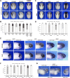
Fig. 3. Sf3b2 knockdown alters neural crest development in Xenopus embryos.
A Unilateral injection of increasing doses of SF3B2MO (10-30 ng) interferes with sox10 expression at the neurula stage, in a manner similar to SF3B4 knockdown. Injection of a control MO (CoMO) did not significantly affect sox10 expression. The SF3B2 knockdown phenotype is efficiently rescued by injection of human SF3B2 (HSF3B2; 10 pg or 100 pg) plasmid DNA. mRNA encoding the lineage tracer ß -galactosidase was co-injected with the MOs to identify the injected side (punctate red staining). The injected side is indicated by an asterisk. Dorsal view, anterior to top. The two rows show examples of the phenotype for each injection condition. Scale bar, 500 μm. B Quantification of the phenotypes. The numbers on the top of each bar indicate the number of embryos analyzed from seven independent experiments. C Unilateral injection of SF3B2MO (20 ng) reduces the expression of other neural crest genes at the neurula stage including tfap2e and snai2, and expanded the neural plate expression domain of sox2 (white brackets) on the injected side. sox9 was not affected in these embryos. The injected side is indicated by an asterisk. Dorsal view, anterior to top. Scale bar, 500 μm. D Quantification of the phenotypes. The numbers on the top of each bar indicate the number of embryos analyzed from three independent experiments. E At the tailbud stage, SF3B2MO (20 ng) injected embryos show a decrease in the length of the NC streams (arrows) as visualized by sox10 (stage 25), sox9, and twist1 (stage 28) expression. CoMO injection did not affect NC streams formation. Lateral view, dorsal to top, anterior to right. Scale bar, 250 μm. F Quantification of the phenotypes. The numbers on the top of each bar indicate the number of embryos analyzed from three independent experiments. G At stage 40, the expression of runx2 in the pharyngeal arches mesenchyme (arrows) is severely downregulated in SF3B2MO-injected tadpoles. The white line indicates the midline (frontal view). Scale bar, 250 μm. The quantification of the runx2 phenotype is shown in panel F. Source data are provided as a source data file.
Fig. 4
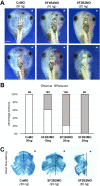
Fig. 4. Sf3b2 knockdown causes craniofacial defects in Xenopus tadpoles.
A Gross morphology of the head of SF3B2MO- and CoMO-injected Xenopus tadpoles. The lineage tracer ß -galactosidase was again co-injected, with the injected side indicted by an asterisk. Dorsal views, anterior to top. The two rows show examples of the phenotype for each injection condition. Scale bar, 600 μm. B The graph is a quantification of the results from three independent experiments. The number of tadpoles analyzed is indicated on the top of each bar. C Alcian blue staining of dissected craniofacial cartilages of CoMO (30 ng) and SF3B2MO (20 ng) injected tadpoles at stage 45. The injected side is indicated by an asterisk. The black lines indicate the midline. Source data are provided as a source data file.

References

    1. Beleza-Meireles A, et al. Oculo-auriculo-vertebral spectrum: a review of the literature and genetic update. J. Med. Genet. 2014;51:635–645. doi: 10.1136/jmedgenet-2014-102476. - DOI - PubMed
    1. Keogh IJ, et al. Isolated microtia as a marker for unsuspected hemifacial microsomia. Arch. Otolaryngol. Head. Neck Surg. 2007;133:997–1001. doi: 10.1001/archotol.133.10.997. - DOI - PubMed
    1. Vendramini-Pittoli S, Kokitsu-Nakata NM. Oculoauriculovertebral spectrum: report of nine familial cases with evidence of autosomal dominant inheritance and review of the literature. Clin. Dysmorphol. 2009;18:67–77. doi: 10.1097/MCD.0b013e328323a7dd. - DOI - PubMed
    1. Rollnick BR, Kaye CI. Hemifacial microsomia and variants: pedigree data. Am. J. Med. Genet. 1983;15:233–253. doi: 10.1002/ajmg.1320150207. - DOI - PubMed
    1. Kaye CI, et al. Oculoauriculovertebral anomaly: segregation analysis. Am. J. Med. Genet. 1992;43:913–917. doi: 10.1002/ajmg.1320430602. - DOI - PubMed

Publication types

LinkOut - more resources