. 2021 Aug 3;11(1):15722.
doi: 10.1038/s41598-021-94495-9.

Cerebral malaria: insight into pathology from optical coherence tomography

Affiliations

Cerebral malaria: insight into pathology from optical coherence tomography

Zhanhan Tu et al. Sci Rep. .

Abstract

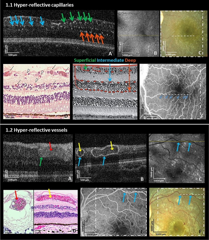
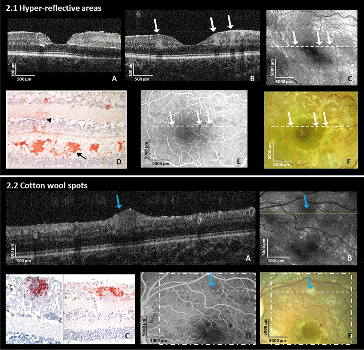
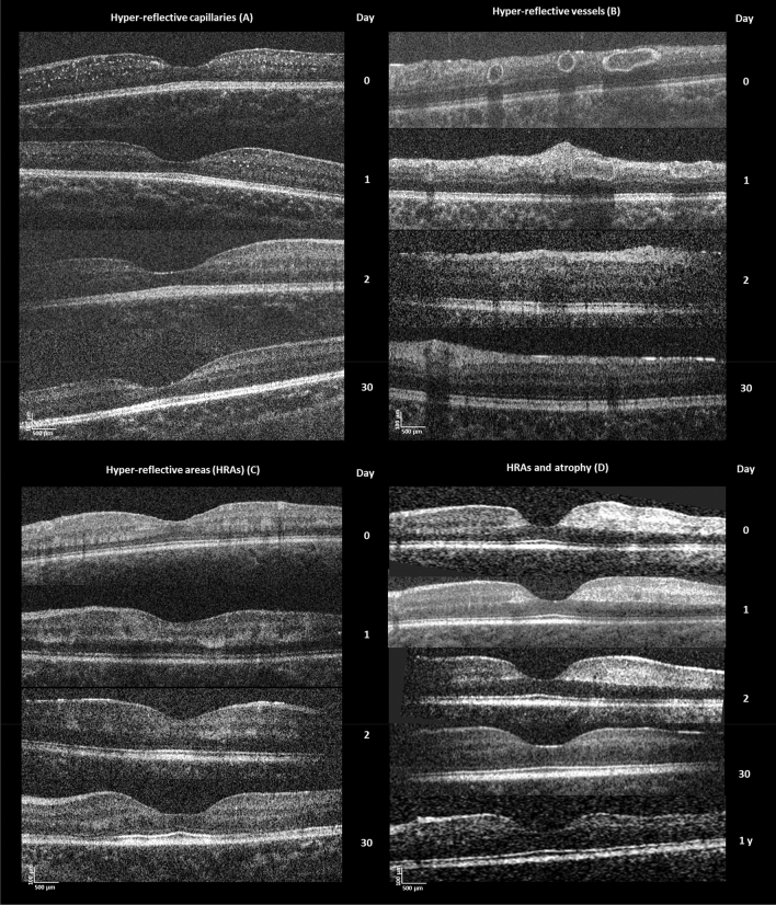
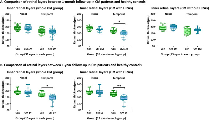
We aimed to investigate structural retinal changes in malarial retinopathy (MR) using hand-held optical coherence tomography (HH-OCT) to assess its diagnostic potential. Children with MR (n = 43) underwent ophthalmoscopy, fluorescein angiography and HH-OCT during admission, 1-month (n = 31) and 1-year (n = 8) post-discharge. Controls were comatose patients without malaria (n = 6) and age/sex-matched healthy children (n = 43). OCT changes and retinal layer thicknesses were compared. On HH-OCT, hyper-reflective areas (HRAs) were seen in the inner retina of 81% of MR patients, corresponding to ischaemic retinal whitening on fundus photography. Cotton wool spots were present in 37% and abnormal hyper-reflective dots, co-localized to capillary plexus, in 93%. Hyper-reflective vessel walls were present in 84%, and intra-retinal cysts in 9%. Vascular changes and cysts resolved within 48 h. HRAs developed into retinal thinning at 1 month (p = 0.027) which was more pronounced after 1 year (p = 0.009). Ischaemic retinal whitening is located within inner retinal layers, distinguishing it from cotton wool spots. Vascular hyper-reflectivity may represent the sequestration of parasitized erythrocytes in vessels, a key CM feature. The mechanisms of post-ischemic retinal atrophy and cerebral atrophy with cognitive impairment may be similar in CM survivors. HH-OCT has the potential for monitoring patients, treatment response and predicting neurological deficits.


Conflict of interest statement

The authors declare no competing interests.

Figures

Figure 1
Figure 1
Hyper-reflective capillaries and vessels. (1.1) Left eye of a 7-month old male CM patient on admission. (A) HH-OCT B-scan showing hyper-reflective dots corresponding to locations of capillaries (green, blue and orange arrows: superficial, intermediate and deep capillary plexus); (B) En-face OCT displays no visible change; (yellow line: location of the OCT B-scan in the image (A)). (C) Fundus photo; dashed line and square correspond to (A, B); (D) Representative retinal cross-section histology from a different fatal patient showing capillaries and venules affected by parasite sequestration (black arrows: intense sequestration; arrowheads: low or no sequestration); Reprinted with permission; (E) Schematic representation of superficial, intermediate and deep capillary plexus collocated to capillaries filled with pRBCs on OCT in (A). Reprinted with permission; (F) Fundus fluorescein angiography (FA) shows perfusion deficits of capillaries (blue arrows) corresponding to parasitized vessels on OCT in the same location of (1.1.A). (1.2) Left eye of a 42-month old male CM patient on admission. (A) Hyper-reflective vessel (blue arrow) corresponding to localization of vessel on fundus photo and FA (not shown) with hyper-reflective lumen (red arrow); and (B) Hyper-reflective vessels (blue arrows) with hypo-reflective lumina (yellow arrows) on OCT B-scans; (C) En-face OCT (yellow line: location of OCT B-scan in B, blue arrows: vessels shown in (B); (D) Representative retinal cross-section histology showing pRBCs in venules from a different fatal patient (red arrow shows vessels with pRBCs filling the lumen; yellow arrow shows a vessel where pRBCs cytoadhere to the vessel wall with red blood cells without parasites in the center of the vessel (similar to hypo-reflective lumina of blood vessels in (1.2.B); (E) Fundus fluorescein angiography and (F) Fundus photo corresponding to OCT in (1.2.B) (blue arrows corresponding to vessels in the image (B); dashed line and square correspond to (B, C).
Figure 2
Figure 2
Hyper-reflective areas and cotton wool spots (Retinal Whitening). (2.1.A, B) shows multiple hyper-reflective areas of various sizes (white arrows) located in the inner nuclear layer level, the outer plexiform layer and outer nuclear layer on OCT B-scans in the right eye of a 22-month old male CM patient and in the left eye of an 84-month old female CM patient at admission, respectively; (C) En-face OCT of the patient in (B); white arrows correspond to the location of white arrows in the image (B) (hyper-reflective areas); The hyper-reflective areas in OCT B-scan (B) show darkening on the en-face image (C) in this patient (white arrows); (D) Representative histology of immuno-histochemical staining for fibrinogen surrounding a small vessel (black arrowhead) mainly located in OPL (black arrow) from the literature (Reprinted with permission). (E) Fundus fluorescein angiography showing hypo-perfusion of capillaries and (F) fundus photo same eye as OCT in (B); white arrows and dashed lines show parafoveal whitening which are corresponding to the same fundus locations in (B, C, E, F). (2.2.A) Cotton wool spot (blue arrow) in the left eye at admission on OCT B-scan in the left eye of a 42-months old male CM patient at admission; (B) En-face OCT with darkening in area of cotton wool spot (blue arrow); fine yellow line: location of the B-scan in the image (A); blue arrow corresponds to blue arrow in image (A); (C) Immunohistochemical staining for b-APP in retinal nerve fiber layer (RNFL) from different CM patients. Reprinted with permission. (D) Fundus fluorescein angiography showing grey area masking underlying capillaries (blue arrow) and (E) fundus photo from patient’s left eye corresponding to OCT showing white cotton wool spot (blue arrow) in (2.2.A). The dashed square and the dashed line correspond to en-face OCT in B and B-scan in (A). Blue arrows show the location of a cotton wool spot. Blue arrows correspond to the same fundus locations in (A, B, D, E).
Figure 3
Figure 3
Longitudinal changes in the retina. (A) Hyper-reflective capillaries: OCT of a 7-month-old male CM patient at admission, 1 day, 2 days and 30 days. The number of hyper-reflective capillaries diminished rapidly 1 day after treatment initiation; hyper-reflective capillaries were almost not detectable on OCT after 2 days. (B) Hyper-reflective vessels: OCT of 42-month-old male CM patient at admission, 1 day, 2 days and 30 days after start of treatment. The abnormal hyper-reflective vessels also diminished rapidly within 24 h. (C) Hyper-reflective areas: OCT of 45-month-old female CM patient at admission, 1 day, 2 days and 30 days after start of treatment. The hyper-reflective areas remained visible, but were smaller and less intense at 1-month follow-up. Retinal atrophy was just evident at this time point. (D) Parafoveal hyper-reflective areas and atrophy: OCT of 22-month-old male CM patient at admission, 1 day, 2 days, 30 days and 1 year. He had relatively large and dense hyper-reflective areas at admission (particularly to the temporal side of the fovea (right)). There is corresponding retinal atrophy at 1-year follow-up with marked thinning compared to the left of the fovea.
Figure 4
Figure 4
Comparisons of thicknesses of retinal layers between follow-up of CM patients with HRAs and healthy controls. (A) Comparison of thicknesses of inner retinal layers between controls and CM patients at 1-month follow-up. (B) Comparisons of thicknesses of inner retinal layers between controls and CM patients at 1-year follow-up. Error bars in the boxplot are the ranges of data. *p < 0.05 and **p < 0.01 show significant differences between the two groups. Control groups are in green and patient groups in blue (Con controls, CM cerebral malaria, 1 M 1-month follow-up, 1Y 1-year follow-up).

References

    1. Seydel KB, et al. Brain swelling and death in children with cerebral malaria. N. Engl. J. Med. 2015;372:1126–1137. doi: 10.1056/NEJMoa1400116. - DOI - PMC - PubMed
    1. Postels DG, Chimalizeni YF, Mallewa M, Boivin MJ, Seydel KB. Pediatric cerebral malaria: A scourge of Africa. Future Neurol. 2013;8:67–85. doi: 10.2217/fnl.12.84. - DOI
    1. WHO . World Malaria Report 2020. WHO; 2020.
    1. Langfitt JT, et al. Neurodevelopmental impairments 1 year after cerebral malaria. Pediatrics. 2019;143:e20181026. doi: 10.1542/peds.2018-1026. - DOI - PubMed
    1. Taylor TE, et al. Differentiating the pathologies of cerebral malaria by postmortem parasite counts. Nat. Med. 2004;10:143–145. doi: 10.1038/nm986. - DOI - PubMed

Publication types