

Cerebral vasospasm and hypoperfusion after traumatic brain injury: Combined CT angiography and CT perfusion imaging study
- PMID: 34345501
- PMCID: PMC8326138
- DOI: 10.25259/SNI_859_2020
Cerebral vasospasm and hypoperfusion after traumatic brain injury: Combined CT angiography and CT perfusion imaging study
Abstract
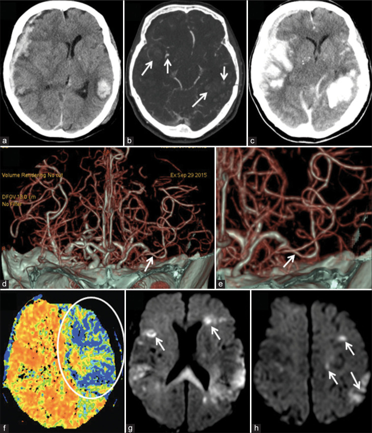
Background: Timely identification of the cerebral perfusion abnormalities after traumatic brain injury (TBI) is highly important. The objective of this study was the evaluation of the post traumatic vasospasm and cerebral hypoperfusion with the serial combined CT angiography (CTA) and CT perfusion (CTP) imaging examinations.
Methods: The case series comprised 25 adult patients with closed TBI accompanied by various types of intracranial hematoma. Emergency surgery was done in 15 cases (60%). Combined CTA and CTP were performed on days 0 (D0) and 7 ± 1 (D7) after trauma.
Results: CTA on D0 did not demonstrate vasospasm in any case but revealed it on D7 in 9 patients (36%). In the multivariate analysis, only the presence of subarachnoid hemorrhage (SAH) on D7 had confirmed a significant association with the development of vasospasm (P = 0.0201). Cerebral hypoperfusion at least in one evaluated brain region was noted on D0 and D7 in 76% and 60% of patients, respectively, and showed highly variable spatial distribution and temporal development. Treatment results were not associated with the presence of vasospasm (P = 0.7337) or the number of brain regions affected by hypoperfusion on D0 (P = 0.2285), but the number of brain regions affected by hypoperfusion on D7 was significantly greater in cases of unfavorable outcome (P = 0.0187).
Conclusion: Vasospasm is merely related to SAH sustained at the subacute stage of TBI, but its spatial and temporary interrelationships with the post traumatic cerebral hypoperfusion are complex. Serial combined CTA and CTP examinations may facilitate monitoring of perfusion abnormalities and treatment guidance.
Keywords: CT angiography; CT perfusion imaging; Cerebral hypoperfusion; Post traumatic vasospasm; Traumatic brain injury.
Copyright: © 2021 Surgical Neurology International.
Conflict of interest statement
There are no conflicts of interest.
Figures
Similar articles
-
Whole brain CT perfusion combined with CT angiography in patients with subarachnoid hemorrhage and cerebral vasospasm.Clin Neurol Neurosurg. 2013 Dec;115(12):2496-501. doi: 10.1016/j.clineuro.2013.10.004. Epub 2013 Oct 16. Clin Neurol Neurosurg. 2013. PMID: 24210268
-
Cerebral watershed hypoperfusion in subarachnoid hemorrhage: computed tomography perfusion analysis.J Neurosurg. 2011 Apr;114(4):961-8. doi: 10.3171/2010.8.JNS091766. Epub 2010 Sep 17. J Neurosurg. 2011. PMID: 20849218
-
Cost-effectiveness of CT angiography and perfusion imaging for delayed cerebral ischemia and vasospasm in aneurysmal subarachnoid hemorrhage.AJNR Am J Neuroradiol. 2014 Sep;35(9):1714-20. doi: 10.3174/ajnr.A3947. Epub 2014 May 8. AJNR Am J Neuroradiol. 2014. PMID: 24812015 Free PMC article.
-
Diagnosing Vasospasm After Subarachnoid Hemorrhage: CTA and CTP.Can J Neurol Sci. 2014 May;41(3):314-9. doi: 10.1017/s031716710001725x. Can J Neurol Sci. 2014. PMID: 24718816 Review.
-
Neuroimaging research on cerebrovascular spasm and its current progress.Acta Neurochir Suppl. 2011;110(Pt 2):233-7. doi: 10.1007/978-3-7091-0356-2_42. Acta Neurochir Suppl. 2011. PMID: 21125477 Review.
Cited by
-
Computed tomography perfusion stroke mimics on RAPID commercial software: A case-based review.Brain Circ. 2023 Jun 30;9(2):68-76. doi: 10.4103/bc.bc_100_22. eCollection 2023 Apr-Jun. Brain Circ. 2023. PMID: 37576575 Free PMC article. Review.
-
Neural control of cerebral blood flow: scientific basis of scalp acupuncture in treating brain diseases.Front Neurosci. 2023 Aug 15;17:1210537. doi: 10.3389/fnins.2023.1210537. eCollection 2023. Front Neurosci. 2023. PMID: 37650106 Free PMC article. Review.
References
-
- Al-Mufti F, Amuluru K, Changa A, Lander M, Patel N, Wajswol E, et al. Traumatic brain injury and intracranial hemorrhage-induced cerebral vasospasm: A systematic review. Neurosurg Focus. 2017;43(5):E14. - PubMed
-
- Al-Mufti F, Amuluru K, Lander M, Mathew M, El-Ghanem M, Nuoman R, et al. Low Glasgow coma score in traumatic intracranial hemorrhage predicts development of cerebral vasospasm. World Neurosurg. 2018;120:e68–71. - PubMed
-
- Bendinelli C, Bivard A, Nebauer S, Parsons MW, Balogh ZJ. Brain CT perfusion provides additional useful information in severe traumatic brain injury. Injury. 2013;44:1208–12. - PubMed
LinkOut - more resources
Full Text Sources