. 2021 Aug:135:104654.
doi: 10.1016/j.compbiomed.2021.104654. Epub 2021 Jul 16.

Investigation of nonsynonymous mutations in the spike protein of SARS-CoV-2 and its interaction with the ACE2 receptor by molecular docking and MM/GBSA approach

Affiliations

Investigation of nonsynonymous mutations in the spike protein of SARS-CoV-2 and its interaction with the ACE2 receptor by molecular docking and MM/GBSA approach

Reem Y Aljindan et al. Comput Biol Med. 2021 Aug.

Abstract

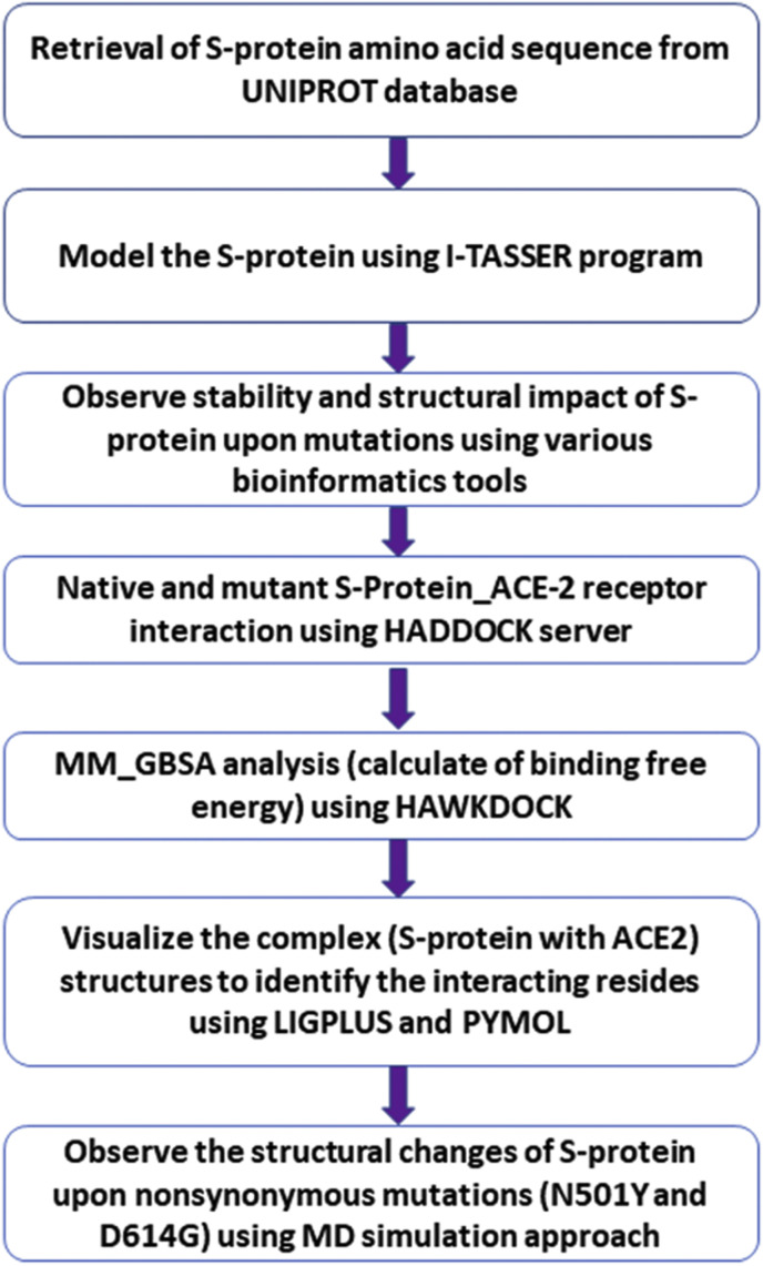
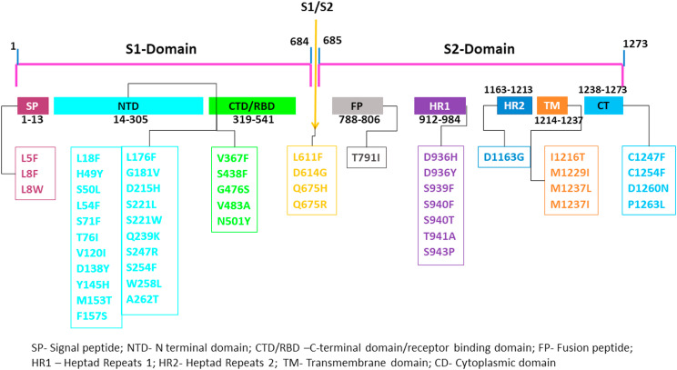
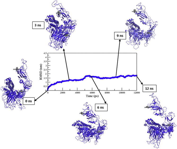
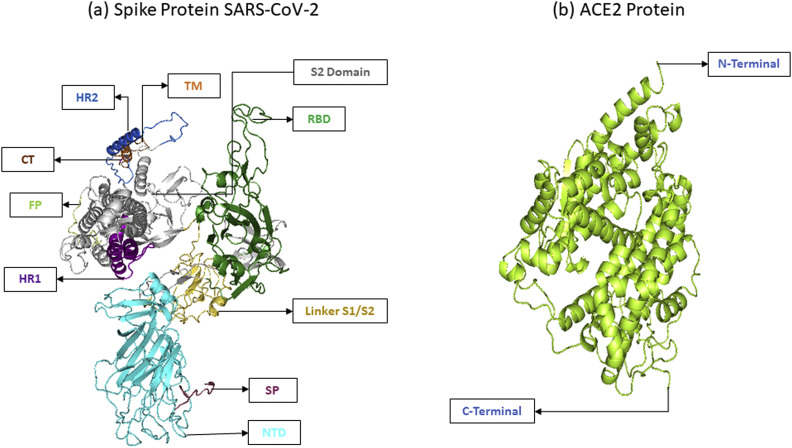
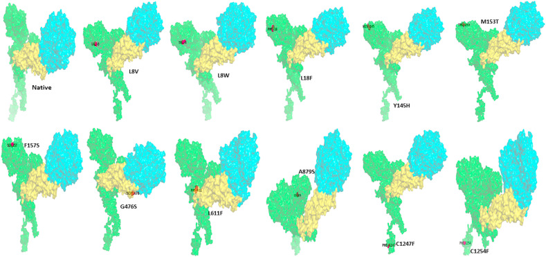
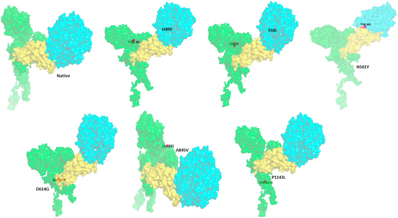
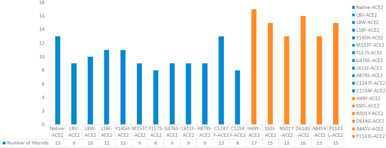
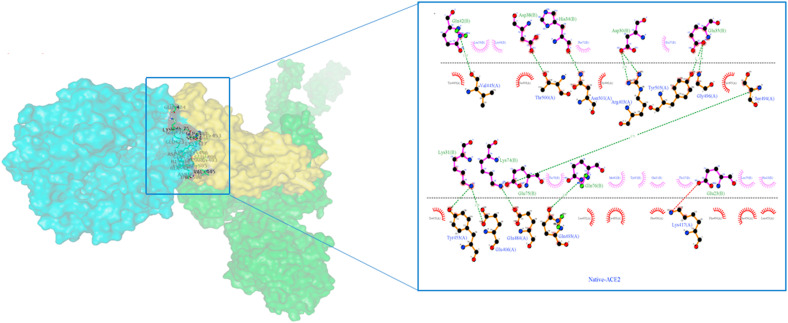
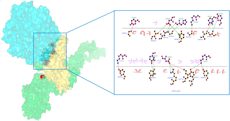
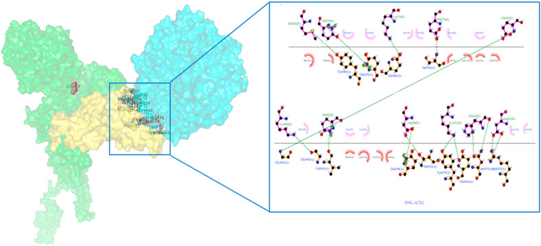
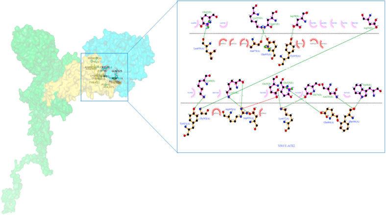
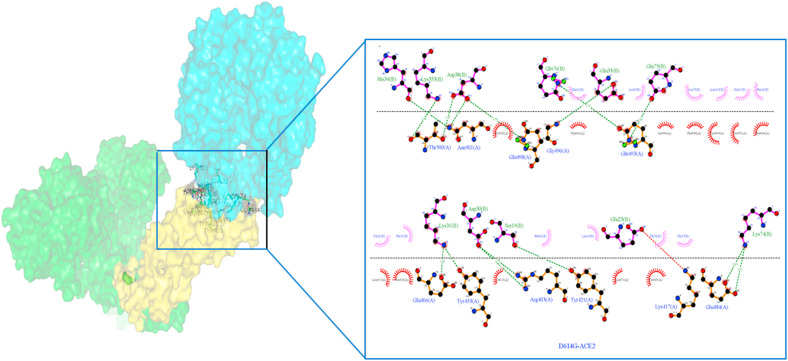
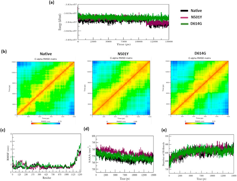
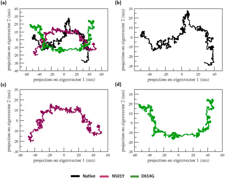
COVID-19 is an infectious and pathogenic viral disease caused by SARS-CoV-2 that leads to septic shock, coagulation dysfunction, and acute respiratory distress syndrome. The spreading rate of SARS-CoV-2 is higher than MERS-CoV and SARS-CoV. The receptor-binding domain (RBD) of the Spike-protein (S-protein) interacts with the human cells through the host angiotensin-converting enzyme 2 (ACE2) receptor. However, the molecular mechanism of pathological mutations of S-protein is still unclear. In this perspective, we investigated the impact of mutations in the S-protein and their interaction with the ACE2 receptor for SAR-CoV-2 viral infection. We examined the stability of pathological nonsynonymous mutations in the S-protein, and the binding behavior of the ACE2 receptor with the S-protein upon nonsynonymous mutations using the molecular docking and MM_GBSA approaches. Using the extensive bioinformatics pipeline, we screened the destabilizing (L8V, L8W, L18F, Y145H, M153T, F157S, G476S, L611F, A879S, C1247F, and C1254F) and stabilizing (H49Y, S50L, N501Y, D614G, A845V, and P1143L) nonsynonymous mutations in the S-protein. The docking and binding free energy (ddG) scores revealed that the stabilizing nonsynonymous mutations show increased interaction between the S-protein and the ACE2 receptor compared to native and destabilizing S-proteins and that they may have been responsible for the virulent high level. Further, the molecular dynamics simulation (MDS) approach reveals the structural transition of mutants (N501Y and D614G) S-protein. These insights might help researchers to understand the pathological mechanisms of the S-protein and provide clues regarding mutations in viral infection and disease propagation. Further, it helps researchers to develop an efficient treatment approach against this SARS-CoV-2 pandemic.

Keywords: ACE2 receptor; Binding affinity; Nonsynonymous mutations; SARS-CoV-2; Spike protein; Stability.


Conflict of interest statement

We have no conflicts of interest to disclose.

Figures

Fig. 1
Fig. 1
The workflow applied in this investigation.
Fig. 2
Fig. 2
S-protein domains and their nonsynonymous mutations list.
Fig. 3
Fig. 3
The backbone RMSD of the native S-protein versus time at 300 K. The average structure of native S-protein has shown in different timescale.
Fig. 4
Fig. 4
(a) Modeled Spike (S) - protein, (b) ACE2 protein (PDB ID: 1R42_A). This figure was prepared by PYMOL.
Fig. 5a
Fig. 5a
Native and destabilizing nonsynonymous mutations (L8V, L8W, L18F, Y145H, M153T, F157S, G476S, L611F, A879S, C1247F, and C1254F) of S-protein interaction with ACE2 receptor. All the proteins were represented in the surface style. The color coding represents the S-protein in green color, the RBD domain of S-protein in yellow color, and ACE2 protein in cyan color. The image was prepared by PYMOL.
Fig. 5b
Fig. 5b
Native and nonsynonymous stabilizing mutations (H49Y, S50L, N501Y, D614G, A845V, and P1143L) of S-protein interaction with ACE2 receptor. All the proteins were represented in the surface style. The color coding represents the S-protein in green color, the RBD domain of S-protein in yellow color, and ACE2 protein in cyan color. The image was prepared by PYMOL.
Fig. 6
Fig. 6
The no. H-bonds formed between the native and nonsynonymous mutants spike receptor and ACE2 receptor.
Fig. 7a
Fig. 7a
Native S-protein residue interaction with ACE-2 receptor prepared by Ligplot. The S-protein is represented by a brown color, while the ACE2 protein is represented by a purple color. The green dashed lines represent hydrogen bonding interactions.
Fig. 7b
Fig. 7b
H49Y nonsynonymous mutant S-protein residue interaction with ACE-2 receptor prepared by Ligplot. The S-protein is represented by a brown color, while the ACE2 protein is represented by a purple color. The green dashed lines represent hydrogen bonding interactions.
Fig. 7c
Fig. 7c
S50L nonsynonymous mutant S-protein residue interaction with ACE-2 receptor prepared by Ligplot. The S-protein is represented by a brown color, while the ACE2 protein is represented by a purple color. The green dashed lines represent hydrogen bonding interactions.
Fig. 7d
Fig. 7d
N501Y nonsynonymous mutant S-protein residue interaction with ACE-2 receptor prepared by Ligplot. The S-protein is represented by a brown color, while the ACE2 protein is represented by a purple color. The green dashed lines represent hydrogen bonding interactions.
Fig. 7e
Fig. 7e
D614G nonsynonymous Mutant S-protein residue interaction with ACE-2 receptor prepared by Ligplot. The S-protein is represented by a brown color, while the ACE2 protein is represented by a purple color. The green dashed lines represent hydrogen bonding interactions.
Fig. 8
Fig. 8
a–e: The total energy, RMSD matrix, RMSF, SASA, and NH-bonds of native and nonsynonymous mutants (N501Y and D614G) of the S-protein versus time at 300 K.
Fig. 9
Fig. 9
The projection of native and nonsynonymous mutant spike proteins motion in dimensional space through the initial two significant eigenvectors at 300 K. (a) The native is depicted in black, the N501Y nonsynonymous mutant is depicted in green, and the D614G nonsynonymous mutant is depicted in yellow. Each trajectory is also illustrated independently in (b), (c), and (d) for clarification.

Similar articles

Cited by

References

    1. Riou J., Althaus C.L. Pattern of early human-to-human transmission of Wuhan 2019 novel coronavirus (2019-nCoV) Euro Surveill. 2020;25:2000058. doi: 10.2807/1560-7917.ES.2020.25.4.2000058. December 2019 to January 2020. - DOI - PMC - PubMed
    1. World Health Organization . World Health Organization; 2020. Coronavirus Disease 2019 (COVID-19): Situation Report; p. 52.https://apps.who.int/iris/handle/10665/331476
    1. Yang P., Wang X. COVID-19: a new challenge for human beings. Cell. Mol. Immunol. 2020;17:555–557. doi: 10.1038/s41423-020-0407-x. - DOI - PMC - PubMed
    1. Chen J. Pathogenicity and transmissibility of 2019-nCoV—a quick overview and comparison with other emerging viruses. Microb. Infect. 2020;22:69–71. doi: 10.1016/j.micinf.2020.01.004. - DOI - PMC - PubMed
    1. Wu A., Peng Y., Huang B., et al. Genome composition and divergence of the novel coronavirus (2019-nCoV) originating in China. Cell Host Microbe. 2020;27:325–328. doi: 10.1016/j.chom.2020.02.001. - DOI - PMC - PubMed

Substances