. 2021 Sep 3;9(1):e0012521.
doi: 10.1128/Spectrum.00125-21. Epub 2021 Aug 4.

Analysis of Cryptococcal Extracellular Vesicles: Experimental Approaches for Studying Their Diversity Among Multiple Isolates, Kinetics of Production, Methods of Separation, and Detection in Cultures of Titan Cells

Affiliations

Analysis of Cryptococcal Extracellular Vesicles: Experimental Approaches for Studying Their Diversity Among Multiple Isolates, Kinetics of Production, Methods of Separation, and Detection in Cultures of Titan Cells

Flavia C G Reis et al. Microbiol Spectr. .

Abstract

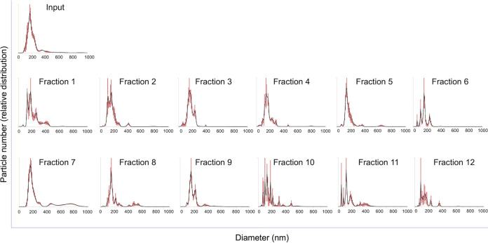
Extracellular vesicles (EVs) produced by members of the Cryptococcus genus are associated with fundamental processes of fungal physiology and virulence. However, several questions about the properties of cryptococcal EVs remain unanswered, mostly because of technical limitations. We recently described a fast and efficient protocol of high-yield EV isolation from solid medium. In this study, we aimed at using the solid medium protocol to address some of the open questions about EVs, including the kinetics of EV production, the diversity of EVs produced by multiple isolates under different culture conditions, the separation of vesicles in a density gradient followed by the recovery of functional EVs, the direct detection of EVs in culture supernatants, and the production of vesicles in solid cultures of Titan cells. Our results indicate that the production of EVs is directly impacted by the culture medium and time of growth, resulting in variable detection of EVs per cell and a peak of EV detection at 24 h of growth. Nanoparticle tracking analysis (NTA) of EV samples revealed that multiple isolates produce vesicles with variable properties, including particles of diverging dimensions. EVs were produced in the solid medium in amounts that were separated on a centrifugation density gradient, resulting in the recovery of functional EVs containing the major cryptococcal capsular antigen. We also optimized the solid medium protocol for induction of the formation of Titan cells, and analyzed the production of EVs by NTA and transmission electron microscopy. This analysis confirmed that EVs were isolated from solid cultures of cryptococcal enlarged cells. With these approaches, we expect to implement simple methods that will facilitate the analysis of EVs produced by fungal cells. IMPORTANCE Fungal extracellular vesicles (EVs) are considered to be important players in the biology of fungal pathogens. However, the limitations in the methodological approaches to studying fungal EVs impair the expansion of knowledge in this field. In the present study, we used the Cryptococcus genus as a model for the study of EVs. We explored the simplification of protocols for EV analysis, which helped us to address some important, but still unanswered, questions about fungal EVs.

Keywords: Cryptococcus; extracellular vesicles; pathogenesis.


Figures

FIG 1
FIG 1
Diversity of EVs in the Cryptococcus genus. (A and B) Growth rates of each of the isolates (see Materials and Methods for strain identification) were determined after cultivation for 12 (A) or 24 (B) h. Although the growth rates were not identical, they were statistically similar (P > 0.05, ANOVA). (C) NTA of EVs produced by the multiple isolates of Cryptococcus. EVs were isolated from each isolate after growth in solid medium and observed by NTA. Although many isolates produced samples with similar properties, some of the C. neoformans strains produced EV populations enriched in larger diameter ranges. Peak areas in each NTA histogram were determined using the Image J software. In each sample, the sum of the areas of all peaks corresponded to 100%, and the percentage values described in green correspond to the relative area of each peak in individual samples. These results illustrate one out of three experiments producing similar results.
FIG 2
FIG 2
Separation of C. deuterogattii EVs on a density gradient. Fungal cells (R265 strain) were cultivated on solid medium for EV isolation. The EVs were applied to the top of an iodixanol gradient and ultracentrifuged for 18 h. The highest fraction numbers represent the densest fractions. Each of 12 fractions was submitted to NTA. Fraction 7 was the one containing the largest population of EVs of higher diameters. This experiment was repeated three times producing similar results.
FIG 3
FIG 3
Quantification of the different types of EVs isolated from solid medium using the Image J software. (A) Quantification of EVs in the ranges of 300 to 400 nm (4%), 400 to 600 nm (3.6%), and 600 to 900 nm combining all samples, with a clear predomination of the 0 to 300 diameter range. (B) The analysis of each diameter range is demonstrated as follows: top left, 0 to 300 nm diameter range; top right, 300 to 400 nm range; bottom left, 400 to 600 nm range; and bottom right, 600 to 900 nm size range. Larger diameters predominated in fractions 7 and 8. The quantification results illustrated here derive from the histograms presented in Fig. 2.
FIG 4
FIG 4
Analysis of particle number (A), RNA detection (B), GXM quantification (C), and the use of gradient fractions by an acapsular mutant of C. neoformans to make a GXM coat (D). (A) The highest particle number was detected in fraction 7. Particle numbers lower that 1 × 108/ml (dashed line) were considered to be at the background levels of EV detection. (B) RNA concentration was normalized in each fraction to the number of particles. The highest concentration of RNA above the background level of EV detection was found in fraction 8. (C) GXM was highly concentrated in fraction 7, even after normalization of the GXM content to the number of particles. (D) Incorporation of GXM from EV fractions by an acapsular mutant of C. neoformans followed by indirect quantification of GXM coating by flow cytometry. Once again, fraction 7 gave the most positive signals of GXM detection. These results illustrate one out of two experiments producing similar results.
FIG 5
FIG 5
Capsule induction and detection of EVs in culture supernatants. (A) Capsule counterstaining of C. deuterogattii (R265) in different culture media. Scale bar, 10 μm. (B) Direct detection of EVs in the supernatants obtained from the culture conditions in A by NTA. In B, the upper panel shows the general NTA aspects of cryptococcal EVs. The lower panel represents the number of EVs detected to the number of cells in the original culture. These results illustrate one out of three experiments producing similar results.
FIG 6
FIG 6
Kinetics of EV formation in solid YPD. (A) Detection of EVs at 12, 18, and 24 h postinoculation. The peak of EV detection (normalized to the cell number) was observed at 24 h of growth. (B) Individual NTA profiles of the samples described in A, revealing that the size of the EVs varied with the time of cultivation. (C) Quantification of smaller (0 to 200 nm) and larger (>200 nm) particles in the samples illustrated in B, using the Image J software. Shorter cultivation times were associated with the increased detection of larger EVs, while the observation of smaller EVs increased in response to the time of growth.
FIG 7
FIG 7
Quantification of EVs produced by isolates of C. gattii (orange diamonds) or C. deuterogattii (blue diamonds) in solid YPD. EV detection varied from less than 1 to more than 20 particles per cell, with average values represented by the red dashes. Data in this figure originated from duplicates or triplicates, with individual values represented by the diamonds.
FIG 8
FIG 8
Analysis of Titan cells cultivated in solid medium. C. neoformans (H99) was inoculated in solid Sabouraud, TCM agar, or TCM broth at 5 × 106 (A and C) or 107 cells per plate (solid medium) or tube (TCM broth, 104 cells/ml in 96-well plates) (B and D). The cells were incubated for 18 or 48 h for measurement of cell body size, as illustrated in panels A and B. The visual analysis of each condition is shown in panels C and D. (E and F) Incubation of an inoculum of 107 cells for 18 h in TCM agar was selected as the condition for EV isolation and analysis by NTA (E) and TEM (F). These results illustrate one out of three experiments producing similar results.

References

    1. Rodrigues ML, Nimrichter L, Oliveira DL, Frases S, Miranda K, Zaragoza O, Alvarez M, Nakouzi A, Feldmesser M, Casadevall A. 2007. Vesicular polysaccharide export in Cryptococcus neoformans is a eukaryotic solution to the problem of fungal trans-cell wall transport. Eukaryot Cell 6:48–59. doi: 10.1128/EC.00318-06. - DOI - PMC - PubMed
    1. Reis F, Borges BS, Jozefowicz L, Sena B, Garcia A, Medeiros LC, Martins S, Honorato L, Schrank A, Vainstein MHMH, Kmetzsch L, Nimrichter L, Alves L, Staats CCCC, Rodrigues MLML. 2019. A novel protocol for the isolation of fungal extracellular vesicles reveals the participation of a putative scramblase in polysaccharide export and capsule construction in Cryptococcus gattii. mSphere 4:e00080-19. doi: 10.1128/mSphere.00080-19. - DOI - PMC - PubMed
    1. Wolf JM, Espadas-Moreno J, Luque-Garcia JL, Casadevall A. 2014. Interaction of cryptococcus neoformans extracellular vesicles with the cell wall. Eukaryot Cell 13:1484–1493. doi: 10.1128/EC.00111-14. - DOI - PMC - PubMed
    1. Bielska E, Sisquella MA, Aldeieg M, Birch C, O'Donoghue EJ, May RC. 2018. Pathogen-derived extracellular vesicles mediate virulence in the fatal human pathogen Cryptococcus gattii. Nat Commun 9:1556. doi: 10.1038/s41467-018-03991-6. - DOI - PMC - PubMed
    1. Rodrigues ML, Nakayasu ES, Oliveira DL, Nimrichter L, Nosanchuk JD, Almeida IC, Casadevall A. 2008. Extracellular vesicles produced by Cryptococcus neoformans contain protein components associated with virulence. Eukaryot Cell 7:58–67. doi: 10.1128/EC.00370-07. - DOI - PMC - PubMed

Publication types

MeSH terms

LinkOut - more resources