. 2022 Jun;142(6):1147-1154.
doi: 10.1007/s00402-021-04009-5. Epub 2021 Aug 4.

Multiple iliopsoas tendons: a cadaveric study and treatment implications for internal snapping hip syndrome

Affiliations

Multiple iliopsoas tendons: a cadaveric study and treatment implications for internal snapping hip syndrome

Benjamin Lin et al. Arch Orthop Trauma Surg. 2022 Jun.

Abstract

Purpose: This cadaveric study aimed at describing the anatomical variations of the iliopsoas complex.

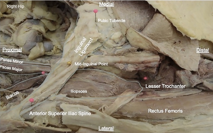
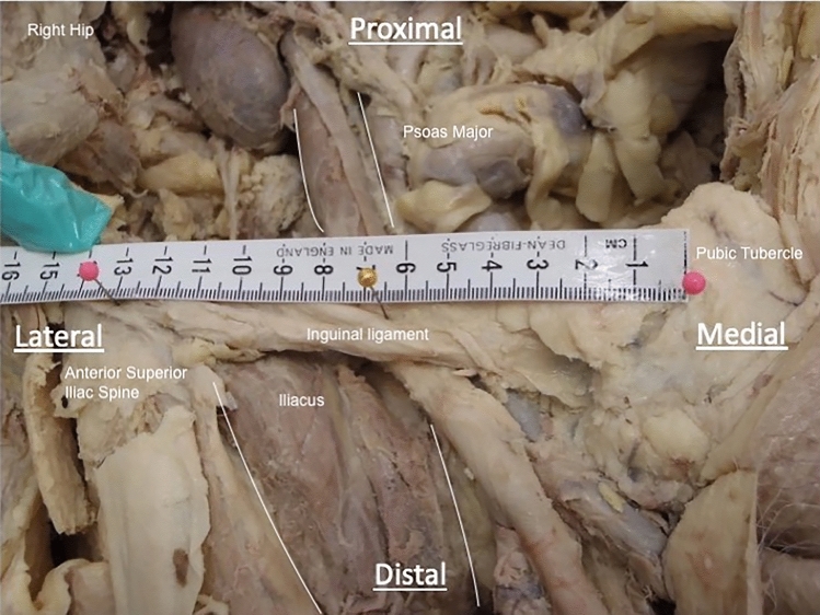
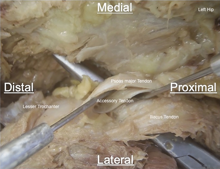
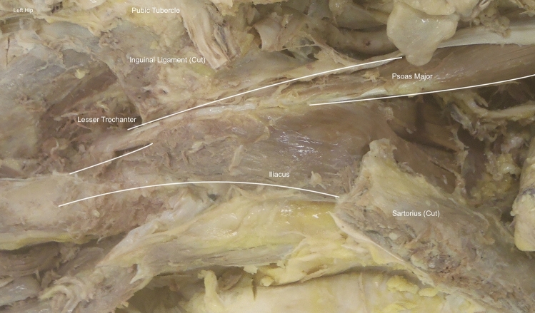
Methods: The iliopsoas complex was dissected unilaterally in 28 formalin-embalmed cadavers-13 males and 15 females with a mean age of 85.6 years. The number, courses and widths of the iliacus and psoas major tendons were determined. Patients with previous hip surgery were excluded. The following measurements were taken from the mid-inguinal point: the distance to the point of union of the psoas major and iliacus tendon; and the distance to the most distal insertion of iliopsoas.

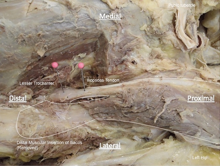
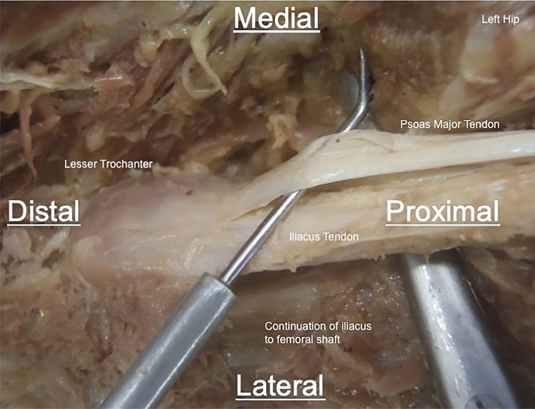
Results: The presence of single, double and triple tendon insertions of iliopsoas were found in 12, 12 and 4 of the 28 specimens, respectively. When present, double and triple tendons inserted separately onto the lesser trochanter. The average length of the iliopsoas tendon from the mid-inguinal point to the most distal attachment at the lesser trochanter was 122.3 ± 13.0 mm. The iliacus muscle bulk merged with psoas major at an average distance of 24.9 ± 17.9 mm proximal to the mid-inguinal point. In all cases, the lateral-most fibres of iliacus yielded a non-tendinous, muscular insertion on to the anterior surface of the lesser trochanter and the femoral shaft, rather than joining onto the main iliopsoas tendon(s). The average total width of the psoas major tendon decreased with an increasing number of tendons: 14.6 ± 2.2 mm (single tendon), 8.2 ± 3.0 mm (2 tendons present) and 5.9 ± 1.1 mm (3 tendons present) (P < 0.001).

Conclusions: The results of this study suggest that multiple tendinous insertions of iliopsoas are present as an anatomical variant in more than 50% of the population. The non-tendinous muscular insertion of the iliopsoas on to the anterior surface of the lesser trochanter and femoral shaft found represents a novel anatomical variant not previously described.

Level of evidence: Level V.

Keywords: Anatomical variation; Hip arthroscopy; Iliopsoas; Iliopsoas tenotomy; Internal snapping hip syndrome.


Conflict of interest statement

The authors report they have no conflict of interests to declare.

Figures

Fig. 1
Fig. 1
Example of iliopsoas dissection. A labelled example dissection of the left iliopsoas complex from the abdomen (proximal, left) through to the insertion of the lesser trochanter (distal, right). Viewed from anterior aspect in coronal plane
Fig. 2
Fig. 2
Example of mid-inguinal point dissection. The location of the right mid-inguinal point (central pin) measured halfway between the anterior superior iliac spine (left pin) and the pubic tubercle (right pin) along the preserved inguinal ligament. Viewed from anterior aspect in coronal plane, perpendicular to body axis
Fig. 3
Fig. 3
Example of point at which iliopsoas tendons were cut. A dissection of the left iliopsoas complex to demonstrate the point at which the iliopsoas tendon was cut (represented by the pin on the right). This was 2 cm proximal to the distal-most insertion of the iliopsoas complex. The location of the non-tendinous, muscular insertion has been highlighted—this was removed in this specimen to enable measurement of the iliopsoas tendon, though the remanent muscle fibres can be seen. Viewed from anterior aspect in coronal plane, perpendicular to body axis
Fig. 4
Fig. 4
Example of dissection of a double iliopsoas tendon variant. A dissection of the left iliopsoas demonstrating a double tendinous insertion of iliopsoas onto the lesser trochanter. Viewed from anterior aspect in coronal plane, perpendicular to body axis
Fig. 5
Fig. 5
Example of dissection of a triple iliopsoas tendon variant. A dissection of the left iliopsoas demonstrating a triple tendinous insertion of iliopsoas onto the lesser trochanter. The medial most tendon originated from the body of psoas, whilst the remaining two originated from iliacus. Viewed from anterior aspect in coronal plane, perpendicular to body axis
Fig. 6
Fig. 6
Example of dissection of accessory anterior insertion of iliacus. A dissection of the left hip to demonstrate the non-tendinous muscular insertion of iliacus onto the femoral shaft (highlighted). Some of the muscle fibres have been removed to allow access to the iliopsoas tendons. Viewed from anterior aspect in coronal plane, perpendicular to body axis

References

    1. Piechota M, Maczuch J, Skupiński J, et al. Internal snapping hip syndrome in dynamic ultrasonography. J Ultrason. 2016;16:296–303. doi: 10.15557/JoU.2016.0030. - DOI - PMC - PubMed
    1. Shibata KR, Matsuda S, Safran MR. Is there a distinct pattern to the acetabular labrum and articular cartilage damage in the non-dysplastic hip with instability? Knee Surg Sports Traumatol Arthrosc. 2017;25:84–93. doi: 10.1007/s00167-016-4342-4. - DOI - PubMed
    1. Harris JD. Editorial commentary: caveat flexor—to release or not to release the iliopsoas, that is the question. Arthroscopy. 2018;34:1851–1855. doi: 10.1016/j.arthro.2018.04.010. - DOI - PubMed
    1. Audenaert EA, Khanduja V, Claes P, Malviya A, Steenackers G (2020) Mechanics of psoas tendon snapping. A virtual population study. Front Bioeng Biotechnol 8:264. 10.3389/fbioe.2020.00264 - PMC - PubMed
    1. Brandenburg JB, Kapron AL, Wylie JD, et al. The functional and structural outcomes of arthroscopic iliopsoas release. Am J Sports Med. 2016;44:1286–1291. doi: 10.1177/0363546515626173. - DOI - PubMed