. 2022 Feb;74(2):284-294.
doi: 10.1002/art.41937. Epub 2021 Dec 28.

Evaluation of Immune Response and Disease Status in Systemic Lupus Erythematosus Patients Following SARS-CoV-2 Vaccination

Affiliations

Evaluation of Immune Response and Disease Status in Systemic Lupus Erythematosus Patients Following SARS-CoV-2 Vaccination

Peter M Izmirly et al. Arthritis Rheumatol. 2022 Feb.

Abstract

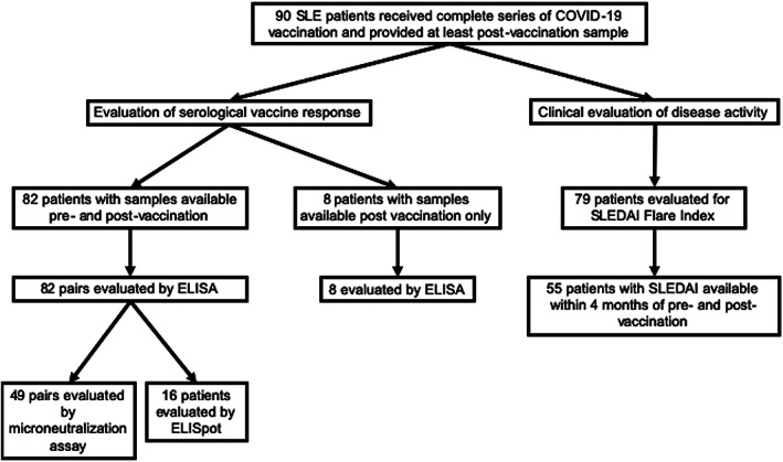
Objective: To evaluate seroreactivity and disease flares after COVID-19 vaccination in a multiethnic/multiracial cohort of patients with systemic lupus erythematosus (SLE).

Methods: Ninety SLE patients and 20 healthy controls receiving a complete COVID-19 vaccine regimen were included. IgG seroreactivity to the SARS-CoV-2 spike receptor-binding domain (RBD) and SARS-CoV-2 microneutralization were used to evaluate B cell responses; interferon-γ (IFNγ) production was measured by enzyme-linked immunospot (ELISpot) assay in order to assess T cell responses. Disease activity was measured by the hybrid SLE Disease Activity Index (SLEDAI), and flares were identified according to the Safety of Estrogens in Lupus Erythematosus National Assessment-SLEDAI flare index.

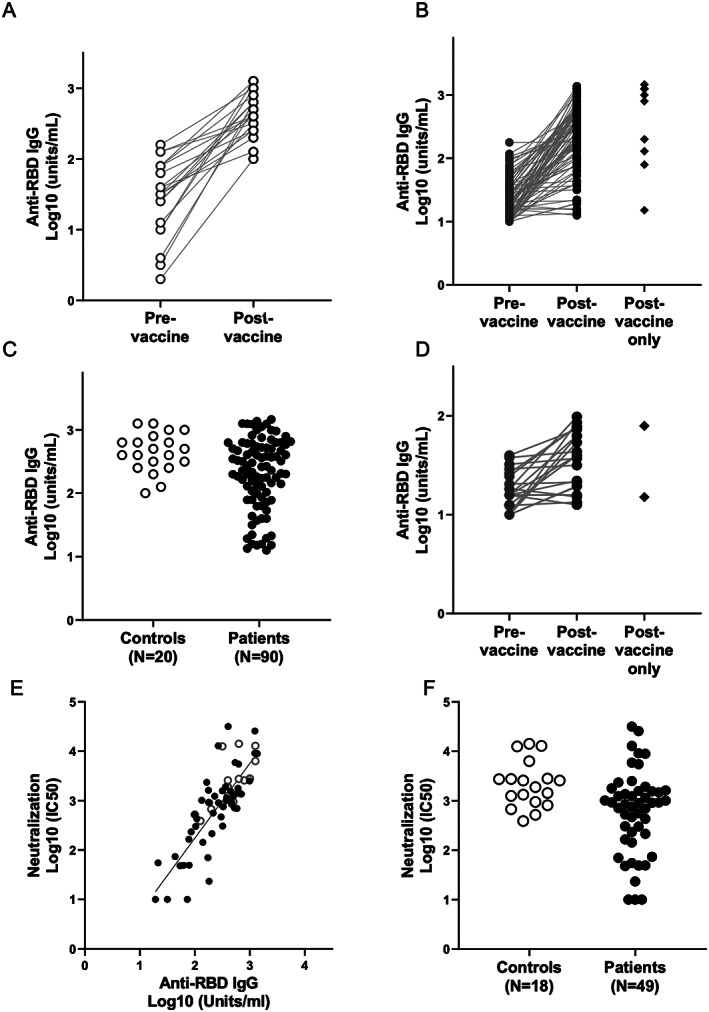
Results: Overall, fully vaccinated SLE patients produced significantly lower IgG antibodies against SARS-CoV-2 spike RBD compared to fully vaccinated controls. Twenty-six SLE patients (28.8%) generated an IgG response below that of the lowest control (<100 units/ml). In logistic regression analyses, the use of any immunosuppressant or prednisone and a normal anti-double-stranded DNA antibody level prior to vaccination were associated with decreased vaccine responses. IgG seroreactivity to the SARS-CoV-2 spike RBD strongly correlated with the SARS-CoV-2 microneutralization titers and correlated with antigen-specific IFNγ production determined by ELISpot. In a subset of patients with poor antibody responses, IFNγ production was similarly diminished. Pre- and postvaccination SLEDAI scores were similar in both groups. Postvaccination flares occurred in 11.4% of patients; 1.3% of these were severe.

Conclusion: In a multiethnic/multiracial study of SLE patients, 29% had a low response to the COVID-19 vaccine which was associated with receiving immunosuppressive therapy. Reassuringly, severe disease flares were rare. While minimal protective levels remain unknown, these data suggest that protocol development is needed to assess the efficacy of booster vaccination.


Figures

Figure 1
Figure 1
Flow diagram of the systemic lupus erythematosus (SLE) patients included in each analysis. ELISA = enzyme‐linked immunosorbent assay; ELISpot = enzyme‐linked immunospot; SLEDAI = SLE Disease Activity Index.
Figure 2
Figure 2
Antibody response to COVID‐19 vaccine in SLE patients. Throughout, open circles represent controls, and solid circles represent SLE patients. Serum IgG titers against the SARS–CoV‐2 spike protein receptor‐binding domain (RBD) were obtained by direct ELISA. Binding of human IgG to recombinant SARS–CoV‐2 RBD was performed as described in Patients and Methods. A, IgG titers of 20 controls pre‐ and postvaccination. B, IgG titers of 81 SLE patients pre‐ and postvaccination and 9 SLE patients with only postvaccination data available. C, Comparison of IgG titers postvaccination between the 2 groups, with controls showing significantly higher titers than SLE patients. D, IgG titers of a subgroup of patients (n = 24) from B with anti‐RBD IgG ≤100 units/ml (the lowest response in controls). E, Correlation of SARS–CoV‐2 virus neutralization titers in the sera of vaccinated subjects with IgG titers. Each dot represents the serum evaluation of a participant with binding of human IgG to recombinant SARS–CoV‐2 RBD (x‐axis) versus live virus neutralization at 50% inhibition concentration (IC50; y‐axis). There is a strong correlation between spike RBD IgG and live virus neutralization (R = 0.76, P < 0.0001). F, Log10 postvaccination neutralization titers for controls compared to SLE patients. See Figure 1 for other definitions.

References

    1. Fernandez‐Ruiz R, Masson M, Kim MY, Myers B, Haberman RH, Castillo R, et al. Leveraging the United States epicenter to provide insights on COVID‐19 in patients with systemic lupus erythematosus. Arthritis Rheumatol 2020;72:1971–80. - PMC - PubMed
    1. Fernandez‐Ruiz R, Paredes JL, Niewold TB. COVID‐19 in patients with systemic lupus erythematosus: lessons learned from the inflammatory disease [review]. Transl Res 2021;232:13–36. - PMC - PubMed
    1. Strangfeld A, Schäfer M, Gianfrancesco MA, Lawson‐Tovey S, Liew JW, Ljung L, et al. Factors associated with COVID‐19‐related death in people with rheumatic diseases: results from the COVID‐19 Global Rheumatology Alliance physician‐reported registry. Ann Rheum Dis 2021;80:930–42. - PMC - PubMed
    1. Gianfrancesco M, Hyrich KL, Al‐Adely S, Carmona L, Danila MI, Gossec L, et al. Characteristics associated with hospitalisation for COVID‐19 in people with rheumatic disease: data from the COVID‐19 Global Rheumatology Alliance physician‐reported registry. Ann Rheum Dis 2020;79:859–66. - PMC - PubMed
    1. Tang W, Askanase AD, Khalili L, Merrill JT. SARS–CoV‐2 vaccines in patients with SLE. Lupus Sci Med 2021;8:e000479. - PMC - PubMed

MeSH terms