. 2021 Aug 19;385(8):707-719.
doi: 10.1056/NEJMoa2028973. Epub 2021 Aug 4.

Variant PNLDC1, Defective piRNA Processing, and Azoospermia

Affiliations

Variant PNLDC1, Defective piRNA Processing, and Azoospermia

Liina Nagirnaja et al. N Engl J Med. .

Abstract

Background: P-element-induced wimpy testis (PIWI)-interacting RNAs (piRNAs) are short (21 to 35 nucleotides in length) and noncoding and are found almost exclusively in germ cells, where they regulate aberrant expression of transposable elements and postmeiotic gene expression. Critical to the processing of piRNAs is the protein poly(A)-specific RNase-like domain containing 1 (PNLDC1), which trims their 3' ends and, when disrupted in mice, causes azoospermia and male infertility.

Methods: We performed exome sequencing on DNA samples from 924 men who had received a diagnosis of nonobstructive azoospermia. Testicular-biopsy samples were analyzed by means of histologic and immunohistochemical tests, in situ hybridization, reverse-transcriptase-quantitative-polymerase-chain-reaction assay, and small-RNA sequencing.

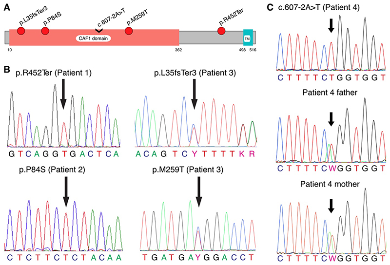
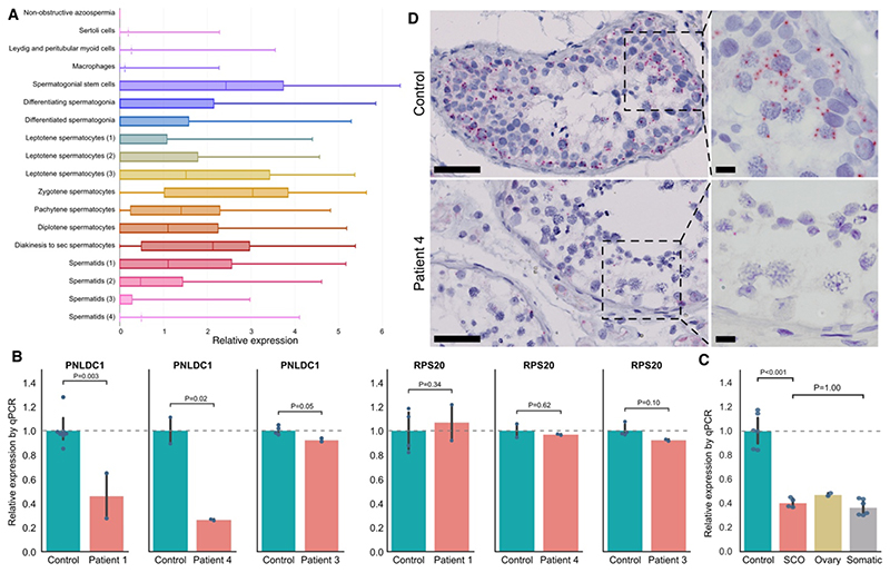
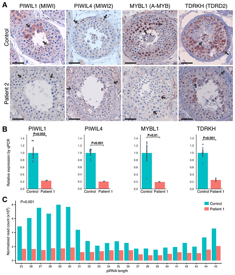
Results: Four unrelated men of Middle Eastern descent who had nonobstructive azoospermia were found to carry mutations in PNLDC1: the first patient had a biallelic stop-gain mutation, p.R452Ter (rs200629089; minor allele frequency, 0.00004); the second, a novel biallelic missense variant, p.P84S; the third, two compound heterozygous mutations consisting of p.M259T (rs141903829; minor allele frequency, 0.0007) and p.L35PfsTer3 (rs754159168; minor allele frequency, 0.00004); and the fourth, a novel biallelic canonical splice acceptor site variant, c.607-2A→T. Testicular histologic findings consistently showed error-prone meiosis and spermatogenic arrest with round spermatids of type Sa as the most advanced population of germ cells. Gene and protein expression of PNLDC1, as well as the piRNA-processing proteins PIWIL1, PIWIL4, MYBL1, and TDRKH, were greatly diminished in cells of the testes. Furthermore, the length distribution of piRNAs and the number of pachytene piRNAs was significantly altered in men carrying PNLDC1 mutations.

Conclusions: Our results suggest a direct mechanistic effect of faulty piRNA processing on meiosis and spermatogenesis in men, ultimately leading to male infertility. (Funded by Innovation Fund Denmark and others.).


Figures

Figure 1
Figure 1. Validation of the Pathogenic PNLDC1 Mutations by Sanger Sequencing.
Panel A shows a schematic diagram of PNLDC1 protein domains, depicted on the basis of the Pfam database (version 32.0), with the locations of the identified mutations. The CAF1 family RNase domain (PFAM:PF04857), common among messenger RNA deadenylases, is thought to contain the RNase activity of PNLDC1. TM denotes potential transmembrane domain. Panel B shows the identified mutations that were validated by means of Sanger sequencing. Homozygous genotypes were confirmed in the case of p.R452Ter (c.1354C→T) in Patient 1 and in the case of p.P84S (c.250C→T) in Patient 2; the compound variants p.L35fsTer3 (c.103dup) and p.M259T (c.776T→C) in Patient 3 were confirmed as being heterozygous. As shown in Panel C, Patient 4 was validated as a homozygous carrier of c.607-2A→T, whereas both of his parents were observed as being heterozygous for the change. In all the panels, the locations of the mutations are indicated by arrows.
Figure 2
Figure 2. Testicular Histologic Findings in Men with PNLDC1 Mutations.
Panel A shows a hematoxylin and eosin stain of a control testicular tissue sample in which seminiferous tubules had complete spermatogenesis. Asterisks indicate the seminiferous tubules with complete spermatogenesis. Panel B shows a hematoxylin and eosin stain of a testicular tissue sample from Patient 2. Hashtags indicate extensive arrest at the late pachytene stage in the tubules. Panels C and D show the sample from Patient 2 at higher magnifications. In Panel C, the arrow indicates round spermatids of stage Sa and the arrowheads pyknotic postmeiotic cells. In Panel D, the arrowhead indicates a spermatogonium that appears abnormal, and the arrow, arrest at diakinesis. The bars in Panels A through D represent 20 μm. Panels E through J also show the sample from Patient 2 at higher magnifications. The arrow in Panel E indicates an abnormal spermatogonium with enlarged nucleus but no signs of malignant change; in Panel F, spermatogonium in mitosis; in panel G, a meiotic spermatocyte that appears pyknotic; and in Panel H, a spermatocyte at diakinesis, the last stage of the meiotic prophase 1. The dashed outline in Panel I defines an area of pyknotic multinucleated cells; and in Panel J, an area of round spermatids of type Sa. The bars in Panels E through J represent 10 μm.
Figure 3
Figure 3. Expression of PNLDC1.
Panel A shows the testicular cell types that express PNLDC1, as determined by interrogation of single-cell RNA sequencing on adult testis. In the horizontal box plots, the vertical solid lines are the medians and the left and right sides of the boxes are the interquartile ranges of the normalized read counts for each cell type. The I bars indicate the minimum and maximum counts, and when the third quartile is zero, the mean is indicated with a dotted line. Each cell type identified by the analysis is marked with a different color, and the number in brackets indicates sequential developmental stages of a given cell type. Panel B shows an in situ hybridization analysis performed with probes targeting PNLDC1 in a testicular-biopsy sample from a control with complete spermatogenesis (top) and a testicular-biopsy sample from Patient 4 (bottom). PNLDC1 shows high expression in pachytene spermatocytes (each red dot represents a single transcript) in the control but is nearly absent in Patient 4. The bars on the left images represent 50 μm, and on the right images 10 μm. Panel C shows a reverse-transcriptase–quantitative-polymerase-chain-reaction (RT-qPCR) analysis performed with primers targeting PNLDC1 on RNA isolated from fixed testis samples with complete spermatogenesis from controls (Table S3 in the Supplementary Appendix) and testicular RNA isolated from Patients 1, 3, and 4 (RNA was not available from Patient 2). The analysis was performed in duplicate. An RT-qPCR analysis of the housekeeping gene RPS20 showed nonsignificant differences in expression between the patients and controls, a finding that indicates that the RNA quality was not compromised. Panel D shows an RT-qPCR analysis performed with primers targeting PNLDC1 on three samples from controls with complete spermatogenesis, two samples of tissue devoid of germ cells (Sertoli cell–only pattern [SCO]), an ovary tissue sample, and different somatic tissue samples (liver, skin, and ductus deferens); the findings indicate that PNLDC1 expression is specific to germ cells. In Panels C and D, the data are presented as relative to the controls, as indicated by the dashed line. Each blue dot represents a measurement (performed in duplicate per sample), and the vertical bars indicate the 95% confidence intervals, which were calculated by means of bootstrapping. NS denotes not significant.
Figure 4
Figure 4. Expression of Key piRNA-Processing Enzymes at Protein and Transcript Levels and Small-RNA Sequencing.
Panel A shows immunohistochemical staining for P-element–induced wimpy testis–interacting RNA (piRNA)–processing proteins PIWIL1, PIWIL4, MYBL1, and TDRKH in a control biopsy sample with complete spermatogenesis and in a sample from Patient 2, who carried the biallelic missense mutation p.P84S in PNLDC1. Arrows indicate the main sites of expression. In the control, we observed expression of PIWIL1 in pachytene spermatocytes, PIWIL4 in spermatogonia, and MYBL1 in spermatocytes, which in all cases were lost or greatly diminished in the patient. TDRKH was observed in the cytoplasm of spermatogonia and spermatocytes and appeared to be less expressed in Patient 2. The expression patterns were confirmed across five controls and also in Patient 1 for PIWIL1 and PIWIL4 and in Patient 4 for PIWIL1, MYBL1, and TDRKH (Fig. S9). A testicular tissue sample from Patient 3 was unavailable for staining. The bars represent 50 μm. Panel B shows that with the use of RNA isolated from a fixed testicular tissue sample from Patient 1, the transcript levels of piRNA- processing enzymes recapitulated the pattern that was observed at the protein level. A significant reduction in transcript levels was observed for PIWIL1, PIWIL4, MYBL1, and TDRKH. The RNA from four controls with complete spermatogenesis was also obtained from fixed testicular tissue and had a similar RNA quality as in Patient 1. The analysis was performed in duplicate. A similar pattern was observed in a sample from Patient 4, but a less pronounced pattern was observed in a sample from Patient 3 (Fig. S10A and S10B). Expression of the housekeeping transcript RPS20 was similar between the patients and their matched controls (Fig. 3C). As shown in Panel C, small-RNA sequencing of RNA isolated from fixed testicular tissue sample from Patient 1 revealed a significantly lower amount of piRNAs than that in the sample from a control with complete spermatogenesis (P<0.001), as well as a major loss of piRNAs with expected lengths of 26 to 31 bases, which were evident in the control samples. Read counts of piRNAs were normalized to the spike-in RNA. Control RNA was isolated from tissue preparations that were similar to those used for the patients. Data from Patient 4 that show a similar significant reduction and from Patient 3 that show a milder effect on piRNA biogenesis are provided in Fig. S10C and S10D.

Comment in

References

    1. Barratt CLR, Björndahl L, De Jonge CJ, et al. The diagnosis of male infertility: an analysis of the evidence to support the development of global WHO guidance-challenges and future research opportunities. Hum Reprod Update. 2017;23:660–80. - PMC - PubMed
    1. Kasak L, Laan M. Monogenic causes of non-obstructive azoospermia: challenges, established knowledge, limitations and perspectives. Hum Genet. 2021;140:135–54. - PubMed
    1. Özata DM, Yu T, Mou H, et al. Evolutionarily conserved pachytene piRNA loci are highly divergent among modern humans. Nat Ecol Evol. 2020;4:156–68. - PMC - PubMed
    1. Dai P, Wang X, Gou L-T, et al. A translation-activating function of MIWI/piRNA during mouse spermiogenesis. Cell. 2019;179(7):1566–1581.:e16. - PMC - PubMed
    1. Gou L-T, Dai P, Yang J-H, et al. Pachytene piRNAs instruct massive mRNA elimination during late spermiogenesis. Cell Res. 2014;24:680–700. - PMC - PubMed

Publication types