. 2021 Aug 3;36(5):109495.
doi: 10.1016/j.celrep.2021.109495.

Severe deficiency of the voltage-gated sodium channel NaV1.2 elevates neuronal excitability in adult mice

Affiliations

Severe deficiency of the voltage-gated sodium channel NaV1.2 elevates neuronal excitability in adult mice

Jingliang Zhang et al. Cell Rep. .

Abstract

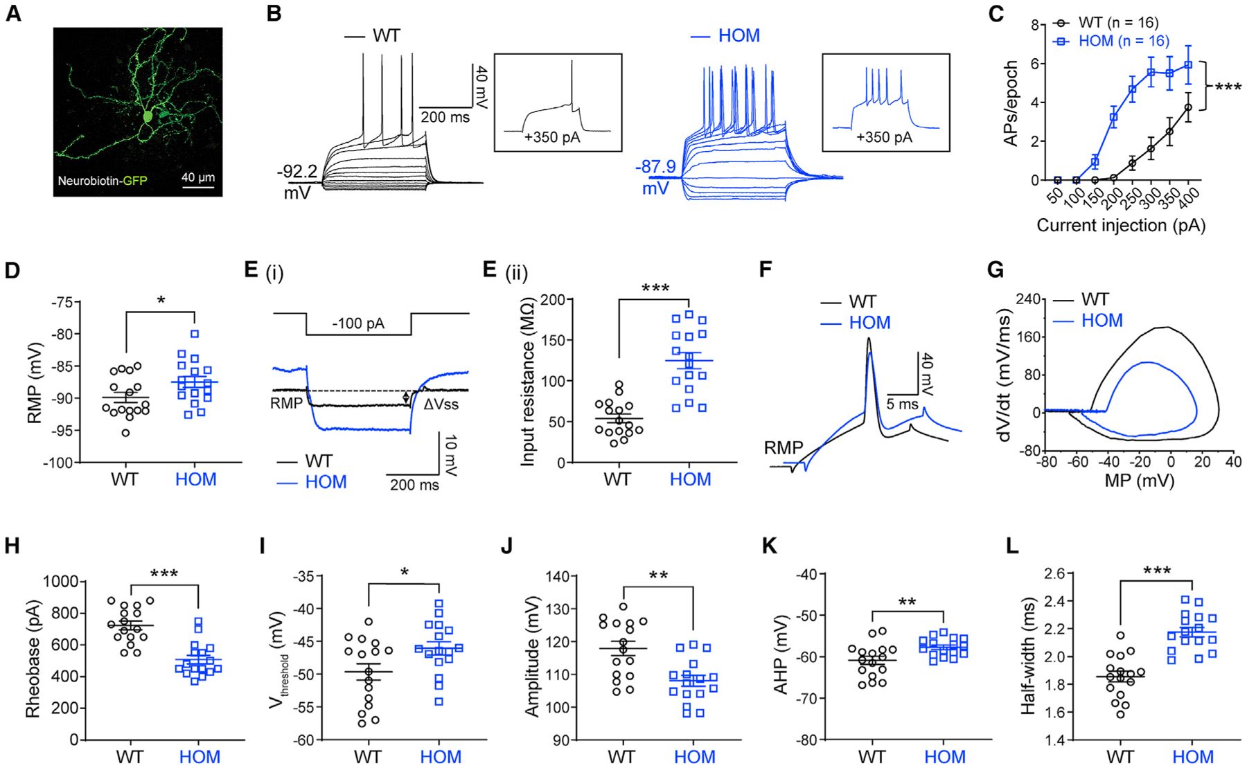
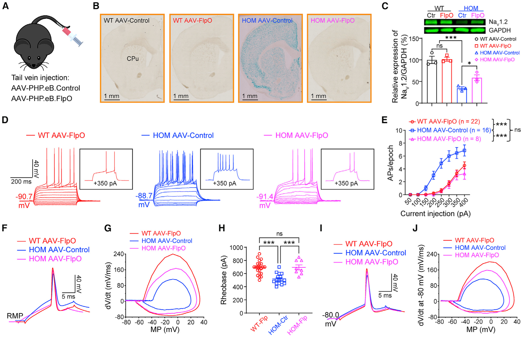
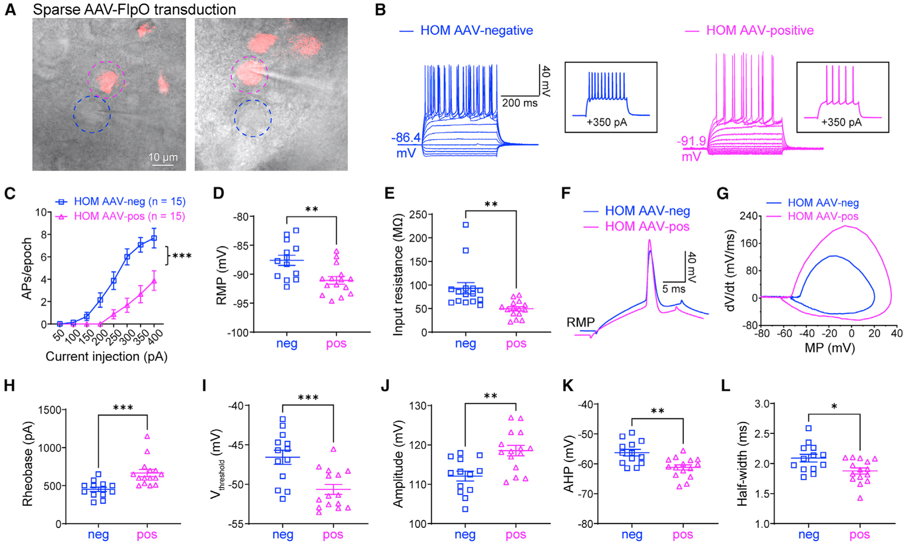
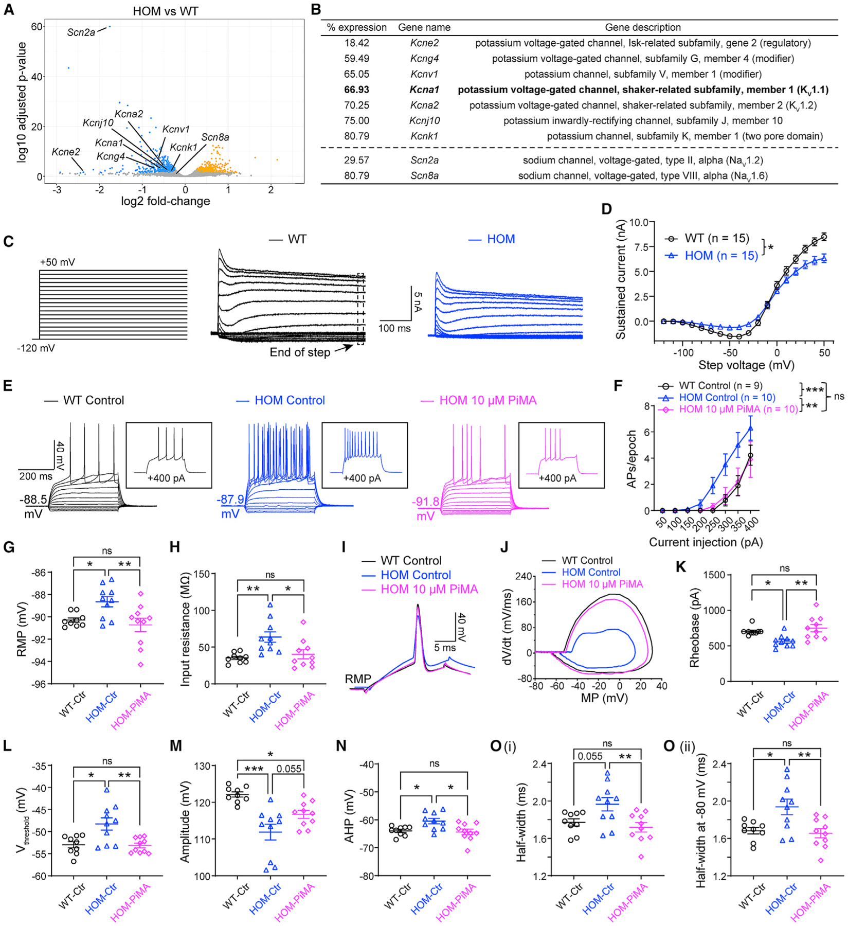
Scn2a encodes the voltage-gated sodium channel NaV1.2, a main mediator of neuronal action potential firing. The current paradigm suggests that NaV1.2 gain-of-function variants enhance neuronal excitability, resulting in epilepsy, whereas NaV1.2 deficiency impairs neuronal excitability, contributing to autism. However, this paradigm does not explain why ∼20%-30% of individuals with NaV1.2 deficiency still develop seizures. Here, we report the counterintuitive finding that severe NaV1.2 deficiency results in increased neuronal excitability. Using a NaV1.2-deficient mouse model, we show enhanced intrinsic excitability of principal neurons in the prefrontal cortex and striatum, brain regions known to be involved in Scn2a-related seizures. This increased excitability is autonomous and reversible by genetic restoration of Scn2a expression in adult mice. RNA sequencing reveals downregulation of multiple potassium channels, including KV1.1. Correspondingly, KV channel openers alleviate the hyperexcitability of NaV1.2-deficient neurons. This unexpected neuronal hyperexcitability may serve as a cellular basis underlying NaV1.2 deficiency-related seizures.

Keywords: K(V)1.1; Na(V)1.2; SCN2A/Scn2a; epilepsy; gene trap; neuronal excitability; potassium channel; voltage-gated sodium channel.


Conflict of interest statement

Declaration of interests The authors declare no competing interests.

Figures

Figure 1.
Figure 1.. Elevated neuronal firing of striatal medium spiny neurons (MSNs) in adult NaV1.2-deficient mice
(A) A typical MSN labeled by neurobiotin. Scale bar, 40 μm. (B) Representative current-clamp recordings of MSNs from wild-type (WT, black) and homozygous (HOM) Scn2agt/gt (blue) mice were obtained at the RMP. Inset: representative trace in response to +350 pA injection. (C) The average number of action potentials (APs) generated in response to depolarizing current pulses. Unpaired two-tailed non-parametric Mann-Whitney U test for each current pulse. (D) Individuals and mean RMP values. Unpaired t test. (Ei) Representative traces in response to −100 pA injection. Vsteady-state (Vss) is the voltage recorded 0–10 ms before the end of the stimulus. (Eii) Individuals and mean input resistance values at the RMP. Unpaired t test. (F) Typical spikes of MSNs from WT (black) and HOM (blue) mice were obtained at the normal RMP. (G) Associated phase-plane plots. (H–L) Individual and mean spike rheobase, voltage threshold, amplitude, fast after-hyperpolarization (AHP), and half-width values. Unpaired t test. Data are shown as mean ± SEM.
Figure 2.
Figure 2.. Elevated neuronal firing is reversible by FlpO-mediated restoration of NaV1.2 expression in adult NaV1.2-deficient mice
(A) Cartoon illustration of mice systemically administered PHP.eB.AAV-control or PHP.eB.AAV-FlpO via tail vein injection. (B) Coronal views of LacZ staining of the striatum from WT and Scn2agt/gt (HOM) mice injected with AAV-control or AAV-FlpO. Blue staining of HOM mice largely disappeared in the AAV-FlpO group. CPu, caudate nucleus and the putamen (dorsal striatum). Scale bar, 1 mm. (C) Western blot analysis showing NaV1.2 protein levels in whole-brain homogenates from HOM mice in the AAV-control or AAV-FlpO group. One-way ANOVA with multiple comparisons. (D) Representative current-clamp recordings of MSNs from WT mice transduced with AAV-FlpO (red), HOM mice transduced with AAV-Control (blue), and HOM mice transduced with AAV-Control (magenta) obtained at the RMP. Inset: representative trace in response to +350 pA injection. (E) The average number of APs generated in response to depolarizing current pulses at the RMP. Unpaired Mann-Whitney U test for each current pulse. (F) Typical spikes of MSNs were obtained at the normal RMP. (G) Associated phase-plane plots. (H) Individuals and average spike rheobase. Unpaired t test. (I) Typical spikes of MSNs at a fixed MP of −80 mV. (J) Associated phase-plane plots at −80 mV. Data were shown as mean ± SEM.
Figure 3.
Figure 3.. Elevated neuronal excitability is autonomous in adult NaV1.2-deficient mice
(A) Scn2agt/gt (HOM) mice were injected with a dilute FlpO virus, sparsely transducing a subset of neurons in the striatum. Dashed circles highlight two neighboring AAV-negative (blue circle) and AAV-FlpO-positive (magenta circle) neurons. Scale bar, 10 μm. (B) Representative current-clamp recordings of AAV-negative (blue) and AAV-FlpO-positive (magenta) MSNs in the CPu of HOM mice were obtained at the RMP. Inset: representative trace in response to +350 pA injection. (C) The average number of APs generated in response to depolarizing current pulses. Unpaired Mann-Whitney U test for each current pulse. (D) Individuals and average RMP values. Unpaired t test. (E) Individuals and average input resistance values at the RMP. Unpaired t test. (F) Typical spikes were obtained at the RMP. (G) Associated phase-plane plots. (H–L) Individual and average spike rheobase, voltage threshold, amplitude, AHP, and half-width values. Unpaired t test. Data are shown as mean ± SEM.
Figure 4.
Figure 4.. Activation of KV channels reverses elevated neuronal firing in adult NaV1.2-deficient mice
(A) Volcano plot displaying Scn2a and Scn8a as well as potassium channels that are statistically downregulated in Scn2agt/gt (HOM) mice compared with WT mice identified by RNA-seq. Significantly upregulated genes are shown in yellow, and downregulated genes are shown in blue. (B) List of potassium channels that are significantly downregulated in HOM mice compared with the WT. Hits were identified from both DESeq2 and edgeR differential expression analysis with a false discovery rate of less than 0.05. “% expression” of WT level (100%). n = 4 mice for each group. (C) Representative whole-cell voltage-clamp recordings of MSNs in brain slices from WT and HOM mice, showing potassium currents at voltage steps from −120 mV to +50 mV. Voltage-gated Ca2+ channels were not blocked to allow activation of Ca2+-dependent K+ channels. (D) Summary of the total sustained current (measured at the end of steps). Two-way ANOVA with repeated measures. (E) Representative current-clamp recordings of MSNs from WT slices perfused with 0.1% DMSO in artificial cerebrospinal fluid (aCSF) (WT control, black), HOM slices perfused with 0.1% DMSO in aCSF (HOM control, blue), and HOM slices perfused with 0.1% DMSO in aCSF containing PiMA (HOM 10 μM PiMA, magenta) at the RMP. Inset: representative trace in response to +400 pA injection. (F) The average number of APs generated in response to depolarizing current pulses at the RMP. Unpaired Mann-Whitney U test for each current pulse. (G) Individuals and average RMP values. Unpaired t test. (H) Individuals and average input resistance values at the RMP. Unpaired t test. (I) Typical spikes of MSNs were obtained at the RMP. (J) Associated phase-plane plots. (K–O) Individuals and average spike rheobase, voltage threshold, amplitude, AHP, and half-width values. Unpaired t test. Data are shown as mean ± SEM.

Comment in

Similar articles

Cited by

References

    1. Alessi C, Raspanti A, and Magistretti J (2016). Two distinct types of depolarizing afterpotentials are differentially expressed in stellate and pyramidal-like neurons of entorhinal-cortex layer II. Hippocampus 26, 380–404. - PubMed
    1. Anders S, Pyl PT, and Huber W (2015). HTSeq–a Python framework to work with high-throughput sequencing data. Bioinformatics 31, 166–169. - PMC - PubMed
    1. Andrews S (2010). FastQC: a quality control tool for high throughput sequence data. https://www.bioinformatics.babraham.ac.uk/projects/fastqc/.
    1. Aupy J, Wendling F, Taylor K, Bulacio J, Gonzalez-Martinez J, and Chauvel P (2019). Cortico-striatal synchronization in human focal seizures. Brain 142, 1282–1295. - PubMed
    1. Bekkers JM (2000). Properties of voltage-gated potassium currents in nucleated patches from large layer 5 cortical pyramidal neurons of the rat. J. Physiol 525, 593–609. - PMC - PubMed

Publication types

Substances