Review
. 2022 Mar:87:100998.
doi: 10.1016/j.preteyeres.2021.100998. Epub 2021 Aug 1.

Molecular regulation of neuroinflammation in glaucoma: Current knowledge and the ongoing search for new treatment targets

Affiliations
Review

Molecular regulation of neuroinflammation in glaucoma: Current knowledge and the ongoing search for new treatment targets

Gülgün Tezel. Prog Retin Eye Res. 2022 Mar.

Abstract

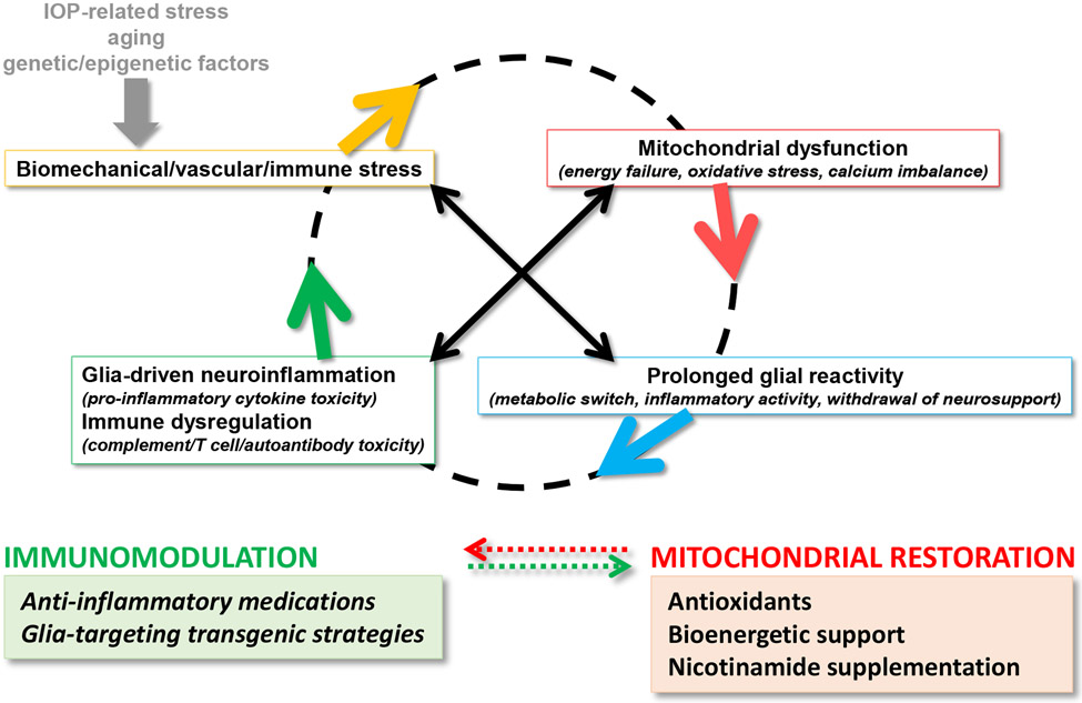
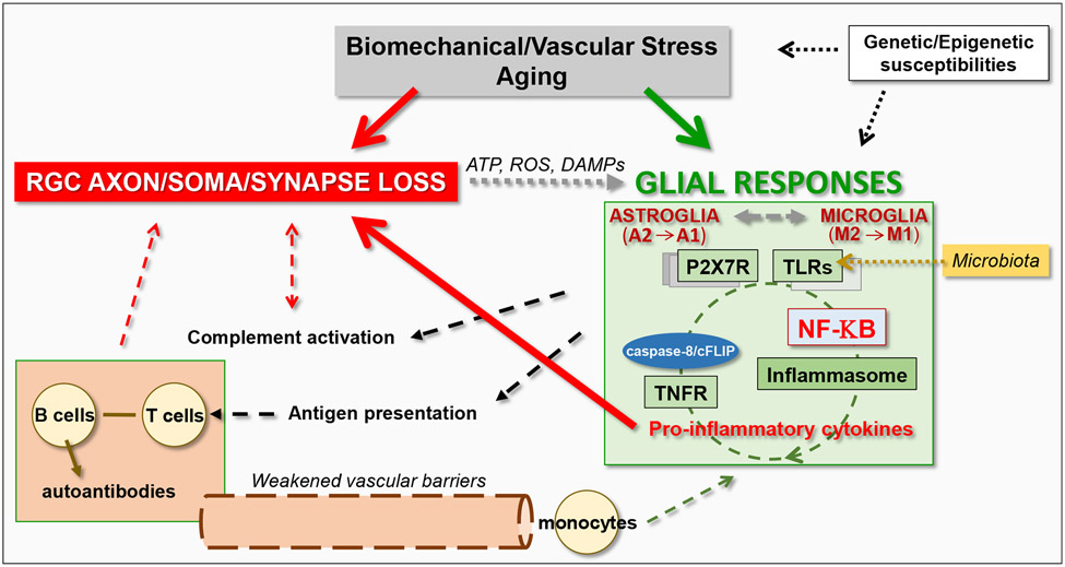
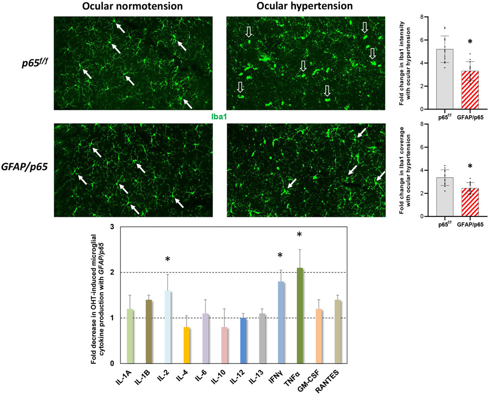
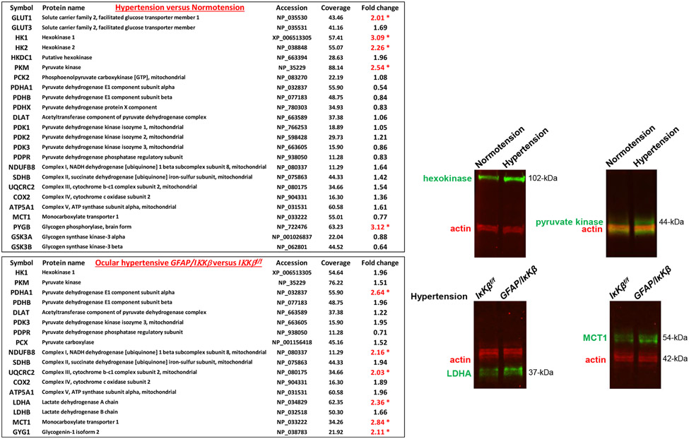
Neuroinflammation relying on the inflammatory responses of glial cells has emerged as an impactful component of the multifactorial etiology of neurodegeneration in glaucoma. It has become increasingly evident that despite early adaptive and reparative features of glial responses, prolonged reactivity of the resident glia, along with the peripheral immune cells, create widespread toxicity to retinal ganglion cell (RGC) axons, somas, and synapses. As much as the synchronized responses of astrocytes and microglia to glaucoma-related stress or neuron injury, their bi-directional interactions are critical to build and amplify neuroinflammation and to dictate the neurodegenerative outcome. Although distinct molecular programs regulate somatic and axonal degeneration in glaucoma, inhibition of neurodegenerative inflammation can provide a broadly beneficial treatment strategy to rescue RGC integrity and function. Since inflammatory toxicity and mitochondrial dysfunction are converging etiological paths that can boost each other and feed into a vicious cycle, anti-inflammatory treatments may also offer a multi-target potential. This review presents an overview of the current knowledge on neuroinflammation in glaucoma with particular emphasis on the cell-intrinsic and cell-extrinsic factors involved in the reciprocal regulation of glial responses, the interdependence between inflammatory and mitochondrial routes of neurodegeneration, and the research aspects inspiring for prospective immunomodulatory treatments. With the advent of powerful technologies, ongoing research on molecular and functional characteristics of glial responses is expected to accumulate more comprehensive and complementary information and to rapidly move the field forward to safe and effective modulation of the glial pro-inflammatory activities, while restoring or augmenting the glial immune-regulatory and neurosupport functions.

Keywords: Glaucoma; Glia; Immunomodulation; Neurodegeneration; Neuroinflammation; Retinal ganglion cell.


Figures

Figure 1.
Figure 1.. Converging etiological paths of glaucomatous neurodegeneration.
Mitochondrial dysfunction and glia-driven neuroinflammation present interdependent pathogenic processes. Major stressors in glaucoma, including intraocular pressure (IOP) elevation and aging, along with genetic/epigenetic susceptibilities, create biomechanical, vascular, and/or immune stress on RGCs. While dysfunctional mitochondria induce glial inflammatory responses, pro-inflammatory mediators further impair mitochondria, thereby feeding into a vicious cycle that amplifies neurodegeneration in glaucoma. As reviewed herein, preclinical studies explore new strategies for immunomodulation in glaucoma (such as those shown in the green box), which are also expected to protect mitochondria against inflammatory injury. By similarly counting on the interdependence between mitochondrial dysfunction and neuroinflammation, inflammatory outcomes can also be modulated by the treatments targeting mitochondrial dysfunction (such as those shown in the red box, which are tested in clinical studies).
Figure 2.
Figure 2.. Induction and amplification of glia-driven neuroinflammation in glaucoma.
Glial cells, including both astroglia and microglia, prominently respond to glaucoma-related tissue stress and RGC injury. By sensing the ATP, reactive oxygen species (ROS), and other damage-associated molecular patterns (DAMPs) released from stressed or dying cells, glial cells stimulate inflammation through purinergic receptors (such as P2X7R), pattern-recognition receptors (such as TLRs), and inflammasome activation. The produced pro-inflammatory neurotoxic cytokines contribute to RGC injury. The glia-driven inflammation through multiple pathways are commonly regulated by the NF-κB-mediated transcriptional program. Besides cytokine-mediated neurotoxicity, inflammatory outcomes of complement activation and adaptive immune responses may also contribute to neurodegeneration.
Figure 3.
Figure 3.. Astrocyte-microglia communication in neuroinflammation.
Similar to microglia-derived factors stimulating inflammatory activation of astrocytes, astroglia-derived factors may shape the inflammatory responses of microglia in experimental glaucoma. Besides decreased inflammatory activity of astroglia after cre/lox-based conditional deletion of p65 in GFAP-expressing astrocytes, some alterations were also detectable in microglia responses. The morphological response of microglia to ocular hypertension (a shift from ramified morphology, shown by white arrows, to ameboid morphology, shown by translucent arrows) was less prominent, and both the intensity and the coverage of Iba1 labeling were ~30% less in ocular hypertensive eyes of GFAP/p65 mice than ocular hypertensive p65f/f controls (*P<0.001; n= >4 mice/group). As presented in the bar graph (mean±SD), ocular hypertension-induced production of NF-κB-regulated pro-inflammatory cytokines, including IL-2, IFNγ, and TNFα, was significantly lower in the isolated samples of microglia (by immunomagnetic cell selection) from ocular hypertensive GFAP/p65 mice than ocular hypertensive p65f/f controls (*P<0.02; n= >20 mice/group). Intraocular pressure elevation was induced by anterior chamber microbead injections, and the ocular hypertensive mice were followed for 12 weeks. This work has recently been presented at ARVO 2021 Meeting (Abstract 3517862).
Figure 4.
Figure 4.. Switched metabolic profile of reactive astrocytes in experimental mouse glaucoma.
Proteomic analysis of the isolated astrocyte proteins (by immunomagnetic cell selection) by isotope labeling-based quantitative mass spectrometry presented upregulation of glycolysis as evident by increased expression of enzymes catalyzing the rate-limiting steps of glycolysis, including hexokinase and phosphofructokinase (*P<0.05). Alterations in the expression of selected proteins were also validated by Western blot analysis of astrocyte proteins. In support of aerobic glycolysis, unchanged or reduced expression of pyruvate dehydrogenase suggested limited entry of pyruvate into the tricarboxylic acid cycle and oxidative phosphorylation. However, there was an accompanying decrease in the expression of monocarboxylate transporter (MCT1) in the glaucomatous astroglia, suggesting decreased metabolite delivery to stressed RGCs. Inhibition of NF-κB activation by IκKβ deletion in GFAP-expressing astrocytes (GFAP/IκKβ) reversed this metabolic profile of reactive astrocytes in mouse glaucoma, as prominent by an increased expression of pyruvate dehydrogenase and MCT1. Intraocular pressure elevation was induced by anterior chamber microbead injections, and the ocular hypertensive mice were followed for 12 weeks. This work has recently been presented at ARVO 2019 Meeting (Abstracts 3788 and 3792).
Figure 5.
Figure 5.. Imbalance of T lymphocyte subsets in the glaucoma patients’ blood.
T lymphocyte subset markers were analyzed in the peripheral blood samples collected from patients with glaucoma (n=32) and age-matched non-glaucomatous controls (n=21). The percentage of CD4+/CD25+/FoxP3+ Treg (CD4-Treg) within the entire CD4+ population (A), the Treg to Th1 (B) or Treg to Th17 (C) ratios were significantly lower in the glaucoma group (P<0.001). The bars on univariate scatterplots present the group mean. Panel D shows representative flow cytometry images. Next, the same samples were analyzed for a proliferation marker after in vitro stimulation with retinal antigens. As presented by bar graphs (mean±SD) and univariate scatterplots in panel E, the percentage of CD4+/EdU+ cells was significantly higher in the glaucomatous samples than controls (P<0.001). Panel F shows representative flow cytometry images. This work has recently been published (Yang et al., 2019b).
Figure 6.
Figure 6.. Inflammatory injury to RGCs in glaucoma.
Various outcomes of glia-driven neuroinflammation promote neurotoxicity to RGC axons, somas, and synapses at dendrites and axon terminals in glaucoma. While pro-inflammatory cytokines produced by reactive glia, including TNFα, can induce RGC apoptosis, axon degeneration, and oligodendrocyte death by activating dead receptor signaling (TNFR), complement cascades mediate the loss of RGC synapses.
Figure 7.
Figure 7.. NF-κB-regulated activation of neurodegenerative inflammation in glaucoma.
A. Ocular hypertension was experimental induced by anterior chamber microbead injections in mice with or without cre/lox-based conditional deletion of IκKβ in GFAP-expressing astroglia. Optic nerve cross-sections presented well preserved structure of RGC axons (A), and the number of remaining axons was significantly higher (B) in ocular hypertensive GFAP/IκKβ mice compared to ocular hypertensive IκKβf/f controls (***P<0.001). As shown in Panel C, deletion of astroglial IκKβ resulted in an approximately 60% protection of RGC axons in ocular hypertensive eyes over 12 weeks. Similarly, retinal whole mounts labeled for RBPMS showed a visible protection of RGC somas (D), and the number of RBPMS-labeled RGCs was significantly higher (E) in ocular hypertensive GFAP/IκKβ mice compared to ocular hypertensive IκKβf/f controls (***P<0.001). As shown in Panel F, deletion of astroglial IκKβ resulted in an approximately 60% protection of RGC somas in ocular hypertensive eyes over 12 weeks. Presented data (mean±SD) represents a minimum of 16 mice per group. This work has recently been published (Yang et al., 2020).
Figure 8.
Figure 8.. Functional recovery by inhibition of glia-driven neuroinflammation in glaucoma.
Ocular hypertension was experimental induced by anterior chamber microbead injections in mice with or without cre/lox-based conditional deletion of IκKβ in GFAP-expressing astroglia. Recording of pattern electroretinography (PERG) responses in GFAP/IκKβ mice and IκKβf/f controls with or without experimentally induced ocular hypertension (A) showed preserved PERG amplitude (***P<0.001) in ocular hypertensive GFAP/IκKβ mice compared to ocular hypertensive IκKβf/f controls (B and C). Presented data (mean±SD) represents a minimum of 16 mice per group. This work has recently been published (Yang et al., 2020).
Figure 9.
Figure 9.. Molecular regulation of the parallel pathways modulating inflammation signaling.
As illustrated by the blue triangles at the bottom, different states of caspase-8 function, including its enzymatic or catalytic activities, are critical for the outcome. While the enzymatic activity of caspase-8 induces apoptosis in RGCs (A), its catalytic activity, in the absence of full proteolytic cleavage, signals towards cell survival in astroglia (B). Alternatively, the lack of caspase-8 activity induces necroptosis (C). Caspase-8 deletion in astroglia changes the state from “B to C”, while the inhibition of caspase-8 cleavage by z-IETD-fmk shifts the signal from “A to B” in RGCs. By interacting with caspase-8, cFLIP inhibits caspase-8-mediated cell death but induces pro-inflammatory outcomes. This regulator molecule that is highly expressed in astroglia but not in RGCs functions as a molecular switch between cell death, survival, and inflammation signals. This work has recently been published (Yang et al., 2021).
Figure 10.
Figure 10.. Epigenetic modulation of immune regulation.
A. Quantitative Western blot analysis of retinal proteins detected a prominent variation in TNFAP3 (A20, a negative regulator of NF-κB-regulated inflammation) expression among ten human donor eyes with glaucoma. As presented by the bar graph, four out of ten of these samples presented an over two-fold increased expression over age-matched controls (*P<0.01); however, TNFAIP3 expression was unchanged or decreased in rest of the human glaucoma samples. B. When the cytosine nucleotide methylation in the TNFAIP3 promoter was studied, in the glaucoma samples with low TNFAIP3 expression (corresponding to donors #4, 5, and 9), three CpG (triangle) and ten non-CpG (circle) methylation sites (filled symbols) were found. In the glaucoma samples exhibiting high expression of TNFAIP3 (corresponding to donors #1 and 7), these sites remained unmethylated. Panel C presents two examples of the bisulfite sequencing results. This work has recently been published (Yang et al., 2011).

References

    1. Ahmad L, Zhang SY, Casanova JL, Sancho-Shimizu V, 2016. Human TBK1: A gatekeeper of neuroinflammation. Trends Mol Med. 22, 511–527. - PMC - PubMed
    1. Aires ID, Ribeiro-Rodrigues T, Boia R, Catarino S, Girao H, Ambrosio AF, Santiago AR, 2020. Exosomes derived from microglia exposed to elevated pressure amplify the neuroinflammatory response in retinal cells. Glia. 68, 2705–2724. - PubMed
    1. Alarcon-Martinez L, Villafranca-Baughman D, Quintero H, et al., 2020. Interpericyte tunnelling nanotubes regulate neurovascular coupling. Nature. 585, 91–95. - PubMed
    1. Albalawi F, Lu W, Beckel JM, Lim JC, McCaughey SA, Mitchell CH, 2017. The P2X7 receptor primes IL-1beta and the NLRP3 inflammasome in astrocytes exposed to mechanical strain. Front Cell Neurosci. 11, 227. - PMC - PubMed
    1. Anderson MA, Burda JE, Ren Y, et al., 2016. Astrocyte scar formation aids central nervous system axon regeneration. Nature. 532, 195–200. - PMC - PubMed

Publication types