

Diagnostic accuracy of coronary computed tomography angiography-derived fractional flow reserve
- PMID: 34348731
- PMCID: PMC8340407
- DOI: 10.1186/s12938-021-00914-3
Diagnostic accuracy of coronary computed tomography angiography-derived fractional flow reserve
Abstract
Background: Fractional flow reserve (FFR) is a widely used gold standard to evaluate ischemia-causing lesions. A new method of non-invasive approach, termed as AccuFFRct, for calculating FFR based on coronary computed tomography angiography (CCTA) and computational fluid dynamics (CFD) has been proposed. However, its diagnostic accuracy has not been validated.
Objectives: This study sought to present a novel approach for non-invasive computation of FFR and evaluate its diagnostic performance in patients with coronary stenosis.
Methods: A total of 54 consecutive patients with 78 vessels from a single center who underwent CCTA and invasive FFR measurement were retrospectively analyzed. The CT-derived FFR values were computed using a novel CFD-based model (AccuFFRct, ArteryFlow Technology Co., Ltd., Hangzhou, China). Diagnostic performance of AccuFFRct and CCTA in detecting hemodynamically significant coronary artery disease (CAD) was evaluated using the invasive FFR as a reference standard.
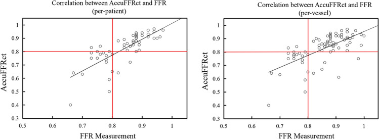
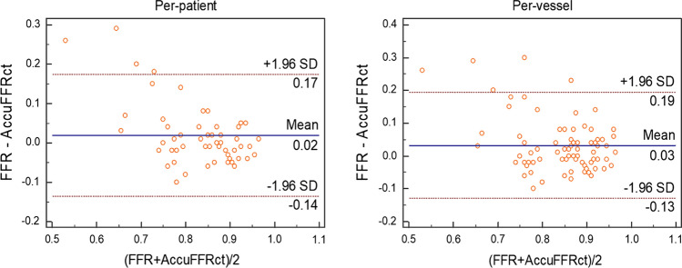
Results: Diagnostic accuracy, sensitivity, specificity, positive predictive value (PPV) and negative predictive value (NPV) for AccuFFRct in detecting FFR ≤ 0.8 on per-patient basis were 90.7, 89.5, 91.4, 85.0 and 94.1%, respectively, while those of CCTA were 38.9, 100.0, 5.71, 36.5 and 100.0%, respectively. The correlation between AccuFFRct and FFR was good (r = 0.76 and r = 0.65 on per-patient and per-vessel basis, respectively, both p < 0.0001). Area under the curve (AUC) values of AccuFFRct for identifying ischemia per-patient and per-vessel basis were 0.945 and 0.925, respectively. There was much higher accuracy, specificity and AUC for AccuFFRct compared with CCTA.
Conclusions: AccuFFRct computed from CCTA images alone demonstrated high diagnostic performance for detecting lesion-specific ischemia, it showed superior diagnostic power than CCTA and eliminated the risk of invasive tests, which could be an accurate and time-efficient computational tool for diagnosing ischemia and assisting clinical decision-making.
Keywords: CT-derived FFR; Computational fluid dynamics; Coronary computed tomography angiography; Fractional flow reserve.
© 2021. The Author(s).
Conflict of interest statement
The authors confirm that no conflict of interest or any financial relationship related to the manuscript's content has been associated with this publication.
Figures


Similar articles
-
Accuracy of intravascular ultrasound-derived virtual fractional flow reserve (FFR) and FFR derived from computed tomography for functional assessment of coronary artery disease.Biomed Eng Online. 2023 Jun 27;22(1):64. doi: 10.1186/s12938-023-01122-x. Biomed Eng Online. 2023. PMID: 37370077 Free PMC article.
-
Prospective comparisons of three interpretation methods of fractional flow reserve derived from coronary computed tomography angiography.Quant Imaging Med Surg. 2025 Mar 3;15(3):2146-2161. doi: 10.21037/qims-24-600. Epub 2025 Feb 26. Quant Imaging Med Surg. 2025. PMID: 40160645 Free PMC article.
-
Is it the Time to Move Towards Coronary Computed Tomography Angiography-Derived Fractional Flow Reserve Guided Percutaneous Coronary Intervention? The Pros and Cons.Curr Cardiol Rev. 2023;19(4):e190123212887. doi: 10.2174/1573403X19666230119115228. Curr Cardiol Rev. 2023. PMID: 36658709 Free PMC article. Review.
-
Diagnostic Performance of Fractional Flow Reserve Derived From Coronary CT Angiography: The ACCURATE-CT Study.JACC Cardiovasc Interv. 2024 Sep 9;17(17):1980-1992. doi: 10.1016/j.jcin.2024.06.027. Epub 2024 Aug 21. JACC Cardiovasc Interv. 2024. PMID: 39177553
-
Meta-Analysis of Diagnostic Performance of Coronary Computed Tomography Angiography, Computed Tomography Perfusion, and Computed Tomography-Fractional Flow Reserve in Functional Myocardial Ischemia Assessment Versus Invasive Fractional Flow Reserve.Am J Cardiol. 2015 Nov 1;116(9):1469-78. doi: 10.1016/j.amjcard.2015.07.078. Epub 2015 Aug 14. Am J Cardiol. 2015. PMID: 26347004 Free PMC article. Review.
Cited by
-
Functional evaluation of intracranial atherosclerotic stenosis by pressure ratio measurements.Heliyon. 2023 Feb 10;9(2):e13527. doi: 10.1016/j.heliyon.2023.e13527. eCollection 2023 Feb. Heliyon. 2023. PMID: 36852079 Free PMC article.
-
Accuracy of intravascular ultrasound-derived virtual fractional flow reserve (FFR) and FFR derived from computed tomography for functional assessment of coronary artery disease.Biomed Eng Online. 2023 Jun 27;22(1):64. doi: 10.1186/s12938-023-01122-x. Biomed Eng Online. 2023. PMID: 37370077 Free PMC article.
-
Prospective comparisons of three interpretation methods of fractional flow reserve derived from coronary computed tomography angiography.Quant Imaging Med Surg. 2025 Mar 3;15(3):2146-2161. doi: 10.21037/qims-24-600. Epub 2025 Feb 26. Quant Imaging Med Surg. 2025. PMID: 40160645 Free PMC article.
-
[Research progress on image-based calculation of coronary artery fractional flow reserve].Sheng Wu Yi Xue Gong Cheng Xue Za Zhi. 2023 Feb 25;40(1):171-179. doi: 10.7507/1001-5515.202206044. Sheng Wu Yi Xue Gong Cheng Xue Za Zhi. 2023. PMID: 36854563 Free PMC article. Chinese.
-
Is it the Time to Move Towards Coronary Computed Tomography Angiography-Derived Fractional Flow Reserve Guided Percutaneous Coronary Intervention? The Pros and Cons.Curr Cardiol Rev. 2023;19(4):e190123212887. doi: 10.2174/1573403X19666230119115228. Curr Cardiol Rev. 2023. PMID: 36658709 Free PMC article. Review.
References
-
- Meijboom WB, Van Mieghem CAG, van Pelt N, Weustink A, Pugliese F, Mollet NR, et al. Comprehensive assessment of coronary artery stenoses: computed tomography coronary angiography versus conventional coronary angiography and correlation with fractional flow reserve in patients with stable angina. J Am Coll Cardiol. 2008;52(8):636–643. doi: 10.1016/j.jacc.2008.05.024. - DOI - PubMed
MeSH terms
Grants and funding
LinkOut - more resources
Full Text Sources
Medical
Miscellaneous
