. 2021 Aug 4;11(1):15849.
doi: 10.1038/s41598-021-95119-y.

Application of alpha1-antitrypsin in a rat model of veno-arterial extracorporeal membrane oxygenation

Affiliations

Application of alpha1-antitrypsin in a rat model of veno-arterial extracorporeal membrane oxygenation

Fabian Edinger et al. Sci Rep. .

Abstract

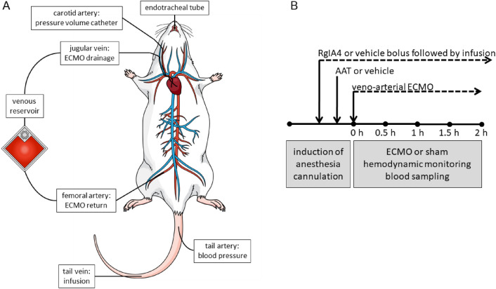
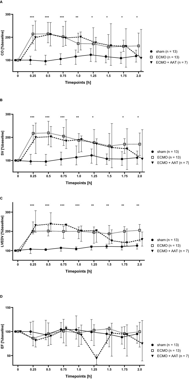
Extracorporeal membrane oxygenation (ECMO) is a life-saving intervention for patients suffering from respiratory or cardiac failure. The ECMO-associated morbidity and mortality depends to a large extent on the underlying disease and is often related to systemic inflammation, consecutive immune paralysis and sepsis. Here we tested the hypothesis that human α1-antitrypsin (SERPINA1) due to its anti-protease and anti-inflammatory functions may attenuate ECMO-induced inflammation. We specifically aimed to test whether intravenous treatment with α1-antitrypsin reduces the release of cytokines in response to 2 h of experimental ECMO. Adult rats were intravenously infused with α1-antitrypsin immediately before starting veno-arterial ECMO. We measured selected pro- and anti-inflammatory cytokines and found, that systemic levels of tumor necrosis factor-α, interleukin-6 and interleukin-10 increase during experimental ECMO. As tachycardia and hypertension developed in response to α1-antitrypsin, a single additional bolus of fentanyl and midazolam was given. Treatment with α1-antitrypsin and higher sedative doses reduced all cytokine levels investigated. We suggest that α1-antitrypsin might have the potential to protect against both ECMO-induced systemic inflammation and immune paralysis. More studies are needed to corroborate our findings, to clarify the mechanisms by which α1-antitrypsin inhibits cytokine release in vivo and to explore the potential application of α1-antitrypsin in clinical ECMO.


Conflict of interest statement

The authors declare no competing interests.

Figures

Figure 1
Figure 1
Design of the study. (A) Cannulation strategy used for experimental veno-arterial extracorporeal membrane oxygenation (ECMO) in the rat. The animals were endotracheally intubated and ventilated. The right atrium was cannulated via the jugular vein for venous blood drainage. Venous blood passed through the ECMO device and the oxygenized and carbon dioxide cleared blood was returned via the femoral artery. A pressure volume catheter was placed into the left ventricle through the right carotid artery. The peripheral blood pressure was measured in the tail artery. The lateral tail vein was punctured for infusion of medicaments and the test substances under investigation. (B) Graphical visualization of the time course of the experiments. Rats were anesthetized and treated with the conopeptide RgIA4 or vehicle, which were given as a bolus followed by continuous infusion. α1-antitrypsin (AAT) or vehicle were applied by syringe pump over 5 min. Thereafter, veno-arterial ECMO was started and hemodynamic monitoring as well as blood sampling was done for 2 h. Thereafter, animals were sacrificed. The same procedure was applied to sham-treated animals, but the ECMO was omitted.
Figure 2
Figure 2
Arterial blood pressure and heart rate (HR) in experimental rats. Animals underwent the sham procedure (n = 13), extracorporeal membrane oxygenation (ECMO, n = 13) or ECMO combined with application of α1-antitrypsin (AAT, n = 7). Values were recorded every 15 min until the end of the experiments. The systolic arterial blood pressure (SAP, (A)), the diastolic arterial blood pressure (DAP, (B)) and the mean arterial blood pressure (MAP, (C)) were significantly elevated in animals connected to ECMO compared to the sham group. These parameters were not changed in rats treated with AAT. The HR did not significantly differ among all groups (D). Data are presented as median and interquartile ranges 25% and 75%; Kruskal–Wallis test followed by pairwise Wilcoxon–Mann–Whitney test; **p ≤ 0.01, ***p ≤ 0.001 sham versus ECMO; #p ≤ 0.05, ECMO versus ECMO + AAT.
Figure 3
Figure 3
Hemodynamic parameters in experimental rats. Animals underwent the sham procedure (n = 13), extracorporeal membrane oxygenation (ECMO, n = 13) or ECMO combined with application of α1-antitrypsin (AAT, n = 7). (A) We found an increased cardiac output (CO) in response to ECMO compared to the sham group, irrespective of treatment with AAT. Similar results were found regarding stroke volume (SV, (B)) and left ventricular end-diastolic volume (LVEDV, (C)). (D) The ejection fraction (EF) was similar among most experimental groups and time points investigated. Only 75 min after starting ECMO, the EF was reduced in ECMO + AAT compared to ECMO alone. Values were recorded every 15 min until the end of the experiments. Data are presented as median and interquartile ranges 25% and 75%; Kruskal–Wallis test followed by pairwise Wilcoxon–Mann–Whitney test; *p ≤ 0.05, **p ≤ 0.01; ***p ≤ 0.001 sham versus ECMO.
Figure 4
Figure 4
Cytokine concentrations in experimental rats. Animals underwent the sham procedure (n = 13), extracorporeal membrane oxygenation (ECMO, n = 13) ECMO combined with application of α1-antitrypsin (AAT, n = 7) or ECMO combined with AAT and the conopeptide RgIA4 (n = 7). (A) Tumor necrosis factor-α (TNF-α) levels increased after 1.5 and 2 h of ECMO compared to sham, treatment with AAT reduced this increase. The effect of AAT was not sensitive to RgIA4. (B) No significant changes in circulating interleukin (IL)-1β levels were seen in none of the experimental groups. IL-6 (C) and IL-10 (D) levels increased after 1.5 and 2 h of ECMO compared to sham and treatment with AAT reduced their increase 2 h after starting ECMO. Again, RgIA4 did not cause any changes (C,D). Cytokines were measured every 30 min until the end of the experiments. Data are presented as median and interquartile ranges 25% and 75%; Kruskal–Wallis test followed by pairwise Wilcoxon-Mann–Whitney test; p-values are indicated in the graphs.
Figure 5
Figure 5
Venous and arterial ECMO cannula. The modified multi-orifice 17 G cannula (1.5 mm, B. Braun, cannula with white connector) was inserted into the internal jugular vein and carefully moved into the right atrium for venous drainage. The oxygenated blood from the ECMO was returned to the femoral artery via a 22 G catheter (0.9 mm, Terumo, cannula with blue connector).

References

    1. Karagiannidis C. Extracorporeal membrane oxygenation: Evolving epidemiology and mortality. Intens. Care Med. 2016;42:889–896. doi: 10.1007/s00134-016-4273-z. - DOI - PubMed
    1. Munshi L, et al. Venovenous extracorporeal membrane oxygenation for acute respiratory distress syndrome: A systematic review and meta-analysis. Lancet Respir. Med. 2019;7(2):162–172. doi: 10.1016/S2213-2600(18)30452-1. - DOI - PubMed
    1. Khorsandi M, et al. Extra-corporeal membrane oxygenation for refractory cardiogenic shock after adult cardiac surgery: A systematic review and meta-analysis. J. Cardiothorac. Surg. 2017;12:55. doi: 10.1186/s13019-017-0618-0. - DOI - PMC - PubMed
    1. Fletcher-Sandersjöö A, Thelin EP, Bartek J, Broman M, Sallisalmi M, Elmi-Terander A, Bellander BM. Incidence, outcome, and predictors of intracranial hemorrhage in adult patients on extracorporeal membrane oxygenation: A systematic and narrative review. Front Neurol. 2018;6(9):548. doi: 10.3389/fneur.2018.00548. - DOI - PMC - PubMed
    1. Millar JE, Fanning JP, McDonald CI, McAuley DF, Fraser JF. The inflammatory response to extracorporeal membrane oxygenation (ECMO): A review of the pathophysiology. Crit. Care. 2016;20:387. doi: 10.1186/s13054-016-1570-4. - DOI - PMC - PubMed

Publication types