Observational Study
. 2021 Aug 4;11(1):15826.
doi: 10.1038/s41598-021-95219-9.

Quantifying the pattern of retinal vascular orientation in diabetic retinopathy using optical coherence tomography angiography

Affiliations
Observational Study

Quantifying the pattern of retinal vascular orientation in diabetic retinopathy using optical coherence tomography angiography

Yanhui Ma et al. Sci Rep. .

Abstract

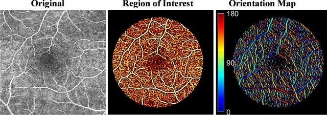
Quantitative imaging using optical coherence tomography angiography (OCTA) could provide objective tools for the detection and characterization of diabetic retinopathy (DR). In this study, an operator combining the second derivative and Gaussian multiscale convolution is applied to identify the retinal orientation at each pixel in the OCTA image. We quantified the pattern of retinal vascular orientation and developed three novel quantitative metrics including vessel preferred orientation, vessel anisotropy, and vessel area. Each of eight 45º sectors of the circular disk centered at the macular region was defined as the region of interest. Significant sectoral differences were observed in the preferred orientation (p < 0.0001) and vessel area (p < 0.0001) in the 34 healthy subjects, whereas vessel anisotropy did not demonstrate a significant difference among the eight sectors (p = 0.054). Differential retinal microvascular orientation patterns were observed between healthy controls (n = 34) and the DR subjects (n = 7). The vessel area characterized from the vascular orientation pattern was shown to be strongly correlated with the traditionally reported vessel density (Pearson R > 0.97, p < 0.0001). With three metrics calculated from the vascular orientation pattern simultaneously and sectorally, our quantitative assessment for retinal microvasculature provides more information than vessel density alone and thereby may enhance the detection of DR. These preliminary results suggest the feasibility and advantage of our vessel orientation-based quantitative approach using OCTA to characterize DR-associated changes in retinal microvasculature.


Conflict of interest statement

The authors declare no competing interests.

Figures

Figure 1
Figure 1
(A) Gray-scale image of a tube-like structure with (B) an intensity profile of 2-dimensional Gaussian with standard deviation s = 1. Zero-crossings of the second derivative (C) correspond to the local maxima in the first derivative (D).
Figure 2
Figure 2
Illustration of vessel orientation extraction from OCTA image. (A) Vessel orientations were calculated by the Hessian matrix-based algorithm. (B) Enlargement of (A): the arrows indicate the vessel directions/orientations. Note the algorithm is demonstrated on large vessels for illustration.
Figure 3
Figure 3
Visualization of retinal microvascular orientation in the region of interest after vesselness filter and binary filter. Dark blue and red indicate 0º and 180º for those horizontal vessels and green indicates 90º for those vertical vessels. The image processing was operated on the full-thickness (non-segmented) en face OCTA image.
Figure 4
Figure 4
Quantification of vascular orientation pattern using preferred orientation, vessel anisotropy, and vessel area. The orientation pattern (middle) for the specific ROI (left) depicts a roughly elliptical shape with a major axis and a minor axis. The preferred orientation is identified by the angle of the major axis. The ratio of major axis length and minor axis length is defined as vessel anisotropy. The vessel area is defined as the area of the shape. Examples of preferred orientation, vessel anisotropy, and vessel area, are illustrated by the dashed ellipse relative to the solid ellipse (right).
Figure 5
Figure 5
Sectoral vascular pattern with preferred vessel orientation (dashed line) unaligned with sector axis. Eight 45º sectors were divided from a circular disk centered at the macula and each sector was defined as the region of interest for the quantitative assessment of retinal vascular orientation pattern. N nasal, S superior, T temporal, I inferior.
Figure 6
Figure 6
Sectoral difference in retinal microvascular pattern in healthy subjects (n = 34). Significant sectoral differences were observed in preferred orientation (p < 0.0001) and vessel area (p < 0.0001), whereas vessel anisotropy did not show a significant difference among the 8 sectors (p = 0.054).
Figure 7
Figure 7
Scatterplot of the relationship between vessel area and vessel density in (A) the IT sector and (B) the average of 8 sectors in healthy subjects. The vessel area quantified from the retinal vascular orientation pattern was strongly correlated with the vessel density with Pearson R = 0.99 for both IT sector and on average (p < 0.0001, n = 34).

References

    1. Congdon N, et al. Causes and prevalence of visual impairment among adults in the United States. Arch. Ophthalmol. (Chicago, Ill. 1960) 2004;122:477–485. doi: 10.1001/archopht.122.4.477. - DOI - PubMed
    1. Resnikoff S, et al. Global data on visual impairment in the year 2002. Bull. World Health Organ. 2004;82:844–851. - PMC - PubMed
    1. Klein R, Klein BEK, Moss SE, Davis MD, DeMets DL. The Wisconsin epidemiologic study of diabetic retinopathy: II. Prevalence and risk of diabetic retinopathy when age at diagnosis is less than 30 years. Arch. Ophthalmol. 1984;102:520–526. doi: 10.1001/archopht.1984.01040030398010. - DOI - PubMed
    1. Control, D. & Group, C. T. R The effect of intensive treatment of diabetes on the development and progression of long-term complications in insulin-dependent diabetes mellitus. N. Engl. J. Med. 1993;329:977–986. doi: 10.1056/NEJM199309303291401. - DOI - PubMed
    1. Cogan DG, Toussaint D, Kuwabara T. Retinal vascular patterns: IV. Diabetic retinopathy. Arch. Ophthalmol. 1961;66:366–378. doi: 10.1001/archopht.1961.00960010368014. - DOI - PubMed

Publication types