. 2022 May;29(5):466-474.
doi: 10.1038/s41417-021-00366-w. Epub 2021 Aug 4.

The application of CRISPR/Cas9 system in cervical carcinogenesis

Affiliations

The application of CRISPR/Cas9 system in cervical carcinogenesis

Chun Gao et al. Cancer Gene Ther. 2022 May.

Erratum in

Abstract

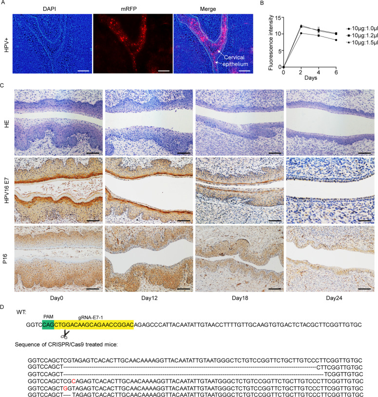
Integration of high-risk HPV genomes into cellular chromatin has been confirmed to promote cervical carcinogenesis, with HPV16 being the most prevalent high-risk type. Herein, we evaluated the therapeutic effect of the CRISPR/Cas9 system in cervical carcinogenesis, especially for cervical precancerous lesions. In cervical cancer/pre-cancer cell lines, we transfected the HPV16 E7 targeted CRISPR/Cas9, TALEN, ZFN plasmids, respectively. Compared to previous established ZFN and TALEN systems, CRISPR/Cas9 has shown comparable efficiency and specificity in inhibiting cell growth and colony formation and inducing apoptosis in cervical cancer/pre-cancer cell lines, which seemed to be more pronounced in the S12 cell line derived from the low-grade cervical lesion. Furthermore, in xenograft formation assays, CRISPR/Cas9 inhibited tumor formation of the S12 cell line in vivo and affected the corresponding protein expression. In the K14-HPV16 transgenic mice model of HPV-driven spontaneous cervical carcinogenesis, cervical application of CRISPR/Cas9 treatment caused mutations of the E7 gene and restored the expression of RB, E2F1, and CDK2, thereby reversing the cervical carcinogenesis phenotype. In this study, we have demonstrated that CRISPR/Cas9 targeting HPV16 E7 could effectively revert the HPV-related cervical carcinogenesis in vitro, as well as in K14-HPV16 transgenic mice, which has shown great potential in clinical treatment for cervical precancerous lesions.


Conflict of interest statement

The authors declare no competing interests.

Figures

Fig. 1
Fig. 1. Efficacies comparison of ZFN, TALEN, and CRISPR/Cas9 on HPV16-positive cell lines.
The representative images of γ-H2AX foci (green signals) in ZFN-treated, TALEN-treated, and CRISPR/Cas9-treated S12 cells (A) and SiHa cells (B). Etoposide (0.25 μM) was used as the positive control, and vector plasmid was used as the negative control. Quantification of γ-H2AX foci in S12 (C) and SiHa (D). T7 endonuclease 1 (T7E1) assay of ZFN-induced, TALEN-induced, and CRISPR/Cas9-induced cleavage at 48 h in S12 cells (E) and SiHa cells (F). Quantification of DNA indel rate in S12 (G) and SiHa (H). ns, no significance; *p < 0.05; **p < 0.01; ***p < 0.001. (n = 3 replications) Scale bars: 50 μm. Each experiment was repeated 3 times.
Fig. 2
Fig. 2. ZFN, TALEN, and CRISPR/Cas9 induced cell growth deficit and cell apoptosis in vitro.
AD Growth curves of ZFN, TALEN, and CRISPR/Cas9-treated S12 (A), SiHa (B), C33A (C), and HeLa (D) cells were constructed using the CCK-8 assay. EH The apoptosis rate of S12 (E), SiHa (F), C33A (G), and HeLa (H) cells 48 h after treatment with ZFN, TALEN, and gRNA-E7-1 + Cas9 plasmids. I The colony-forming assay of SiHa and S12 cells after treatment with ZFN, TALEN, and gRNA-E7-1 + Cas9 plasmids. J Quantification of the number of colonies in S12 and SiHa cells of different treatment groups. KL HPV16 E7/RB/CDK2/E2F1 expression of S12 (K) and SiHa (L) cells 48 h after treatment with ZFN, TALEN, and gRNA-E7-1 + Cas9 plasmids. ns, no significance; *p < 0.05; **p < 0.01; ***p < 0.001. (n = 3 replications) The experiments were repeated 3 times.
Fig. 3
Fig. 3. CRISPR/Cas9 inhibits S12 cell growth in vivo.
Balb/c-nu mice were injected subcutaneously in the right flanks with 5 × 106 of S12 cells. Then, the CRISPR/Cas9 plasmids complexed with in vivo transfection reagents were injected intratumorally when the xenografts reached approximately 50 mm3. A The xenografts were measured every 6 days after treatment with gRNA-E7-1 + Cas9, gRNA-E7-2 + Cas9, gRNA-GFP + Cas9, and PBS. B The photograph of S12 xenografts in different treatment groups. C The estimated tumor size of S12 xenografts in different treatment groups. D Representative pictures of HE staining and IHC staining of HPV16 E7, Caspase-3, CD31, and PCNA in gRNA-E7-1 + Cas9, gRNA-E7-2 + Cas9, gRNA-GFP + Cas9, and PBS treated S12 xenografts. Scale bars: 50 μm. E The average necrosis area and protein expression of HPV16 E7, Caspase-3, CD31, and PCNA in different groups. **p < 0.01. (n = 4 replications).
Fig. 4
Fig. 4. Establishment and application of CRISPR/Cas9 system in K14-HPV16 transgenic mice.
A The expression of mRFP was localized in the cervical epithelia of transgenic mice. Scale bars: 50 μm. B Transfection efficiency was optimized at the DNA-to-polymer ratio of 10 μg:1.0 μl, 10 μg:1.2 μl, and 10 μg:1.5 μl. The exfoliation of cervical cells was collected at 2, 4, and 6 days after vaginal transfection. C Representative HE staining and IHC staining of HPV16 E7 and p16 of gRNA-E7-1 + Cas9 treated K14-HPV16 transgenic mice at days 0,12,18, and 24. N = 3, Scale bars: 50 μm. D The cervical DNA sequencing of the gRNA-E7-1 targeted region of HPV16 E7 gene in gRNA-E7-1 + Cas9 treated K14-HPV16 transgenic mice.
Fig. 5
Fig. 5. Histopathological and protein expression changes in cervical epithelia of K14-HPV16 transgenic mice treated with HPV16 E7 targeting CRISPR/Cas9.
A Representative images of the HE staining and IHC staining of HPV16 E7, RB, Ki67, E2F1, and CDK2 in cervical epithelia of HPV-, gRNA-GFP + Cas9-treated, and gRNA-E7-1 + Cas9-treated mice. B Quantification of the protein expression of HPV16 E7, RB, Ki67, E2F1, and CDK2 in these 3 groups. **p < 0.01; ***p < 0.001. (N = 3 replications) Scale bars: 50 μm.
Fig. 6
Fig. 6. The conduction of regional plasmid transfection showed no influence on other organs.
The HE staining of different organs in gRNA-E7-1 + Cas9 and gRNA-GFP + Cas9-treated K14-HPV16 transgenic mice. Scale bars: 50 μm.

Similar articles

Cited by

References

    1. Bray F, Ferlay J, Soerjomataram I, Siegel RL, Torre LA, Jemal A. Global cancer statistics 2018: GLOBOCAN estimates of incidence and mortality worldwide for 36 cancers in 185 countries. CA Cancer J Clin. 2018;68:394–424. doi: 10.3322/caac.21492. - DOI - PubMed
    1. Vintermyr OK, Andersland MS, Bjørge T, Skar R, Iversen OE, Nygård M, et al. Human papillomavirus type specific risk of progression and remission during long-term follow-up of equivocal and low-grade HPV-positive cervical smears. Int J Cancer. 2018;143:851–60. doi: 10.1002/ijc.31390. - DOI - PubMed
    1. Wang SS, Zuna RE, Wentzensen N, Dunn ST, Sherman ME, Gold MA, et al. Human papillomavirus cofactors by disease progression and human papillomavirus types in the study to understand cervical cancer early endpoints and determinants. Cancer Epidemiol Biomark Prev. 2009;18:113–20. doi: 10.1158/1055-9965.EPI-08-0591. - DOI - PMC - PubMed
    1. Chesson HW, Dunne EF, Hariri S, Markowitz LE. The estimated lifetime probability of acquiring human papillomavirus in the United States. Sex Transm Dis. 2014;41:660–4. doi: 10.1097/OLQ.0000000000000193. - DOI - PMC - PubMed
    1. Kjaer SK, Frederiksen K, Munk C, Iftner T. Long-term absolute risk of cervical intraepithelial neoplasia grade 3 or worse following human papillomavirus infection: role of persistence. J Natl Cancer Inst. 2010;102:1478–88. doi: 10.1093/jnci/djq356. - DOI - PMC - PubMed

Publication types

MeSH terms

Substances