. 2021 Aug;73(4):555-569.
doi: 10.1007/s10616-021-00477-0. Epub 2021 May 26.

Establishment of human airway epithelial cells with doxycycline-inducible cell growth and fluorescence reporters

Affiliations

Establishment of human airway epithelial cells with doxycycline-inducible cell growth and fluorescence reporters

Ai Orimoto et al. Cytotechnology. 2021 Aug.

Abstract

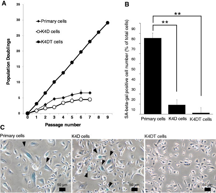
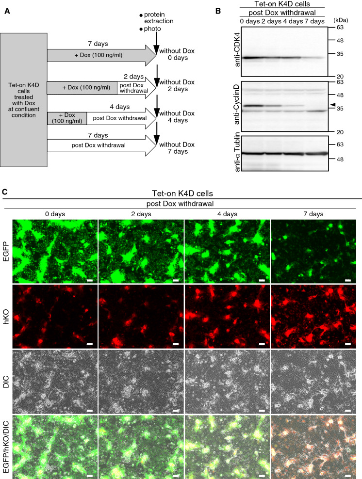
We previously reported the successful establishment of multiple immortalized cell lines that preserved the original nature of the primary cells via co-expression of R24C mutant cyclin-dependent kinase 4 (CDK4R24C), Cyclin D1, and telomerase reverse transcriptase (TERT). However, as these genes are kind of oncogenes, tools to control their expression levels are favorable. In this study, we describe a new polycistronic lentiviral vector expressing proliferation factors, CDK4R24C and Cyclin D1 along with enhanced green fluorescence protein (EGFP) under the control of doxycycline (Dox)-dependent transactivator (rtTA) and tetracycline response element (TRE). By introducing the Dox-inducible lentiviral vector into human airway epithelial cells, we established a novel human airway epithelial cell line harboring polycistronic Dox-inducible CDK4R24C and Cyclin D1, referred to as Tet-on K4D cells. We showed that the cell growth of Tet-on K4D cells could be controlled by Dox. Furthermore, expression of K4D genes and rtTA gene can be independently monitored by fluorescent imaging. Cultured airway epithelial cells are useful as a tool for studying the pathogenesis of lung disorders. Altogether, our established human airway epithelial cells could be used for a variety of studies such as lung pathology and biology underlying the differentiation process to form the complex pseudostratified multicellular layers.

Supplementary information: The online version contains supplementary material available at 10.1007/s10616-021-00477-0.

Keywords: CDK4R24C; Cyclin D1; Doxycycline-inducible lentiviral system; Fluorescence reporters human airway epithelial cells.


Figures

Fig. 1
Fig. 1
Establishment of cells using recombinant retrovirus. a Structure of recombinant retrovirus vectors used to establish the K4D and K4DT cells. LTR, long terminal repeat; CMV, cytomegalovirus promoter; Neo, Neomycin-resistant gene; Puro, Puromycin-resistant gene; Hygro, Hygromycin-resistant gene. The regions marked X represent deletion in the 3’LTR enhancer region (U3). b The morphologies of the human airway epithelial cells, K4D cells, and K4DT cells. Upper panels display cell morphologies viewed using a differential interference contrast (DIC) microscope. Middle panels display fluorescence images. Lower panels display merged images of DIC and fluorescence images. Enhanced green fluorescence protein (EGFP) fluorescence was detected in K4D cells transduced with the polycistronic retrovirus vector carrying CDK4R24C, Cyclin D1, and EGFP. Scale bar, 100 μm. (Color figure online)
Fig. 2
Fig. 2
Detection of the genomic cassette and protein expression in the established cells. a Detection of genomic integration of the expression cassette by polymerase chain reaction (PCR). PCR amplification of the expression cassette containing CDK4, Cyclin D1, TERT, and CDK4-Cyclin D1 as well as the internal control gene (TSC2 gene; Tuberous Sclerosis Type II) was performed. b Western blot analysis of primary cells, K4D cells, and K4DT cells. The results obtained from anti-Cyclin D1, anti-CDK4, and anti-αtubulin antibody staining are shown
Fig. 3
Fig. 3
Growth characteristics and cellular senescence of primary cells, K4D cells, and K4DT cells. a Cell growth and sequential passaging of primary cells, K4D, and K4DT cells. Cell growth is represented by the cumulative population doubling value. b Quantification of SA-beta-Gal-positive cell numbers in primary, K4D, and K4DT cells. The microscopic fields used for counting positively stained cells were randomly selected. Comparisons between the primary cells and K4D cells or K4DT cells were made using the Steel–Dwass test. Bars indicate means ± SE. n = 6. **p < 0.01. c Representative results of senescence-associated β-galactosidase staining of primary, K4D, and K4DT cells at passage 5. The arrows indicate positive staining that denote cellular senescence. Scale bar, 50 μm
Fig. 4
Fig. 4
Characteristics of primary, K4D, and K4DT cells. a Cell cycle histogram of representative results obtained from primary, K4D, and K4DT cells using the Muse Cell Analyzer. b Percentages of primary, K4D, and K4DT cells in each cell cycle phase. Data are presented as the mean ± standard error of the ratio of each cell cycle stage (n = 6). The Steel–Dwass method was used for comparisons. *p < 0.05, **p < 0.01. c G-banding metaphase spread of K4DT cells by karyotype analysis. d Aligned chromosome of K4DT cells
Fig. 5
Fig. 5
Establishment of cells using a Dox-inducible lentiviral system. a Structure of the tet-inducible lentiviral vector carrying cDNA for CDK4R24C, Cyclin D1, and enhanced green fluorescence protein (EGFP) that was used to establish the Tet-on K4D cells. The diagonal arrow indicates the tetracycline transactivator (rtTA) binding to the tetracycline response element (TRE). LTR, long terminal repeat; CMV, cytomegalovirus promoter; hKO, Humanized Kusabira-Orange fluorescence. b Detection of EGFP and hKO fluorescence in Tet-on K4D cells. Tet-on K4D cells were infected with a Dox-inducible lentiviral vector carrying CDK4R24C, Cyclin D1, and EGFP and then cultured for 3 weeks with (+ Dox) or without (No Dox) 100 ng/ml Dox in culture medium. The top panels, second panels, third panels, and bottom panels display EGFP fluorescence images, hKO fluorescence images, DIC images, and merged images of DIC, EGFP, and hKO images, respectively, of the primary cells as a control and Tet-on K4D cells. In the presence of Dox, both EGFP and hKO fluorescence were detected in Tet-on K4D cells. All images were captured using constant exposure times and identical camera settings on the BZ-X810 microscope (Keyence, Osaka, Japan). Scale bar, 100 μm. (Color figure online)
Fig. 6
Fig. 6
Evaluation of Tet-on K4D cells using a Dox-inducible lentiviral system. a Experimental protocol for the evaluation of Tet-on K4D cells. Tet-on K4D cells were split into two dishes and cultured in the presence of Dox until the cells reached confluent. After the cells reached confluent, cells were further incubated for 8 days with or without Dox. The experimental schedule after Dox withdrawal is indicated. b Western blot analysis of Tet-on K4D cells to detect CDK4 and Cyclin D1 under the control of Dox. Total cell lysates extracted from Tet-on K4D cells on day 0 and day 8 after Dox withdrawal were used for Western blot analysis. Bands marked by arrows 1 and 2 would derived from exogenous Cyclin D1 and endogenous Cyclin D1, respectively (see the text). c Fluorescence images of the Tet-on K4D cells under the control of Dox. Fluorescence images of Tet-on K4D cells on days 0, 3, 5, and 8 after Dox withdrawal were captured using constant exposure times and identical camera settings on the BZ-X810 microscope (Keyence, Osaka, Japan). We also captured images of Tet-on K4D cells cultured in the presence of the Dox for 8 days (+ Dox, Day 8). Scale bar, 100 μm
Fig. 7
Fig. 7
Effect of Dox on Tet-on K4D cells using a Dox-inducible lentiviral system. a Experimental protocol for evaluation of the effect of Dox on Tet-on K4D cells. Tet-on K4D cells were split into four dishes and cultured in the presence of Dox until confluency. After the cells reached confluency, Dox was withdrawn at different time points, and the cells were subsequently cultured in the absence of the Dox until day 7. All plates were subjected to western blot analysis and fluorescence imaging at day 7. b Western blot analysis of CDK4 and Cyclin D in Tet-on K4D cells. To determine the effect of Dox withdrawal on the expression levels of CDK4 and Cyclin D, total cell lysates extracted from Tet-on K4D cells at various time points after Dox withdrawal were used for western blot analysis using anti-CDK4, anti-CyclinD, and anti-α-tubulin antibodies. Arrow indicates exogenous Cyclin D1. c Fluorescence images of the Tet-on K4D cells at different time points after Dox withdrawal. Fluorescence images of Tet-on K4D cells that were initially grown in the presence of Dox and then had Dox withdrawal at different time points were captured using constant exposure times and identical camera settings on the BZ-X810 microscope (Keyence, Osaka, Japan)
Fig. 8
Fig. 8
Effect of Dox on cell proliferation in Tet-on K4D cells. a Experimental protocol for evaluation of the effect of Dox on cell proliferation in Tet-on K4D cells. Tet-on K4D cells treated with 100 ng/ml Dox were seeded into 12-well plates at a density of 4.0 × 104 cells. We prepared three culture groups. Dox was withdrawn at three different time points (6, 9, and 12 days) and the total number of cells with or without Dox was counted at each time point. b Cell growth of Tet-on K4D cells in Dox-dependent manner. Data is represented as the mean ± standard deviation of cell number at each time point (n = 4)

Similar articles

References

    1. Brambrink T, Foreman R, Welstead GG, Lengner CJ, Wernig M, Suh H, Jaenisch R. Sequential expression of pluripotency markers during direct reprogramming of mouse somatic cells. Cell Stem Cell. 2008 doi: 10.1016/j.stem.2008.01.004. - DOI - PMC - PubMed
    1. Cozens AL, Yezzi MJ, Kunzelmann K, Ohrui T, Chin L, Eng K, Gruenert DC. CFTR expression and chloride secretion in polarized immortal human bronchial epithelial cells. Am J Respir Cell Mol Biol. 1994 doi: 10.1165/ajrcmb.10.1.7507342. - DOI - PubMed
    1. Crystal RG, Randell SH, Engelhardt JF, Voynow J, Sunday ME. Airway epithelial cells: current concepts and challenges. Proc Am Thorac Soc. 2008 doi: 10.1513/pats.200805-041HR. - DOI - PMC - PubMed
    1. Donai K, Kuroda K, Guo Y, So K-H, Sone H, Kobayashi M, Fukuda T. Establishment of a reporter system to monitor silencing status in induced pluripotent stem cell lines. Anal Biochem. 2013;443:104–112. doi: 10.1016/j.ab.2013.08.014. - DOI - PubMed
    1. Donai K, Kiyono T, Eitsuka T, Guo Y, Kuroda K, Sone H, Fukuda T. Bovine and porcine fibroblasts can be immortalized with intact karyotype by the expression of mutant cyclin dependent kinase 4, cyclin D, and telomerase. J Biotechnol. 2014;176:50–57. doi: 10.1016/J.JBIOTEC.2014.02.017. - DOI - PubMed