. 2021 Jul 24:2021:5587297.
doi: 10.1155/2021/5587297. eCollection 2021.

Astaxanthin Inhibits Interleukin-6 Expression in Cerulein/Resistin-Stimulated Pancreatic Acinar Cells

Affiliations

Astaxanthin Inhibits Interleukin-6 Expression in Cerulein/Resistin-Stimulated Pancreatic Acinar Cells

Min Seung Kwak et al. Mediators Inflamm. .

Abstract

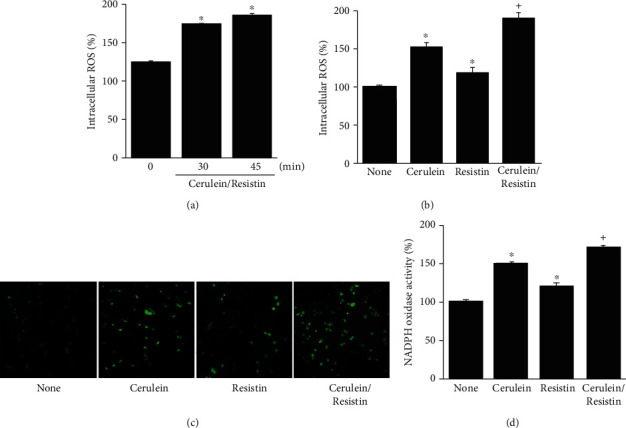
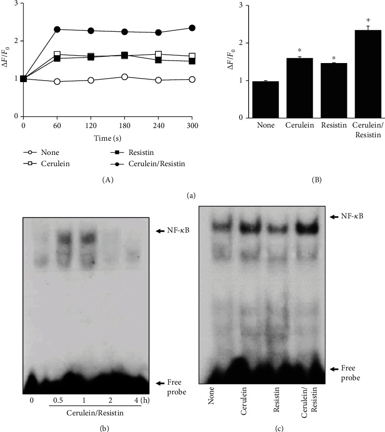
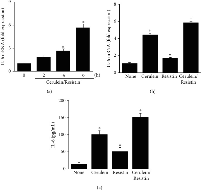
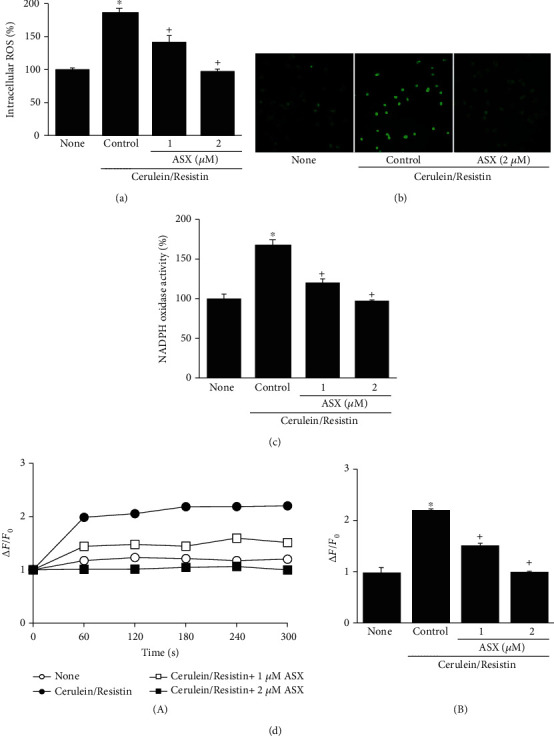
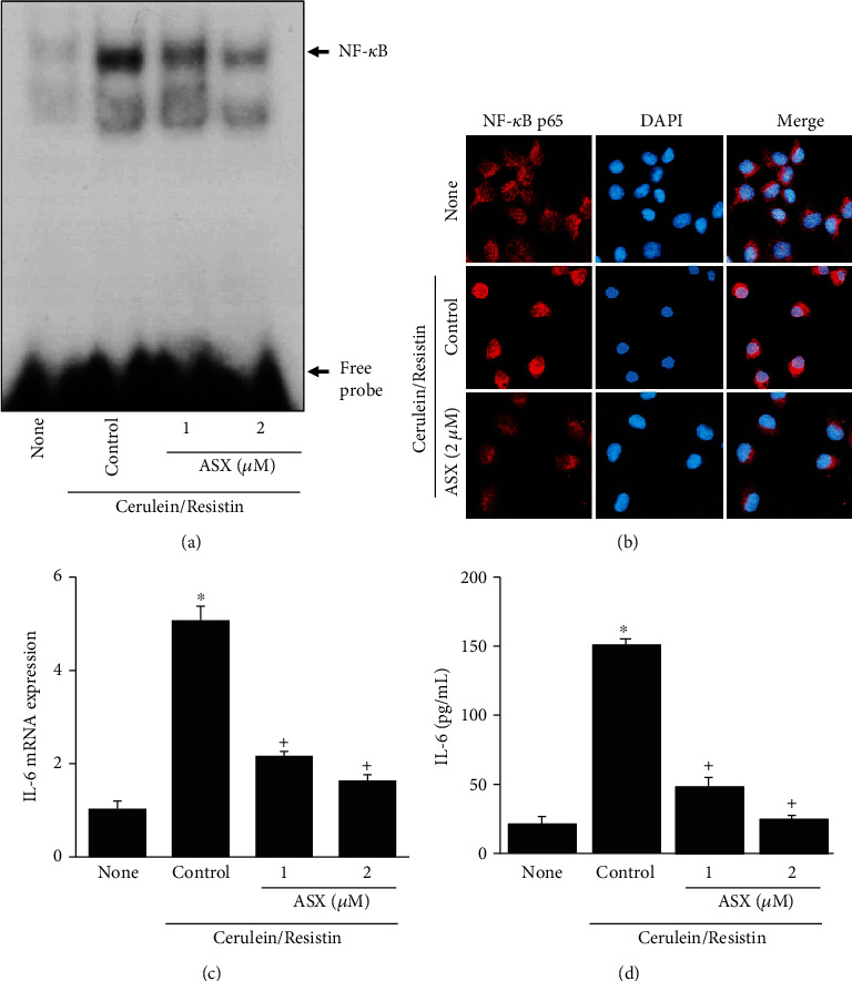
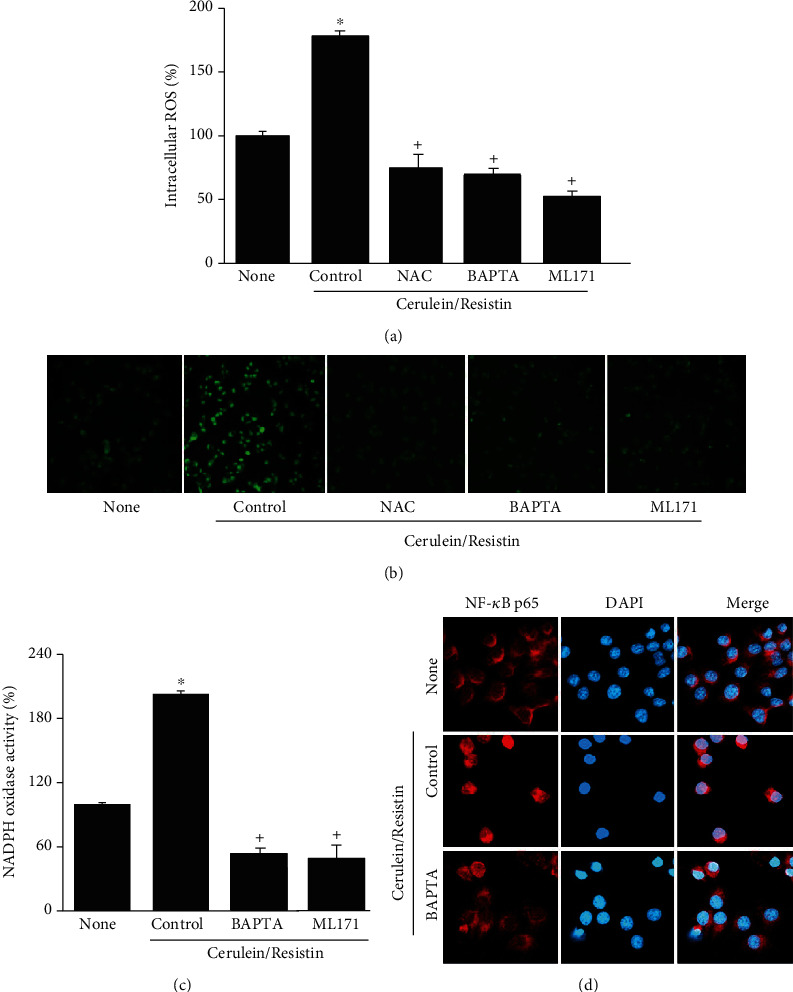
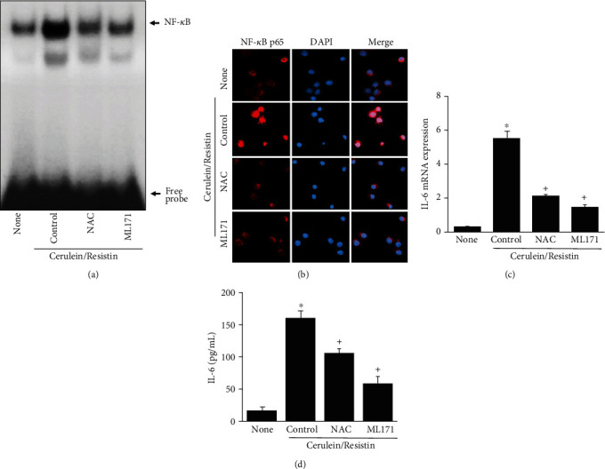
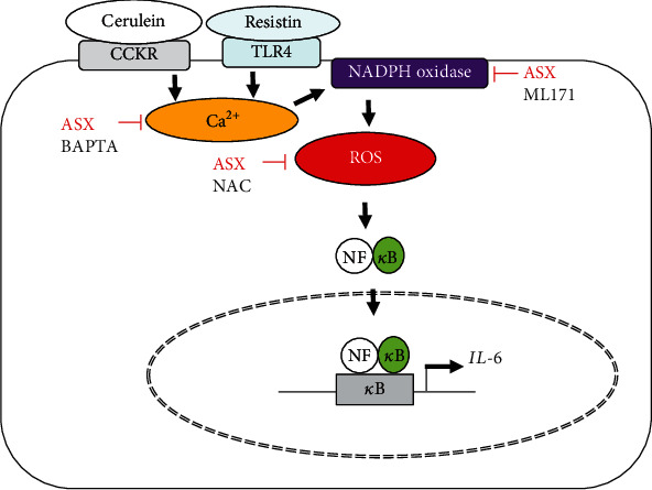
Acute pancreatitis is a common clinical condition with increasing the proinflammatory mediators, including interleukin-6 (IL-6). Obesity is a negative prognostic factor in acute pancreatitis. Obese patients with acute pancreatitis have a higher systemic inflammatory response rate. Levels of serum resistin, an adipocytokine secreted by fat tissues, increase with obesity. Cerulein, a cholecystokinin analog, induces calcium (Ca2+) overload, oxidative stress, and IL-6 expression in pancreatic acinar cells, which are hallmarks of acute pancreatitis. A recent study showed that resistin aggravates the expression of inflammatory cytokines in cerulein-stimulated pancreatic acinar cells. We aimed to investigate whether resistin amplifies cerulein-induced IL-6 expression and whether astaxanthin (ASX), an antioxidant carotenoid with anti-inflammatory properties, inhibits ceruelin/resistin-induced IL-6 expression in pancreatic acinar AR42J cells. We found that resistin enhanced intracellular Ca2+ levels, NADPH oxidase activity, intracellular reactive oxygen species (ROS) production, NF-κB activity, and IL-6 expression in cerulein-stimulated AR42J cells, which were inhibited by ASX in a dose-dependent manner. The calcium chelator BAPTA-AM inhibited cerulein/resistin-induced NADPH oxidase activation and ROS production. Antioxidant N-acetyl cysteine (NAC) and ML171, a specific NADPH oxidase 1 inhibitor, suppressed cerulein/resistin-induced ROS production, NF-κB activation, and IL-6 expression. In conclusion, ASX inhibits IL-6 expression, by reducing Ca2+ overload, NADPH oxidase-mediated ROS production, and NF-κB activity in cerulein/resistin-stimulated pancreatic acinar cells. Consumption of ASX-rich foods could be beneficial for preventing or delaying the incidence of obesity-associated acute pancreatitis.


Conflict of interest statement

The authors declare no conflict of interest.

Figures

Figure 1
Figure 1
Resistin enhances cerulein-induced reactive oxygen species (ROS) production and NADPH oxidase activation in AR42J cells. (a) The cells were stimulated with cerulein/resistin for the indicated times. (b–d) The cells were stimulated with cerulein with or without resistin for 45 min. (a, b) Intracellular ROS levels were measured by dichlorofluorescein diacetate (DCF-DA) fluorescence. ROS levels were expressed as the relative increase. (c) Representative images of ROS-induced fluorescence response of DCF-DA. (d) NADPH oxidase activity in membrane fractions was measured by the lucigenin assay. Data are expressed as the mean ± S.E. of three different experiments. The value for cells without cerulein stimulation in the absence of resistin treatment (none) is set as 100%. p < 0.05 vs. 0 min (a) or none (untreated cells; b, c); +p < 0.05 vs. cerulein (cells treated with cerulein alone).
Figure 2
Figure 2
Resistin enhances cerulein-induced Ca2+ overload and NF-κB activation in AR42J cells. (a) The cells were stimulated with cerulein in the presence or absence of resistin for the indicated times. Ca2+ level was determined by measuring the fluorescence changes of fluo-4 AM at excitation and emission wavelengths of 494 nm and 525 nm, respectively. Ca2+ levels were expressed as ∆F/F0, where F0 is the resting background fluorescence, and ∆F is fluorescence change over time after treatment with or without cerulein in the presence or absence of resistin. Fluorescence transient changes (obtained within 1-5 min) were plotted (A). (B) Ca2+ levels, expressed as ∆F/F0, of the cells. Data are expressed as the mean ± S.E. of three different experiments. Ca2+ level in none (untreated cells) was set as 1. p < 0.05 vs. 0 min (a) or none (untreated cells; b, c); +p < 0.05 vs. cerulein (cells treated with cerulein alone). (b) The cells were stimulated with cerulein/resistin for the indicated times. (c) The cells were stimulated with cerulein in the presence and absence of resistin for 1 h. NF-κB-DNA binding activity was determined by electrophoretic mobility shift assay (EMSA).
Figure 3
Figure 3
Resistin enhances the cerulein-induced IL-6 expression in AR42J cells. (a) The cells were stimulated with cerulein/resistin for the indicated times. (b, c) The cells were stimulated with cerulein in the presence and absence of resistin for 6 h (for mRNA level (b)) or 24 h (for protein level (c)). The mRNA level of IL-6 was determined by real-time PCR analysis and normalized to β-actin (a, b). The protein level of IL-6 in the media was determined by enzyme-linked immunosorbent assay (ELISA) (c). Data are expressed as the mean ± S.E. of three different experiments. p < 0.05 vs. 0 min (a) or none (untreated cells; b, c); +p < 0.05 vs. cerulein (cells treated with cerulein alone).
Figure 4
Figure 4
Astaxanthin (ASX) inhibits cerulein/resistin-induced increases in ROS, NADPH oxidase activity, and Ca2+ level in AR42J cells. The cells were pretreated with the indicated concentrations of ASX for 3 h and then stimulated with cerulein/resistin for 45 min (for ROS levels, ROS fluorescence imaging, and NADPH oxidase activity (a–c)), and for the indicated period (for Ca2+ level (d)). (a) Intracellular ROS levels were measured by dichlorofluorescein diacetate (DCF-DA) fluorescence. (b) Representative images of ROS-induced fluorescence response of DCF-DA. (c) NADPH oxidase activity was measured by the lucigenin assay. (d) Ca2+ level was determined by measuring the fluorescence changes of fluo-4 AM at excitation and emission wavelengths of 494 nm and 525 nm, respectively. Ca2+ levels were expressed as ∆F/F0, where F0 is the resting background fluorescence, and ∆F is fluorescence change over time after treatment with cerulein/resistin in the presence or absence of ASX. Fluorescence transient changes (obtained within 1-5 min) were plotted (A). (B) Ca2+ levels were expressed as ∆F/F0 of the cells. Data are expressed as the mean ± S.E. of three different experiments. ROS level, NADPH oxidase activity, and Ca2+ level in none (untreated cell) were set as 100 (a, c) or 1 (d). p < 0.05 vs. none (untreated cells); +p < 0.05 vs. cerulein (cells treated with cerulein alone).
Figure 5
Figure 5
Astaxanthin (ASX) inhibits cerulein/resistin-induced NF-κB activation and IL-6 expression in AR42J cells. The cells were pretreated with the indicated concentrations of ASX for 3 h and then stimulated with cerulein/resistin for 1 h (for NF-κB-DNA binding activity and nuclear level of NF-κB p65 by confocal images (a, b)), for 6 h (for IL-6 mRNA level (c)), and 24 h (for IL-6 protein level (d)). (a) NF-κB-DNA binding activity was determined by electrophoretic mobility shift assay (EMSA). (b) The immunoreactive NF-κB p65 was visualized using a rhodamine-conjugated mouse anti-rabbit IgG antibody (red) with DAPI counterstaining (blue) of the same field. (c) The mRNA expression level of IL-6 was determined by real-time PCR analysis and normalized to that of β-actin. (d) The protein level of IL-6 in the media was determined by enzyme-linked immunosorbent assay (ELISA). Data are expressed as the mean ± S.E. of three different experiments. p < 0.05 vs. none (untreated cells); +p < 0.05 vs. cerulein (cells treated with cerulein alone).
Figure 6
Figure 6
NAC, ML171, or BAPTA-AM inhibits cerulein/resistin-induced increases in ROS, NADPH oxidase activity, and nuclear translocation of NF-κB p65 in AR42J cells. The cells were pretreated with NAC (1 mM) or ML171 (2 μM) or BAPTA-AM (5 μM) for 1 h and then stimulated with cerulein/resistin for 45 min (for ROS levels, ROS fluorescence imaging, and NADPH oxidase activity (a–c)), and for 1 h (for nuclear level of NF-κB p65 by confocal images (d)). (a) Intracellular ROS levels were measured by dichlorofluorescein diacetate (DCF-DA) fluorescence. (b) Representative images of ROS-induced fluorescence response of DCF-DA. (c) NADPH oxidase activity in the membrane fraction was measured by the lucigenin assay. Data are expressed as the mean ± S.E. of three different experiments. ROS level and NADPH oxidase activity in none (untreated cells) were set as 100%. p < 0.05 vs. none (untreated cells); +p < 0.05 vs. cerulein (cells treated with cerulein alone). (d) The immunoreactive NF-κB p65 was visualized using a rhodamine-conjugated mouse anti-rabbit IgG antibody (red) with DAPI counterstaining (blue) of the same field.
Figure 7
Figure 7
NAC and BAPTA-AM inhibits cerulein/resistin-induced NF-κB activation and IL-6 expression in AR42J cells. The cells were pretreated with NAC (1 mM) or ML171 (2 μM) for 1 h and then stimulated with cerulein/resistin for 1 h (for NF-κB-DNA binding activity (a) and nuclear translocation of NF-κB p65 (b)), for 6 h (for IL-6 mRNA level (c)), and 24 h (for IL-6 protein level (d)). (a) NF-κB-DNA binding activity was determined by electrophoretic mobility shift assay (EMSA). (b) The immunoreactive NF-κB p65 was visualized using a rhodamine-conjugated mouse anti-rabbit IgG antibody (red) with DAPI counterstaining (blue) of the same field. (c) The mRNA expression level of IL-6 was determined by real-time PCR analysis and normalized to that of β-actin. (d) The protein level of IL-6 in the media was determined by enzyme-linked immunosorbent assay (ELISA). Data are expressed as the mean ± S.E. of three different experiments. p < 0.05 vs. none (untreated cells); +p < 0.05 vs. cerulein (cells treated with cerulein alone).
Figure 8
Figure 8
The proposed mechanism by which astaxanthin (ASX) inhibits interleukin-6 (IL-6) expression in cerulein/resistin-stimulated pancreatic acinar cells. Binding of cerulein to cholecystokinin receptor (CCKR) increases intracellular Ca2+ level while binding of resistin to Toll-like receptor 4 (TLR4) initiates Ca2+ overload. High level of Ca2+ activates NADPH oxidase to produce reactive oxygen species (ROS). ROS induce NF-κB activation and the expression of IL-6 in pancreatic acinar AR42J cells. ASX reduces Ca2+ overload and inhibits NADPH oxidase-mediated ROS production, NF-κB activation, and IL-6 expression. The calcium chelator BAPTA-AM, an antioxidant N-acetyl cysteine (NAC), and ML171, a specific NADPH oxidase 1 inhibitor, suppress cerulein/resistin-induced ROS production, NF-κB activation, and IL-6 expression in AR42J cells.

Similar articles

Cited by

References

    1. Boxhoorn L., Voermans R. P., Bouwense S. A., et al. Acute pancreatitis. Lancet. 2020;396(10252):726–734. doi: 10.1016/S0140-6736(20)31310-6. - DOI - PubMed
    1. Heckler M., Hackert T., Hu K., Halloran C. M., Büchler M. W., Neoptolemos J. P. Severe acute pancreatitis: surgical indications and treatment. Langenbeck's Archives of Surgery. 2021;406(3):521–535. doi: 10.1007/s00423-020-01944-6. - DOI - PMC - PubMed
    1. Bhatia M., Wong F. L., Cao Y., et al. Pathophysiology of acute pancreatitis. Pancreatology. 2005;5(2-3):132–144. doi: 10.1159/000085265. - DOI - PubMed
    1. Li N., Wang B.-M., Cai S., Liu P.-L. The role of serum high mobility group box 1 and interleukin-6 levels in acute pancreatitis: a meta-analysis. Journal of Cellular Biochemistry. 2018;119(1):616–624. doi: 10.1002/jcb.26222. - DOI - PubMed
    1. Zhang H., Neuhöfer P., Song L., et al. IL-6 trans-signaling promotes pancreatitis-associated lung injury and lethality. The Journal of Clinical Investigation. 2013;123(3):1019–1031. doi: 10.1172/JCI64931. - DOI - PMC - PubMed

MeSH terms

LinkOut - more resources